| Catalog No. | HB116036 |
| Host species | Humanized |
| Species reactivity | Human |
| Form | Liquid |
| Storage buffer | 0.01M PBS, pH 7.4. |
| Purity | >95% purity as determined by SDS-PAGE. |
| Clonality | Monoclonal |
| Isotype | IgG1-kappa |
| Applications | ELISA, Bioactivity: FACS, Functional assay, Research in vivo |
| Target | ERBB3, HER3, Proto-oncogene-like protein c-ErbB-3, Tyrosine kinase-type cell surface receptor HER3, Receptor tyrosine-protein kinase erbB-3 |
| Purification | Protein A/G purified from cell culture supernatant. |
| Endotoxin level | Please contact the lab for this information. |
| Expression system | Mammalian Cells |
| Accession | P21860 |
| Stability and Storage | Use a manual defrost freezer and avoid repeated freeze-thaw cycles. Store at 4°C for short-term storage (1-2 weeks). Store at -20°C for up to 12 months. For long-term storage, store at -80°C. |
| Alternative name | MEHD7945A, RG-7597, 1314238-96-4 |
| Note | For research use only. Not suitable for clinical or therapeutic use. |
Research Grade Duligotuzumab
Overview
Images
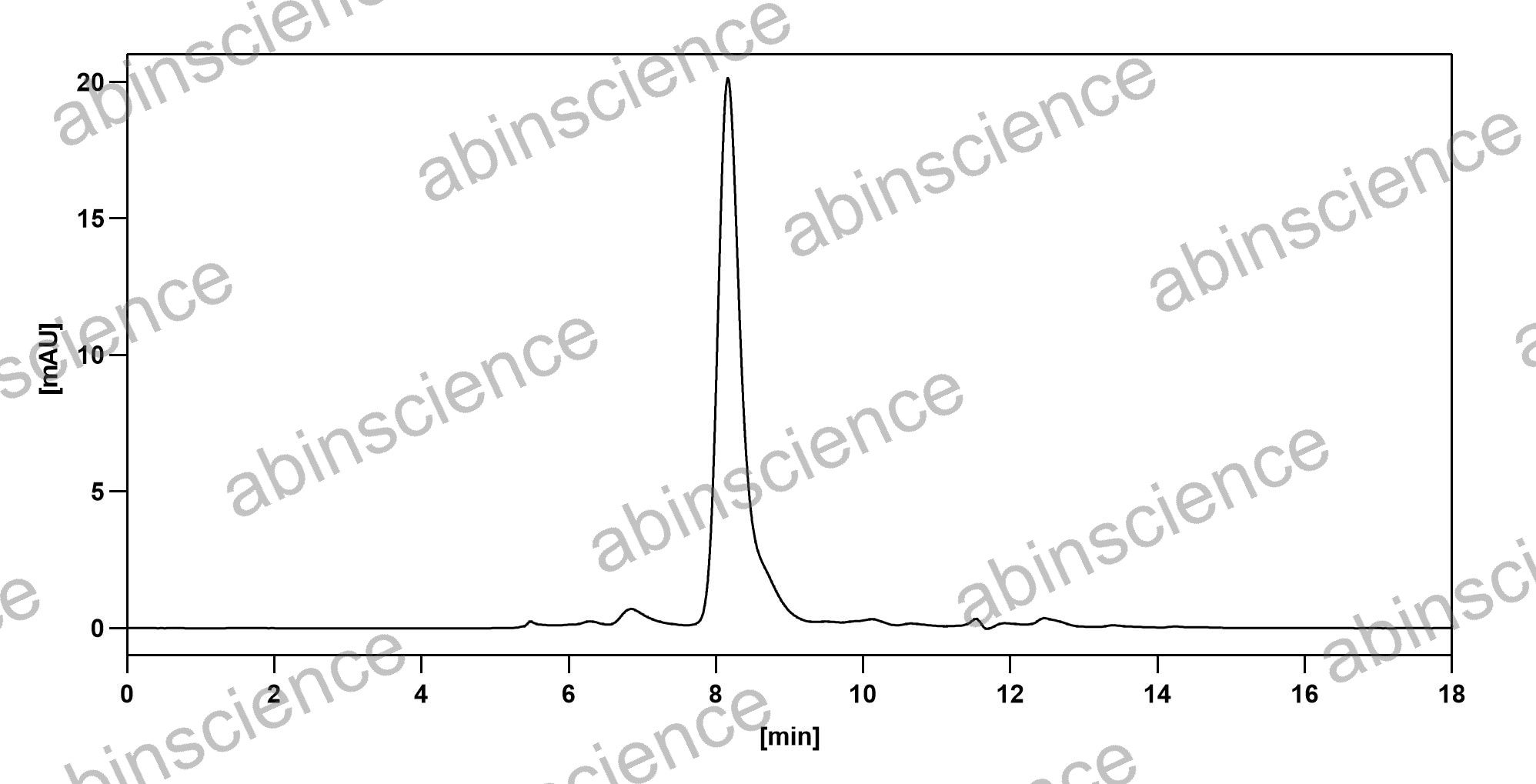 Bioactivity |
SEC-HPLC detection for Research Grade Duligotuzumab. |
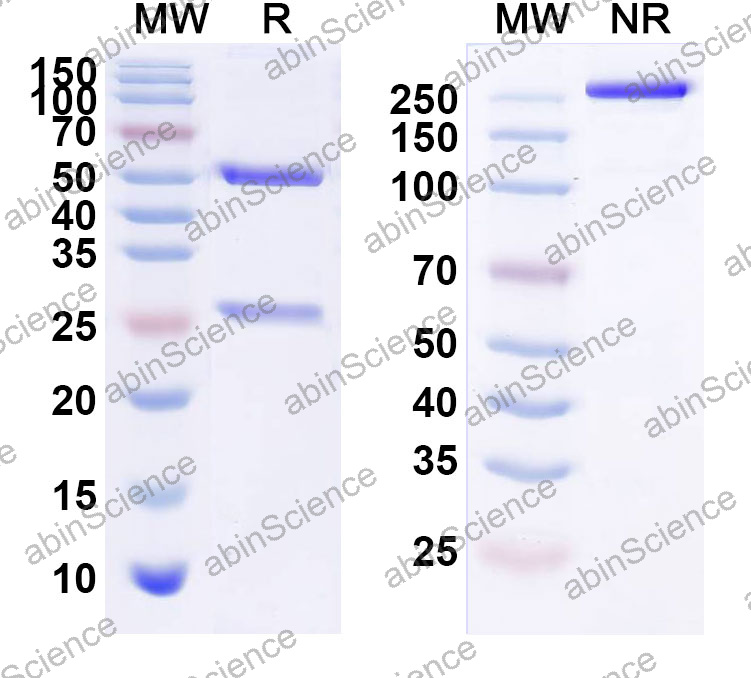 SDS-PAGE |
SDS-PAGE for Research Grade Duligotuzumab. |
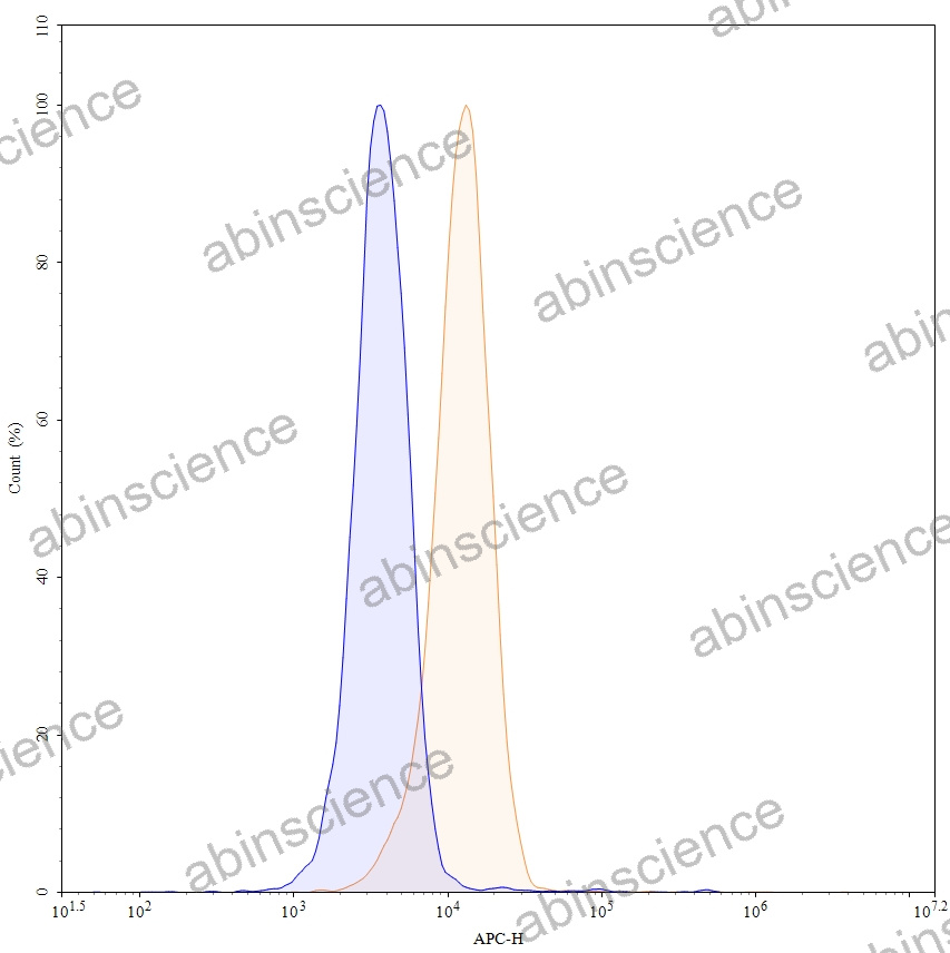 Flow CytoMetry |
Flow-cytometry using APC anti-human ERBB3 antibody. MCF-7 cells were stained with an irrelevant antibody (Blue Histogram) or an APC anti-human ERBB3 monoclonal antibody (Catalog: HB116036, Yellow Histogram) at a concentration of 5 µg/ml for 30 mins at RT. After washing, and cells analysed on a NovoCyte Flow Cytometer. |
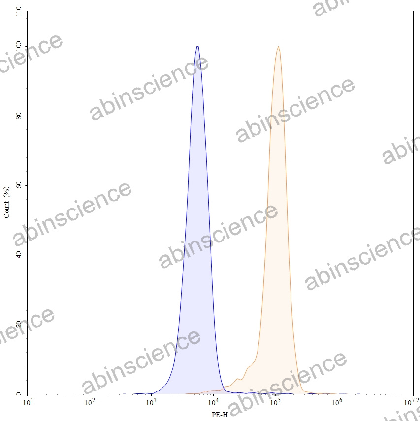 Flow CytoMetry |
Flow-cytometry using PE anti-human ERBB3 antibody. MCF-7 cells were stained with an irrelevant antibody (Blue Histogram) or an PE anti-human ERBB3 monoclonal antibody (Catalog: HB116036, Yellow Histogram) at a concentration of 5 µg/ml for 30 mins at RT. After washing, and cells analysed on a NovoCyte Flow Cytometer. |
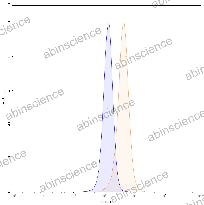 Flow CytoMetry |
Flow-cytometry using FITC anti-human ERBB3 antibody. HeLa cells were stained with an irrelevant antibody (Blue Histogram) or an FITC anti-human ERBB3 monoclonal antibody (Catalog: HB116036, Yellow Histogram) at a concentration of 5 µg/ml for 30 mins at RT. After washing, and cells analysed on a NovoCyte Flow Cytometer. |
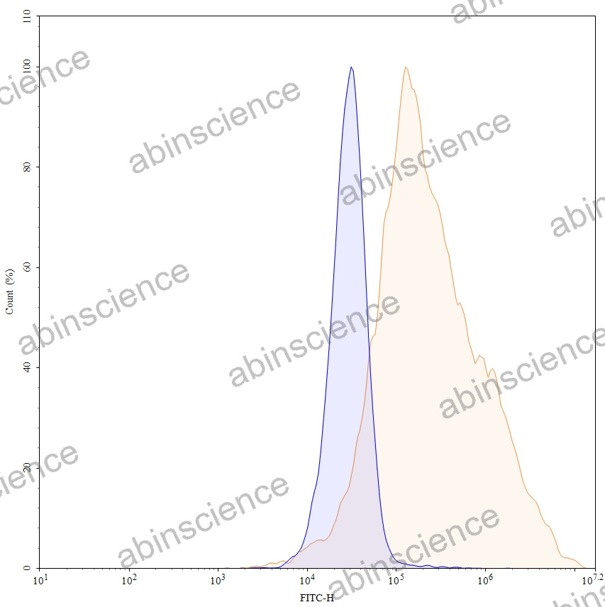 Flow CytoMetry |
Flow-cytometry using anti-human ERBB3 antibody. Untransfected cells (blue Histogram) and Transfected cells (Yellow Histogram) were stained with an anti-human ERBB3 monoclonal antibody (Catalog: HB116036) at a concentration of 5 µg/ml for 30 mins at RT. After washing, bound antibody was detected using a Goat Anti-Human IgG H&L Polyclonal Antibody, FITC (abinScience: HF690414) and cells analysed on a NovoCyte Flow Cytometer. |