| Catalog No. | HY515010 |
| Host species | Human |
| Species reactivity | Human |
| Form | Liquid |
| Storage buffer | 0.01M PBS, pH 7.4. |
| Purity | >95% as determined by SDS-PAGE. |
| Clonality | Monoclonal |
| Isotype | IgG1, kappa |
| Applications | ELISA, FCM, Neutralization |
| Target | CSF1, Macrophage colony-stimulating factor 1, CSF-1, Lanimostim, M-CSF, MCSF |
| Purification | Protein A/G purified from cell culture supernatant. |
| Endotoxin level | Please contact with the lab for this information. |
| Accession | P09603 |
| Stability and Storage | Use a manual defrost freezer and avoid repeated freeze-thaw cycles. Store at 4°C short term (1-2 weeks). Store at -20°C 12 months. Store at -80°C long term. |
| Clone ID | Iv0052 |
| Note | For research use only. Not suitable for clinical or therapeutic use. |
InVivoMAb Anti-Human CSF1/M-CSF (Iv0052)
Overview
Description
InVivoMAb Anti-Human CSF1/M-CSF (Iv0052) [Iv0052] (HY515010) is a human monoclonal antibody detecting CSF1 in ELISA, FCM, Neutralization. Suitable for Human.
Highlights
- ●Multi-Application — Validated across multiple applications.
- ●Flow Cytometry — Validated for flow cytometry applications.
Images
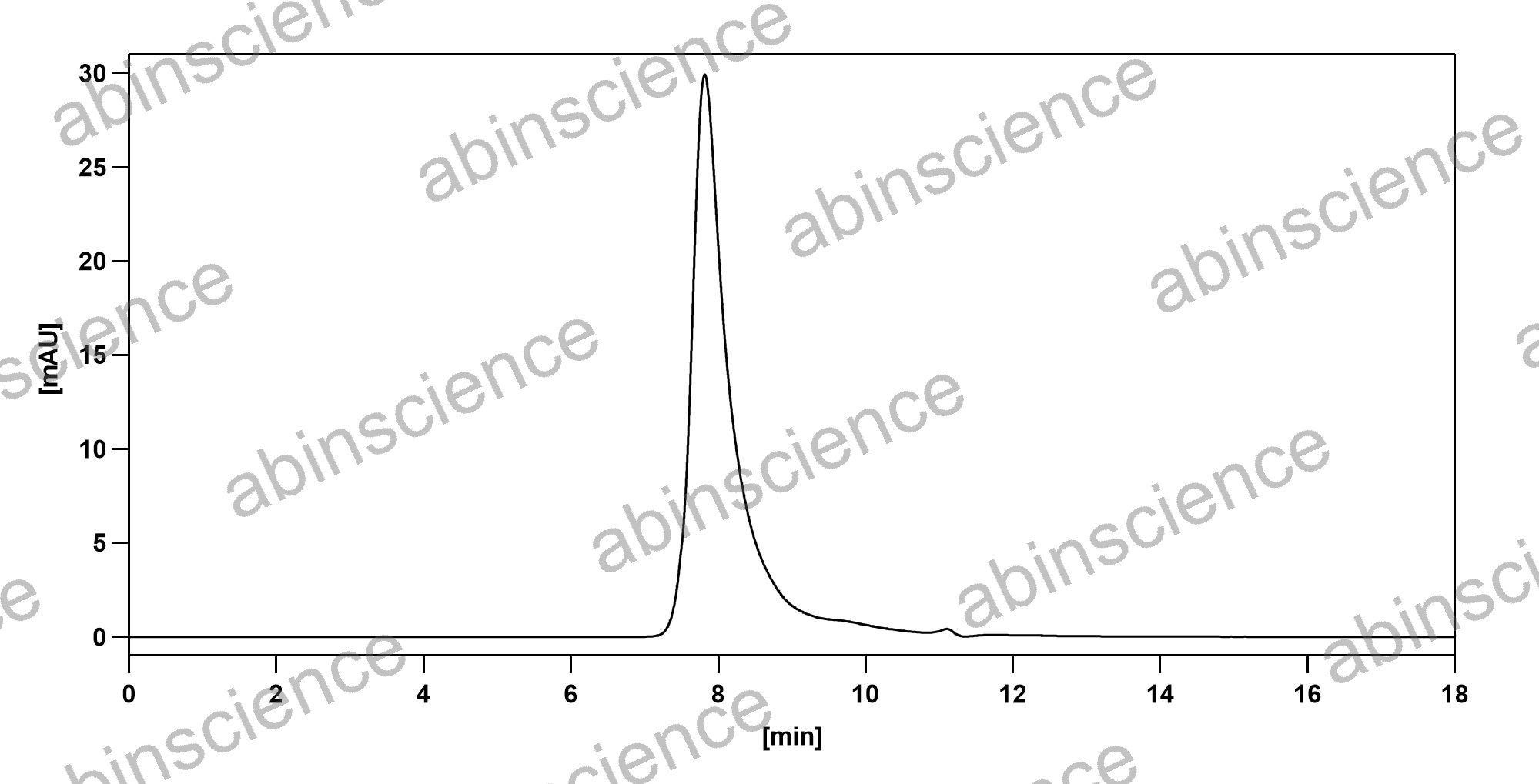 Bioactivity |
SEC-HPLC detection for InVivoMAb Anti-Human CSF1/M-CSF (ABV0114). |
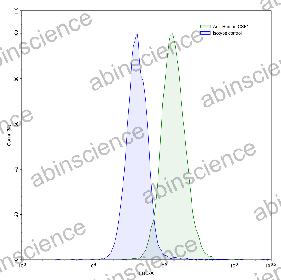 Flow Cytometry |
Flow-cytometry using anti-human CSF1 antibody.MG-63 cells were stained with an irrelevant antibody (Blue Histogram) or an anti-human CSF1 antibody monoclonal antibody (Catalog # HY515010 ,Green Histogram) at a concentration of 5 µg/ml for 30 mins at RT. After washing, bound antibody was detected using a FITC conjugated goat anti-human antibody (Catalog # HF690414) and cells analysed on a NovoCyte Flow Cytometer. |
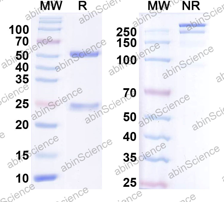 SDS-PAGE |
SDS-PAGE for InVivoMAb Anti-Human CSF1/M-CSF (Iv0052). |
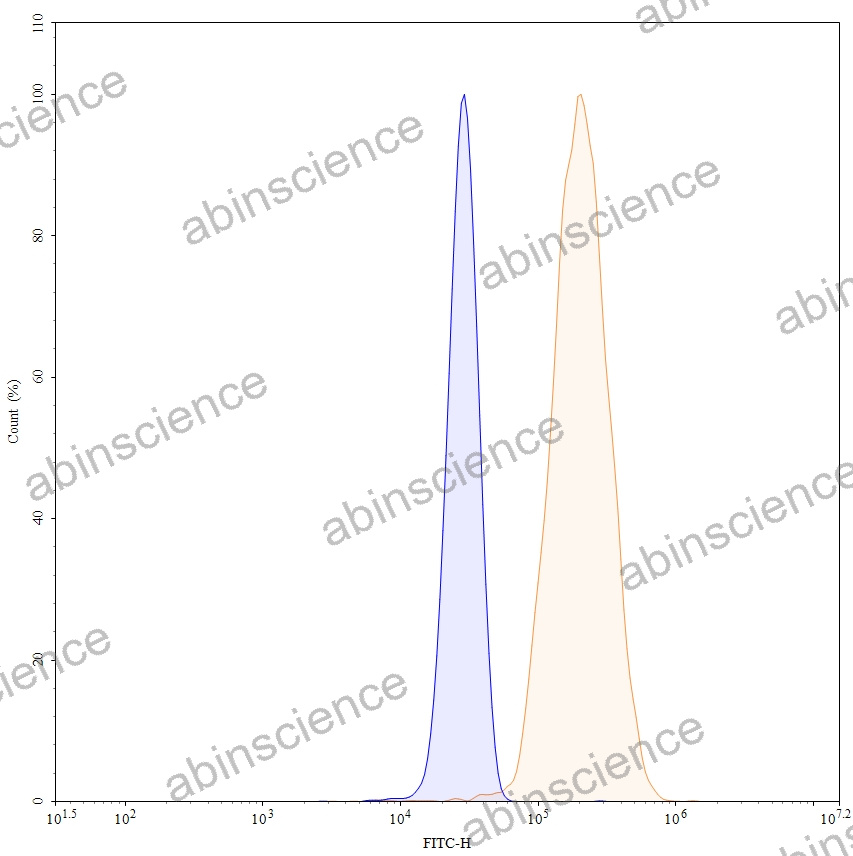 Flow CytoMetry |
Flow-cytometry using FITC anti-human CSF1 antibody. MG-63 cells were fixed and permeabilized, then stained with an irrelevant antibody (Blue Histogram) or an FITC anti-human CSF1 monoclonal antibody (Catalog: HY515010, Yellow Histogram) at a concentration of 5 µg/ml for 30 mins at RT. After washing, cells analysed on a NovoCyte Flow Cytometer. |
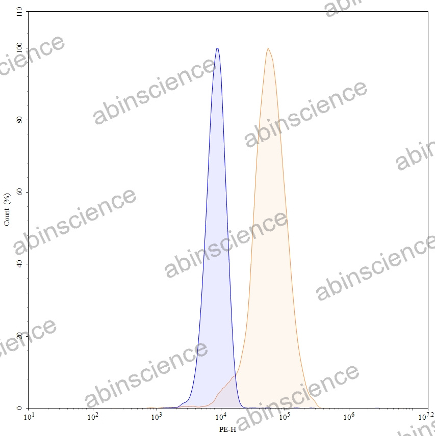 Flow CytoMetry |
Flow-cytometry using PE anti-human CSF1 antibody. MG-63 cells were fixed and permeabilized, then stained with an irrelevant antibody (Blue Histogram) or an PE anti-human CSF1 monoclonal antibody (Catalog: HY515010, Yellow Histogram) at a concentration of 5 µg/ml for 30 mins at RT. After washing, cells analysed on a NovoCyte Flow Cytometer. |
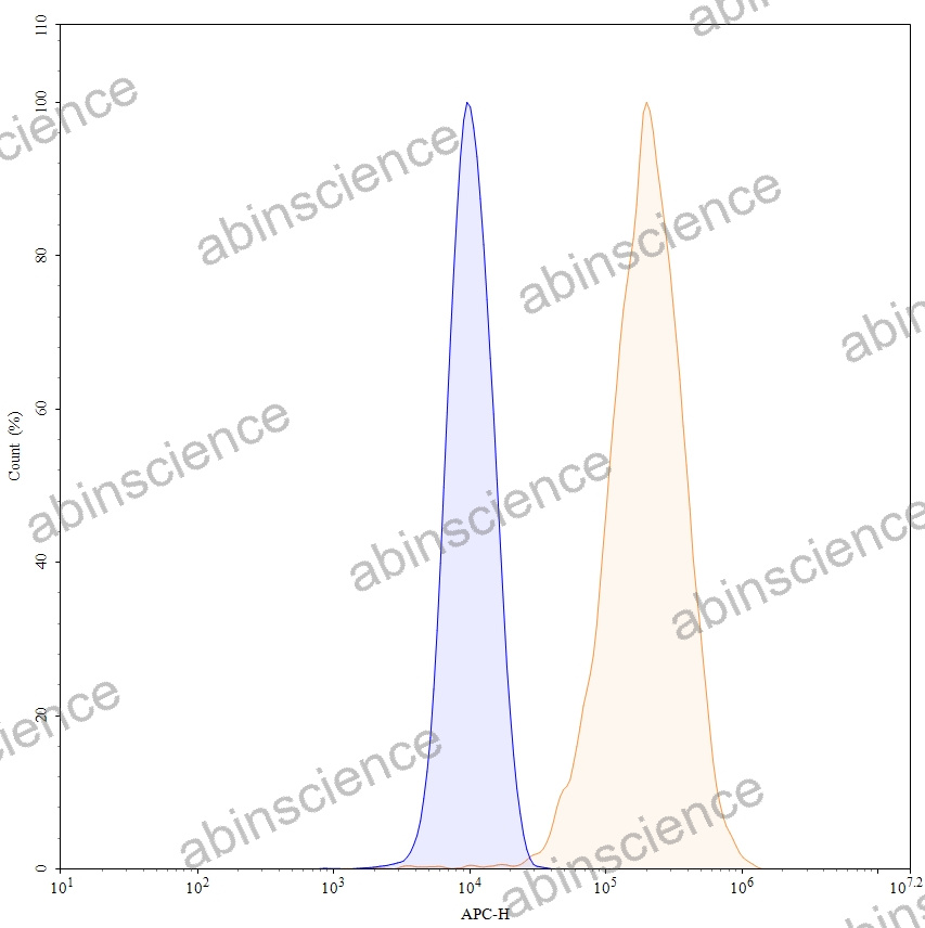 Flow CytoMetry |
Flow-cytometry using APC anti-human CSF1 antibody. MG-63 cells were fixed and permeabilized, then stained with an irrelevant antibody (Blue Histogram) or an APC anti-human CSF1 monoclonal antibody (Catalog: HY515010, Yellow Histogram) at a concentration of 5 µg/ml for 30 mins at RT. After washing, cells analysed on a NovoCyte Flow Cytometer. |