| Catalog No. | HF998014 | ||||||||
| Host species | Rabbit | ||||||||
| Species reactivity | Human, Cercocebus atys, Chlorocebus aethiops, Erythrocebus patas, Macaca fascicularis, Macaca fuscata fuscata, Macaca mulatta, Macaca nemestrina, Pan troglodytes | ||||||||
| Immunogen | E. coli - derived recombinant Human CD4 (Lys26-Trp390). | ||||||||
| Form | Liquid | ||||||||
| Storage buffer | 0.01M PBS, pH 7.4, 50% Glycerol, 0.05% Proclin 300. | ||||||||
| Clonality | Polyclonal | ||||||||
| Isotype | IgG | ||||||||
| Applications | ELISA, IHC, WB | ||||||||
| Target | T-cell surface antigen T4/Leu-3, T-cell surface glycoprotein CD4, CD4 | ||||||||
| Purification | Purified by antigen affinity column. | ||||||||
| Accession | P01730 | ||||||||
| Stability and Storage | Use a manual defrost freezer and avoid repeated freeze thaw cycles. Store at 2 to 8°C for frequent use. Store at -20 to -80°C for twelve months from the date of receipt. | ||||||||
| Product Usage Information |
|
||||||||
| Note | For research use only. |
Anti-CD4 Polyclonal Antibody
Overview
Images
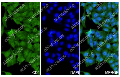 Immunofluorescence |
HepG2 cells were fixed with 4% paraformaldehyde for 20 minutes, followed by blocking with 5% goat serum for 1 hour. The cells were then incubated with a primary antibody targeting CD4 (HF998014) at a concentration of 10.6 ug/ml overnight at 4°C. Afterward, the cells were incubated with a secondary antibody, Goat Anti-Rabbit IgG (Alexa Fluor-488) for 50 minutes at room temperature. The localization of CD4 was visualized (shown in green), and nuclear DNA was stained with DAPI (shown in blue). |