Challenges of Flow Cytometry in Analyzing Rare Samples
Flow cytometry is an indispensable tool in the diagnosis of hematological diseases and clinical immunology research, enabling precise “identity identification” of cells. However, when dealing with rare samples such as cerebrospinal fluid or fine needle aspirates, the sensitivity and accuracy of traditional techniques often fall short. The innovative 10-color, 15-antibody flow cytometry panel proposed by OMIP-010 overcomes the limitations of conventional methods, providing a precise diagnostic tool for rare samples.
Sophisticated Fluorescence Channel Design
OMIP-010 does not simply aim to stack parameters but employs a unique “complementary” fluorescence sharing strategy to maximize the use of each fluorescence channel. This approach allows for more cellular information to be captured within the limited flow cytometry detection channels. Not only does this enhance analytical precision, but it also ensures sufficient diagnostic information can be obtained from minimal cell samples.
High Reliability and Clinical Validation
This panel has undergone rigorous clinical validation, ensuring stable signals and a high signal-to-noise ratio even in low-cell-number samples, addressing the challenge of insufficient cell numbers that previously hindered effective analysis. The innovative design and validation of OMIP-010 provide reliable support for clinical applications.
Traditional flow cytometry techniques face numerous challenges when processing low-cell-count samples, particularly in rare samples such as cerebrospinal fluid and bone marrow, where the number of cells often fails to generate sensitive detection signals. The OMIP-010 study, published in Cytometry Part A, introduces an innovative 10-color, 15-antibody flow cytometry panel, whose sophisticated design and rigorous validation system have led to a breakthrough in the precise diagnosis of small sample sizes.
1. OMIP-010 Panel
2. Gate Logic
1
T Cell Subpopulation Selection
Exclude cell debris, dead cells, and non-target cells. Gate the T cell population, then use CD4 and CD8 markers to isolate CD4+ T cells and CD8+ T cells.
2
Functional Analysis
Analyze cytokine secretion in T cells using markers for IFN-γ, IL-2, and TNF.
3
B Cell and Progenitor Cell Analysis
Analyze B cell subsets using markers CD15, CD19, CD20, and Ig kappa/Ig lambda. Assess progenitor cell populations using CD34 and CD117 markers.
4
NK and NKT Cell Analysis
Analyze the distribution and function of NK and NKT cells using the CD56 marker.
3. Experimental Results
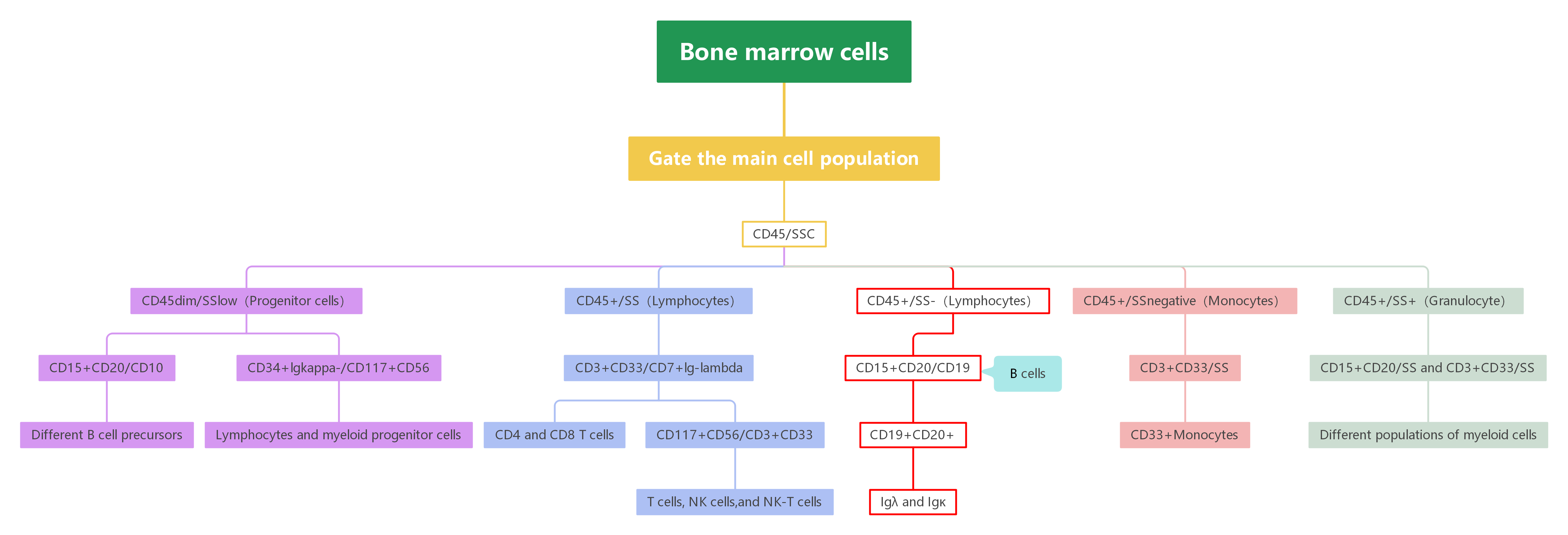
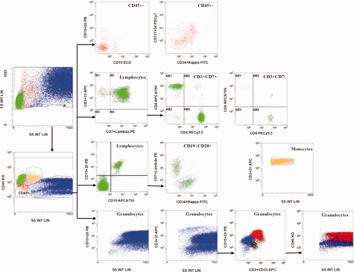
Excluding Cell Debris Using FS/SS Plot, Gating Cell Populations Using CD45/SS:
- CD45dim/SSlow (Progenitor Cells):
- CD15+CD20/CD10: Different B cell progenitors.
- CD34+Ig kappa-/CD117+CD56: Lymphoid and myeloid progenitor cells.
- CD45+/SS- (Lymphocytes):
- CD3+CD33/CD7+Ig lambda: T cell subsets.
- Further, within CD3+CD33, gate for CD4+ T and CD8+ T cells.
- CD117+CD56/CD3+CD33: T cells, NK cells, and NKT cells.
- CD45+/SS- (Lymphocytes):
- CD15+CD20/CD19: B cells.
- Further, within CD19+CD20+, analyze Ig lambda and Ig kappa B cell subsets.
- CD45+/SSdim (Monocytes):
- CD3+CD33/SS: CD33++ monocytes.
- CD45±/SS+ (Granulocytes):
- CD15+CD20/SS and CD3+CD33/SS: Different myeloid populations.
4. Panel Interpretation
4.1 “Complementary” Fluorescence Channel Sharing: Maximizing Information in One Tube
OMIP-010 overcomes the challenge of low cell numbers by introducing a “complementary” fluorescence sharing strategy. Instead of the common “exclusion” method, it uses the same fluorophore for mutually exclusive markers, such as CD34 (hematopoietic progenitors) and Ig kappa (B cell clonality) with FITC, and CD7 (T/NK cells) and Ig lambda (B cell light chain) with PE. This allows for 15 parameters to be detected in a single tube without adding extra fluorescence channels, revolutionizing small sample diagnostics, especially for leukemia and lymphoma.
4.2 Clinical Validation and High Reliability
OMIP-010 was rigorously validated, ensuring non-specific binding of shared antibody combinations was below 1%. Detailed titration experiments optimized signal-to-noise ratios, ensuring that all 15 antibodies could function together effectively, even in low-cell-number samples, allowing accurate detection with minimal interference.
4.3 Clinical Impact: Overcoming Traditional Limitations
OMIP-010 addresses the challenges of low-cell-number samples (e.g., cerebrospinal fluid, bone marrow) that traditional methods struggle to analyze. Its innovative design enables effective detection of aberrant cells, offering sensitivity equal to or greater than traditional methods for these rare samples.
5. Applications
Rare Sample Immune Profiling Precision Diagnosis of Blood Disorders B Cell and T Cell Function Profiling Immune Therapy Response Monitoring Autoimmune Disease Immune Profiling Chronic Infection Immune Response Monitoring
6. Conclusion
The essence of the OMIP-010 panel lies in its “design ingenuity” to break through the physical limitations of technology. Faced with the challenge of small sample sizes, it does not focus on increasing detection channels but rethinks how to more efficiently utilize each existing channel. Its “complementary” fluorescence sharing strategy is essentially a resource optimization algorithm, cleverly integrating information to maximize the value extracted from limited resources.
Get OMIP-010 Compatible Flow Cytometry Antibodies
abinScience provides validated Flow Cytometry Antibodies covering key targets in this panel, supporting your Immunophenotyping research
References
[1] Preijers, F.W.M.B., Huys, E. and Moshaver, B. (2012), OMIP-010: A new 10-color monoclonal antibody panel for polychromatic immunophenotyping of small hematopoietic cell samples. Cytometry, 81A: 453-455.

About Us
As a strategic venture of AtaGenix (established 2011), abinScience was founded in 2023 to deliver premium life science reagents that accelerate discovery. Our flow cytometry antibody products cover commonly used detection markers, with a wide variety to meet the research needs of multiple species (Human/Mouse/Rat/Dog/Hamster/Monkey, etc.). We provide stable and reliable support for scientific research.
Explore abinScience Flow Cytometry Antibodies

 中文
中文 English
 한국어
한국어 日本語
日本語 Español
 Français
 Русский