OMIP-022 addresses the core need in tuberculosis (TB) vaccine and infectious immunity research: the comprehensive evaluation of antigen-specific T cell responses. This assay integrates a panel of functionally relevant markers into a single multicolor flow cytometry panel, encompassing indicators for intracellular bacterial clearance (IFN-γ, TNF), cytotoxicity (CD107a), T cell activation and persistence (CD154, IL-2), and mucosal defense (IL-17A, IL-22). Coupled with classic memory differentiation markers (CD45RO, CCR7), it enables one-stop, single-cell resolution of T cell "functional output" and "differentiation status" upon TB antigen stimulation.
Compared to earlier OMIP protocols, OMIP-022 stands out for its thoughtful "technical adaptation" to the unique challenges of clinical samples (e.g., PBMCs) and the detection of low-abundance cytokines. Rather than a simple marker combination, it maximizes signal-to-noise ratio and minimizes compensation errors from the outset through strategies such as channel integration (combining Live/Dead, CD14, and CD19 in the V500 channel), scientific fluorochrome pairing (selecting high-brightness PE-Cy7 for TNF and low-background V450 for IFN-γ), and proactive resolution of spectral overlap (swapping fluorochromes for CD8 and IL-17A). These optimizations ensure the reliability and reproducibility of data in complex clinical samples.
OMIP-022 provides a validated, standardized framework for evaluating multidimensional antigen-specific T cell responses. Its biology-driven marker selection logic and data quality-focused optimization strategies offer a high-quality methodological template that can be directly adapted for T cell function assays in research on other intracellular pathogens (e.g., HIV, Listeria), tumor immunomonitoring, and even autoimmune diseases. This significantly enhances data comparability and conclusion reliability across different studies.
OMIP-022 establishes a specialized assay for antigen-specific T cells in Mycobacterium tuberculosis infection. Through mechanism-guided marker selection and sample-adapted technical optimizations, it delivers a standardized experimental framework for TB immunology research and clinical diagnostic evaluation. By translating general high-dimensional flow cytometry design principles into a robust, practical tool for TB vaccine evaluation and infectious immunity research—via channel integration, targeted fluorochrome matching, and proactive spectral overlap resolution—OMIP-022 provides a reproducible roadmap for developing robust multicolor flow cytometry panels in complex disease settings.
| Target | Fluorochrome | Function | abinScience Recommendation |
|---|---|---|---|
| Live/Dead | ECD | Exclude dead cells, B cells, and monocytes | — |
| CD14 | V500 | View CD14 antibody | |
| CD19 | V500 | View CD19 antibody | |
| CD3 | ECD | T cell lineage marker | View CD3 antibody |
| CD4 | APC-eFluor 780 | View CD4 antibody | |
| CD8 | Alexa Fluor 700 | View CD8 antibody | |
| CCR7 | BV605 | Memory cell marker | View CCR7 antibody |
| CD45RO | BV785 | View CD45RO antibody | |
| IFN-γ | V450 | Cytokines | View IFN-γ antibody |
| IL-2 | PE | View IL-2 antibody | |
| TNF | PE-Cy7 | View TNF antibody | |
| IL-17A | PerCP-Cy5.5 | View IL-17A antibody | |
| IL-22 | APC | View IL-22 antibody | |
| CD107a | Alexa Fluor 488 | Degranulation marker | View CD107a antibody |
| CD154 | PE-Cy5 | Activation/B-Cell Help | View CD154 antibody |
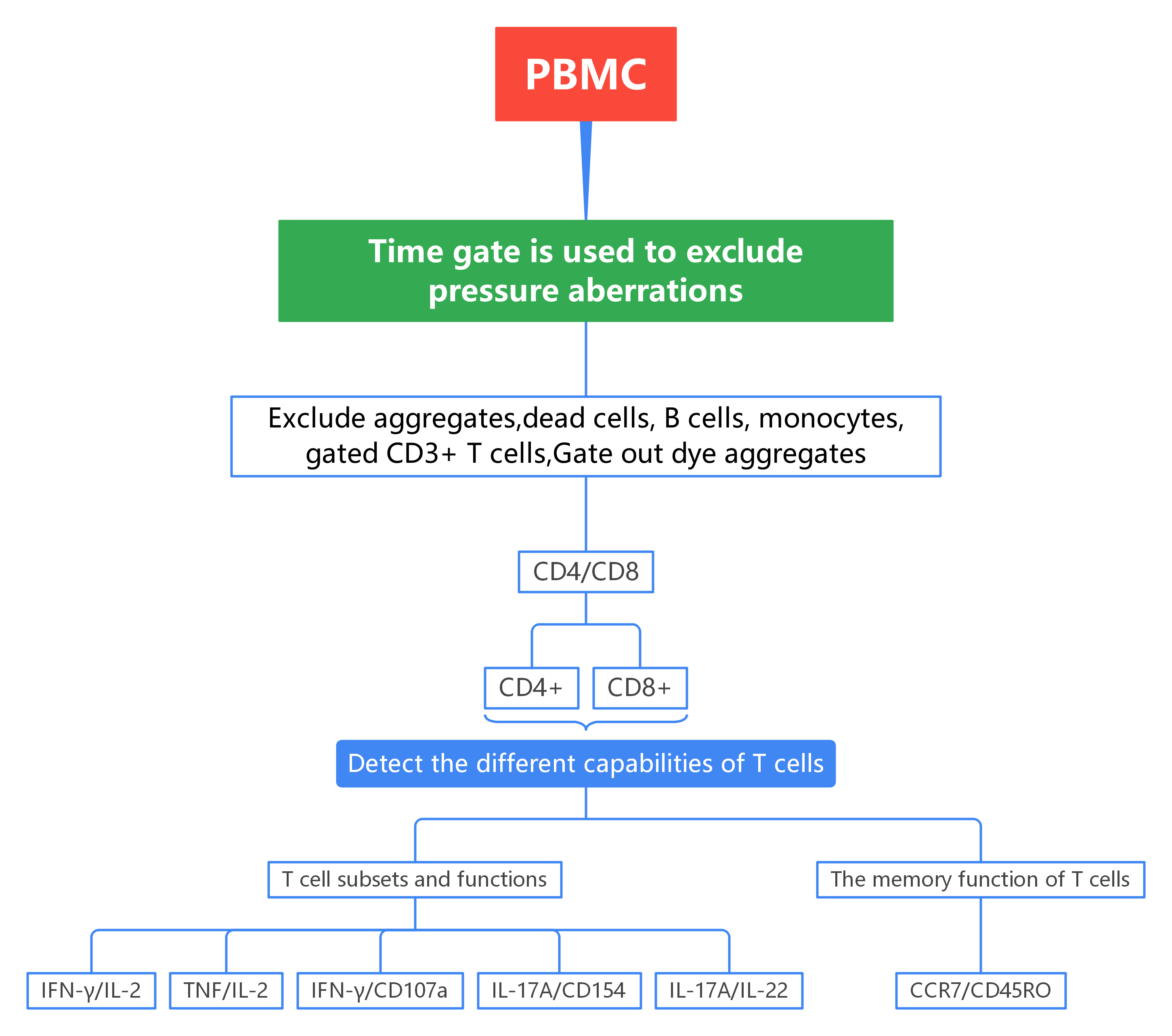
Figure 1. Overview of OMIP-022Gating Strategy
Exclude dead cells, B cells, monocytes, and dye aggregates; gate on viable CD3+ T cells, followed by gating on CD4+ and CD8+ T cell subsets.
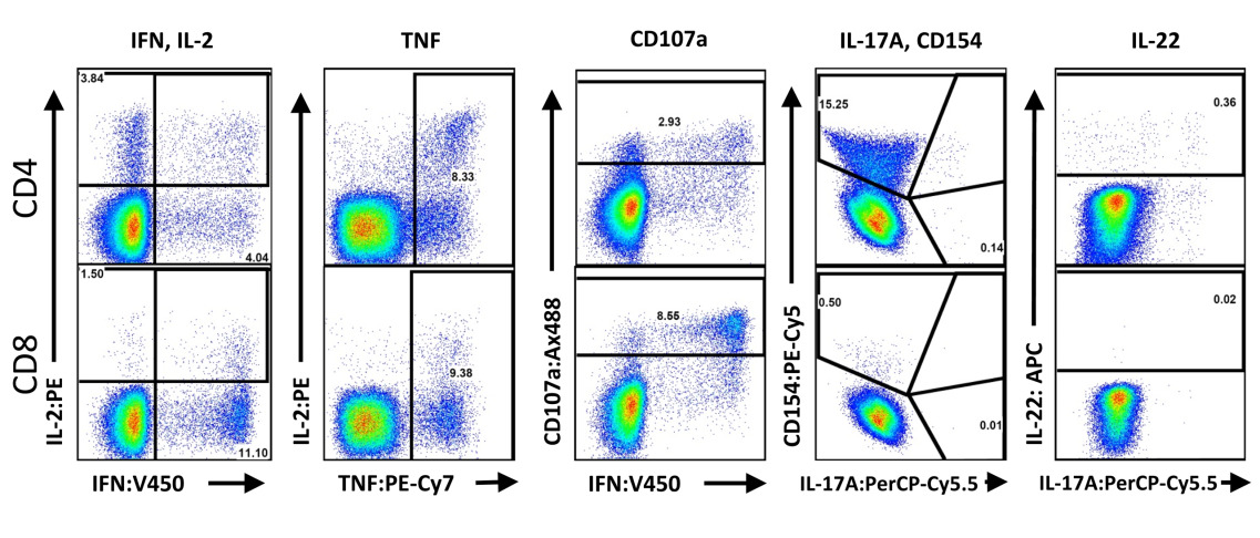
Evaluate T cell function using IFN-γ, IL-2, TNF, IL-17A, IL-22, CD107a, and CD154.
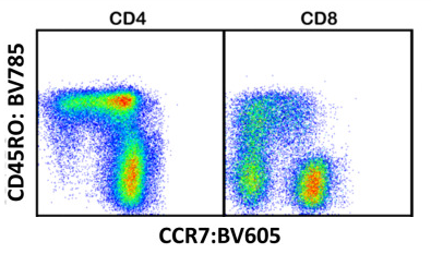
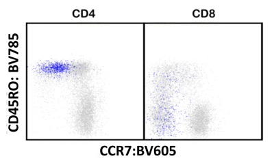
Characterize T cell memory phenotypes using CD45RO and CCR7.
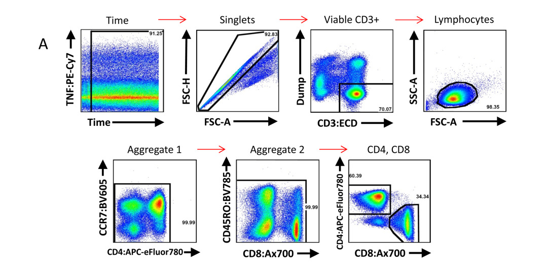
1). Exclude signals from unstable fluid flow using a Time gate; subsequently exclude dead cells, B cells, monocytes, and dye aggregates. Gate on viable CD3+ T cells, then gate CD4+ and CD8+ T cell subsets.
2). Analyze functional subpopulations of CD4+ and CD8+ T cells.
3). Characterize memory phenotypes of CD4+ and CD8+ T cells.
4). Distribution of memory phenotypes in CD4+ and CD8+ T cells (gray) versus all IFN-γ-producing cells (blue) following PBMC stimulation with CMV pp65 peptide pool.
4.1 Adhering to the fundamental flow cytometry principle of "marker selection driven by experimental objectives," focusing on core TB detection needs
Purpose-driven inclusion of core indicators: In flow cytometry design, core markers must be directly aligned with research goals. Targeting the core requirements of TB immunity—intracellular bacterial clearance, inflammatory recruitment, and mucosal defense—OMIP-022 incorporates classic functional and pathway markers: Th1 pathway (IFN-γ, TNF; key pathways for TB immune clearance), proliferation indicator (IL-2; T cell response persistence), activation marker (CD154; CD4+ T cell-specific response), and degranulation marker (CD107a; cytotoxicity associated with intracellular bacterial infection). Additionally, it introduces TB immunity-relevant Th17 (IL-17A) and Th22 (IL-22) pathway markers, ensuring full alignment between indicators and experimental objectives such as TB vaccine evaluation and infectious immune response analysis.
4.2 Implementing the core flow cytometry principle of "fluorochrome-channel optimization" to maximize data quality
"Minimizing spectral compensation and optimizing fluorochrome-channel pairing" is the cornerstone of flow cytometry panel design, aimed at reducing signal interference and improving data accuracy. OMIP-022 achieves maximum data quality through channel selection, fluorochrome adaptation, and spectral overlap resolution:
• Channel Optimization: Following the logic of "integrating non-core functions" in flow channel design, OMIP-022 combines "viable cell selection (ViViD viability dye)" and "non-T cell exclusion (CD14, CD19)" into a single V500 channel. This integration preserves the basic function of eliminating dead cells and non-target cells while freeing up violet laser channels, providing interference-free detection space for low-abundance core indicators such as IL-17A and IL-22, and reducing cross-compensation pressure across multiple channels at the source.
• Fluorochrome Pairing: Guided by the principle of "matching brightness, background, and stability," OMIP-022 selects high-brightness, photostable PE-Cy7 for the low-abundance core factor TNF (replacing low-brightness FITC to amplify weak response signals). For IFN-γ, it replaces high-background BV421 with low-background V450 to balance sensitivity and specificity. The degranulation marker CD107a is substituted with more stable Alexa Fluor 488 to ensure consistency in frozen sample detection—all choices tailored to the characteristics of TB samples.
• Spectral Overlap Resolution: Addressing the core need of "minimizing compensation," OMIP-022 eliminates severe compensation interference from highly expressed CD8 (originally PerCP-Cy5.5) on low-expressed IL-17A (originally Alexa Fluor 700) through a "fluorochrome swap" strategy: CD8 is switched to Alexa Fluor 700, and IL-17A to PerCP-Cy5.5. Additionally, CD45RO is paired with BV785 based on the "minimum spectral overlap" principle to further reduce compensation requirements.
The value of OMIP-022 lies in providing a validated, optimized design example. It demonstrates how to combine functional and phenotypic markers based on a clear scientific question (TB immunity) and resolve common challenges in multicolor flow cytometry—such as compensation interference and background noise—through detailed optimizations in fluorochrome pairing and reagent selection. This enables more reliable data acquisition from clinical samples, offering direct reference for designing similar high-dimensional flow cytometry panels.
abinScience provides validated flow cytometry antibodies covering key targets in this panel, supporting your T cell function and memory research
[1] Graves AJ, Padilla MG, Hokey DA. OMIP-022: Comprehensive assessment of antigen-specific human T-cell functionality and memory. Cytometry A. 2014 Jul;85(7):576-9.

As a strategic venture of AtaGenix (established 2011), abinScience was founded in 2023 to deliver premium life science reagents that accelerate discovery. Our flow cytometry antibody products cover commonly used detection markers, with a wide variety to meet the research needs of multiple species (Human/Mouse/Rat/Dog/Hamster/Monkey, etc.). We provide stable and reliable support for scientific research.
+86-027-65523339
中国武漢市深敦寺路666号C棟、武漢、430206


 中文
中文 English
 한국어
한국어 日本語
日本語 Español
 Français
 Русский