In flow cytometry, fixation is a critical step across numerous experimental workflows. Whether utilized to preserve cellular states, extend sample processing windows, or facilitate the detection of intracellular proteins, transcription factors, and cytokines, fixation is widely relied upon in immunological and cell biology research.
However, researchers frequently encounter a perplexing issue during routine assays: A given marker yields crisp, robust staining in unfixed cells, but its fluorescence intensity drops dramatically post-fixation. Alternatively, when detecting the same marker using different antibody clones, post-fixation performance varies significantly.
This phenomenon is often prematurely dismissed as "poor antibody quality" or "operator error". In reality, the fixation process itself simultaneously impacts multiple parameters:
Consequently, a shift in post-fixation signal strength does not necessarily indicate a change in target protein expression. Rather, it represents the combined biochemical outcome of fixation conditions, antibody clone characteristics, and fluorophore stability.
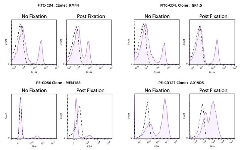
Figure 1. Alterations in antibody staining performance post-fixation
The foundation of flow cytometry rests upon specific binding between a fluorophore-conjugated antibody and its target antigen. Crucially, antibodies do not quantify the total intracellular abundance of a protein; rather, they detect specific, physically accessible antigenic epitopes.For any target protein, successful antibody binding depends not merely on whether the protein exists, but on whether the specific epitope retains an optimal three-dimensional spatial conformation.Fixation fundamentally alters this spatial environment.
In flow cytometry, common fixation strategies fall into two major categories: aldehyde-based crosslinkers and organic solvents. Paraformaldehyde (PFA) and formaldehyde represent classic aldehyde fixatives that stabilize cellular architecture primarily through protein crosslinking.This crosslinking strategy effectively preserves cell morphology and spatial protein distribution, rendering it the standard choice for surface marker post-fixation, intracellular cytokine staining (ICS), and nuclear transcription factor profiling.
However, protein crosslinking can simultaneously alter epitope topology. As inter- and intramolecular crosslinks form, target epitopes may undergo conformational rearrangement or become sterically hindered, thereby diminishing antibody binding efficiency.Thus, even if intracellular expression levels of a target protein remain unchanged, the antibody may display a significant loss in fluorescence signal simply because it can no longer recognize the altered epitope.
Beyond aldehydes, organic solvents such as methanol and ethanol are employed in specialized workflows, including phosphoprotein detection.Unlike aldehydes, organic solvents modify cell states through protein precipitation and dehydration, resulting in a distinct mechanism of structural modification. Epitopes that remain stable under PFA fixation may be denatured or destroyed by methanol. Conversely, targets tailored for organic solvent precipitation may exhibit suboptimal resolution within aldehyde crosslinking systems.
In short, fixation is not a passive "sample preservation" step—it is an active biochemical variable that directly redefines antigen structure and antibody recognition.
Table 1. Comparison of Common Fixatives in Flow Cytometry
| Fixative Type | Typical Usage & Applications | Impact on Antigens & Fluorescent Signals |
|---|---|---|
|
Paraformaldehyde (PFA) |
Concentration: 1%–2% Applications: Post-fixation of surface markers, intracellular cytokines, transcription factors, batch sample storage |
Antigen Impact: Moderate. Preserves most surface epitopes, though conformational epitopes may be masked via crosslinking. Fluorophore Compatibility: Excellent compatibility with PE, APC, and Alexa Fluor dyes. Tandem dyes may degrade under prolonged storage. FITC detection is more susceptible to interference from increased cellular autofluorescence induced by fixation. |
| Formaldehyde Solutions | Concentration: 1%–4% Applications: Clinical sample batch fixation |
Antigen Impact: Similar to PFA. Fluorophore Compatibility: Slightly inferior to pure PFA; methanol stabilizers present in solution may accelerate signal decay. |
|
Methanol |
Concentration: 80%–100% ice-cold Applications: Phospho-protein profiling, nuclear antigens, cell cycle analysis |
Antigen Impact: High. Destroys conformational epitopes of most surface markers while excellently preserving phospho-epitopes. Fluorophore Compatibility: Poor compatibility with phycobiliproteins (PE/APC) due to structural denaturation. Synthetic small-molecule dyes show better tolerance. |
|
Ethanol |
Concentration: 70%–75% ice-cold Applications: Cell cycle and apoptosis DNA staining |
Antigen Impact: Slightly less aggressive than methanol, though surface marker loss remains significant. Fluorophore Compatibility: Similar to methanol. |
Researchers frequently observe that different antibody clones directed against the same marker exhibit markedly distinct post-fixation performance. This variation is not random; it is governed by the specific epitope recognized by each clone. A single protein molecule contains multiple epitope domains, and different clones target different structural regions. When fixation modifies protein topology, distinct epitopes experience varying degrees of disruption.
A classic example of this phenomenon is FoxP3 detection. FoxP3 is a key intranuclear transcription factor. Because it resides inside the nucleus, detection requires fixation and permeabilization to allow antibodies to traverse both the plasma and nuclear membranes. However, different FoxP3 antibody clones exhibit distinct compatibility profiles across commercial Fix/Perm buffer systems.
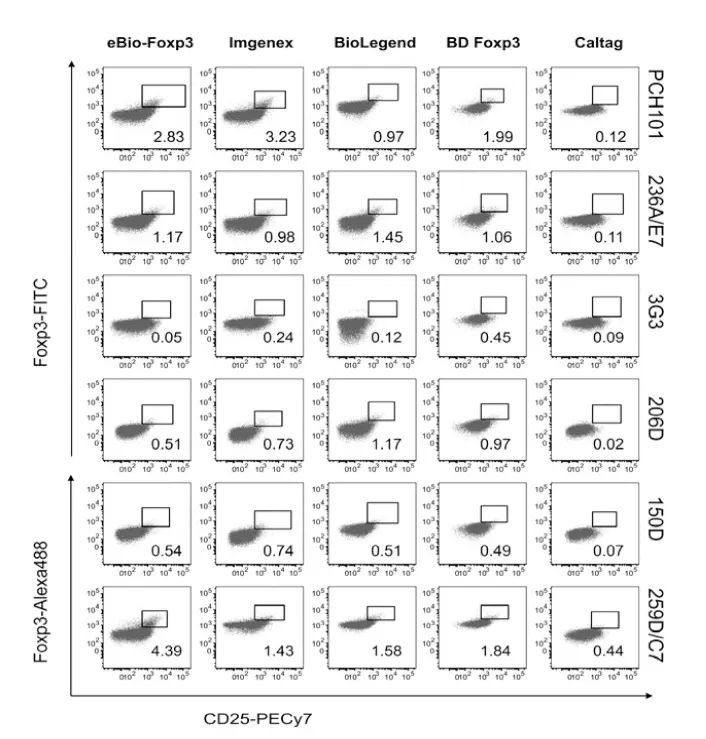
Figure 2. Differential post-fixation performance among distinct FoxP3 antibody clones (DOI: 10.1002/cyto.a.20815)
Similar clone-dependent disparities occur with common lineage markers such as CD3, CD4, and CD8. Although widely utilized in routine panels, distinct clones bind different epitope locations on these molecules, leading to variable degrees of signal alteration post-fixation.
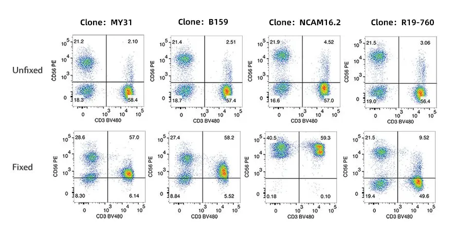
Figure 3. Differential post-fixation performance among distinct CD56 antibody clones
Key Takeaway: Sharing a common target marker name does not mean all antibody clones possess identical fixation compatibility. In experiments involving fixation or permeabilization, clone selection and validation parameters are just as critical as the choice of marker itself.
Table 2. Antibody Clone Performance Following Fixation/Permeabilization
| Antigen | Clone | Before IC fixation | After IC fixation and perm wash | After IC fixation/ methanol |
|---|---|---|---|---|
| Human | ||||
| CD3 | OKT3 | +++ | ++ | + |
| UCHT1 | +++ | +++ | +++ | |
| SK7 | +++ | ++ | +++ | |
| CD4 | OKT4 | +++ | + | +/- |
| RPA-T4 | +++ | +++ | + | |
| SK3 | +++ | +++ | + | |
| CD8 | OKT8 | +++ | +/- | - |
| RPA-T8 | +++ | +++ | +/- | |
| SK1 | +++ | +++ | +/- | |
| Mouse | ||||
| CD3 | 145-2C11 | ++ | + | +/- |
| 500A2 | +++ | +++ | +++ | |
| 17A2 | +++ | +++ | +++ | |
| CD4 | GK1.5 | +++ | ++ | +++ |
| RM4-5 | +++ | +++ | +++ | |
| CD8 | 53-6.7 | +++ | ++ | ++ |
Fluorescence intensity in flow cytometry depends on two sequential conditions: efficient antibody-antigen binding, and stable photon emission from the conjugated fluorophore. Even if antibody binding remains unhindered, fix/perm buffers that alter fluorophore integrity will compromise final signal intensity. Fluorophores vary considerably in chemical stability depending on their structural composition.
In fixed flow experiments, tandem dyes (e.g., PE-Cy7, APC-Cy7, PE-Cy5.5) require careful monitoring. These fluorophores generate long-Stokes-shift signals via Förster Resonance Energy Transfer (FRET) between donor and acceptor molecules. Because FRET relies on precise spatial coupling between fluorophores, tandem dyes are chemically more sensitive to ambient conditions than monomeric fluorophores like FITC, PE, or APC.
Fixation, extended storage, or harsh permeabilization can uncouple energy transfer, leading to decreased target fluorescence and increased donor-channel spillover. Therefore, panel designs incorporating tandem dyes for intracellular staining require rigorous validation.
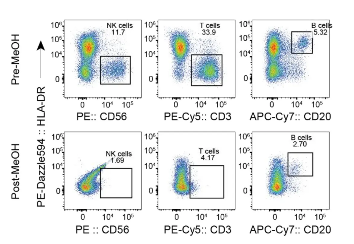
Figure 4. Fixation-induced loss of tandem dye signal intensity (DOI: 10.1002/cyto.a.24904)
Large phycobiliprotein fluorophores (PE and APC) generally exhibit good stability. However, under extended fixation or harsh perm conditions, structural denaturation can occur, leading to signal loss.
Conversely, traditional small-molecule dyes (e.g., FITC, Alexa Fluor synthetics) offer superior chemical robustness under fixation. Nevertheless, fixative concentration, exposure time, and sample autofluorescence still dictate overall resolution.
Fluorophore compatibility is determined holistically by the interplay between fluorophore structure, antibody clone, and the specific Fix/Perm buffer system. Signal decay after fixation is rarely monofactorial—it reflects the net outcome of epitope alteration, clone mismatch, and dye instability.
The standard ICS workflow follows a strict sequence: Stimulation → Protein Transport Inhibition → Fixation → Permeabilization → Intracellular Staining. Fixation must preserve overall cellular architecture while keeping cytokine epitopes intact. Suboptimal fixation leads to epitope alteration, poor antibody penetration, or sample-to-sample variability. Clones used in ICS should be explicitly validated for the intended Fix/Perm buffer system.
Detecting nuclear targets such as FoxP3, T-bet, GATA3, or RORγt requires deep permeabilization to breach both plasma and nuclear membranes. Because different Fix/Perm systems alter nuclear protein structures differently, clone performance varies considerably across buffer formulations. Clone selection and Fix/Perm buffer selection must be treated as a single, interdependent decision.
Phosphorylation states of signaling proteins (e.g., pSTAT, pERK, pAKT) are highly transient. Immediate, rapid fixation is essential to freeze phosphorylation dynamics post-stimulation. Methanol fixation is widely used in phospho-flow protocols to preserve phosphorylated epitopes, but it can significantly impair surface marker detection. Balancing surface marker clones with methanol-based phospho-staining is critical for accurate multiparameter analysis.
Fact: Flow antibodies measure accessible epitopes, not total protein abundance. Fixation-induced conformational changes can lower fluorescence signals even when target protein levels remain unchanged. Post-fixation MFI shifts should not be directly interpreted as altered biological expression.
Fact: Clones recognize distinct topological regions on an antigen. As shown with FoxP3, CD4, CD8, and CD62L, clone selection is just as critical as target selection when working with fixed samples.
Fact: While pairing dim markers with bright fluorophores is standard practice, fixative compatibility must take priority. Bright tandem dyes like PE-Cy7 or APC-Cy7 can undergo energy uncoupling under fixation and storage. Signal stability under assay conditions is more critical than absolute brightness.
Fixation is a powerful tool in flow cytometry, but it is not a neutral process. It actively modifies target epitopes, impacts antibody binding kinetics, and can affect fluorophore stability.
To obtain reliable and reproducible flow cytometry data, experimental workflows should evaluate antibody clone properties, epitope structure, fluorophore stability, and Fix/Perm buffer chemistry as an integrated system.
abinScience was founded in 2023 as a strategic venture of AtaGenix (established 2011), dedicated to delivering premium life science reagents that accelerate discovery.
abinScience flow cytometry antibody products cover commonly used detection markers, with a wide variety to meet the research needs of multiple species(Human, Mouse, Rat, Dog, Hamster, Monkey, etc.)We provide stable and reliable support for scientific research.
+86-027-65523339
中国武漢市深敦寺路666号C棟、武漢、430206


 中文
中文 English
 한국어
한국어 日本語
日本語 Español
 Français
 Русский