October is Breast Cancer Awareness Month, a period dedicated not only to raising public awareness but also to focusing ongoing attention on the biological questions surrounding the initiation, progression, and treatment of breast cancer that require deeper understanding. As research advances, the complexity of breast cancer becomes increasingly apparent: tumors are not composed of groups of entirely identical cells; different cell populations within a tumor may possess distinct molecular features and functional states, which continue to evolve as the disease progresses, therapeutic interventions are applied, and the surrounding environment changes.
Breast cancer characteristics are not fixed once formed. Pre‑existing cellular differences within a tumor may influence how various cell populations respond to therapeutic pressure, while treatment can alter the composition of cell populations or prompt some cells to adjust their states. Simultaneously, microenvironmental components such as cancer‑associated fibroblasts (CAFs), immune cells, and the extracellular matrix (ECM) influence tumor cell behavior through local signaling and undergo corresponding changes during tumor progression.
Consequently, understanding breast cancer cannot be limited to molecular features at a single time point; it requires examining how internal tumor differences influence subsequent evolution, how therapeutic pressure alters cell survival and adaptation, and how tumor cells and the microenvironment interact. Together, these processes constitute vital components of the dynamic evolution of breast cancer and serve as key factors in understanding variations in disease progression and therapeutic response.
Tumor Heterogeneity: Intrinsic Diversity Drives Breast Cancer Evolution
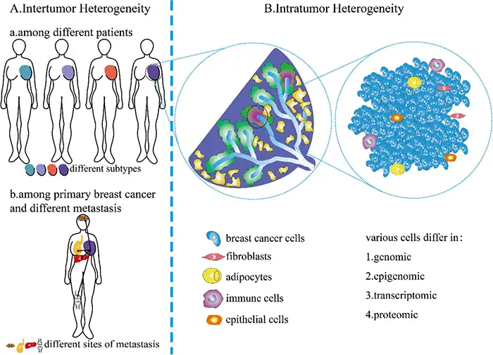
Figure 1. Inter‑ and intratumoral heterogeneity in breast cancer (DOI: 10.1186/s40164‑022‑00363‑1)
Breast cancer heterogeneity encompasses more than molecular variations among different patients; distinct cell populations with diverse biological features can coexist within the same tumor. These differences may stem from genetic alterations accumulated across different cells or manifest as variations in receptor expression, signaling pathway activity, proliferation status, and stress responses. Even among cells with similar genetic backgrounds, local microenvironments and transcriptional regulatory states can drive distinct functional profiles.
Human epidermal growth factor receptor 2 (HER2)‑positive breast cancer exemplifies how such heterogeneity affects the assessment of overall tumor characteristics. While HER2‑positive denotes a specific molecular attribute of a tumor, it does not imply that all tumor cells exhibit identical HER2 protein levels or ERBB2 gene amplification status. Significant regional variations exist within some tumors, and this heterogeneity correlates with poorer therapeutic responses under specific HER2‑targeted therapies. When cell populations differ in their dependency on the same signal, the overall positive status of a tumor fails to capture the biological characteristics of all internal cells.
Such variations extend beyond receptor expression levels. Tumor cells may reside in different proliferative states, exhibit varying signaling pathway activities, or display divergent responses to external stimuli such as hypoxia and inflammation. Furthermore, spatial architecture within the tumor influences the matrix, nutrient conditions, and immune signals encountered by cells, causing cells with similar molecular backgrounds to exhibit distinct states across various regions.
It is also essential to distinguish between genetic differences among cell populations and cellular phenotypic plasticity. The former typically accumulates through the formation and expansion of distinct subclones, whereas the latter reflects the ability of cells to adjust their gene expression and functional states under specific conditions. Both phenomena can coexist and may play distinct roles at various stages of the disease.
Thus, tumor heterogeneity not only describes the complexity of a tumor at a specific time point but also determines its biological foundation when facing subsequent changes. When therapy imposes novel selective pressures, pre‑existing cellular differences may be further amplified, while cellular plasticity provides a mechanism for the tumor to generate new adaptive strategies.
Treatment Adaptation: How Therapeutic Pressure Alters Tumor Composition and State
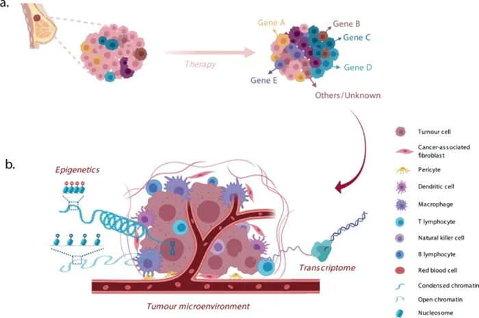
Figure 2. Clonal selection and drug resistance evolution under therapeutic pressure (DOI: 10.1038/s41523‑021‑00363‑0)
Treatment does not simply eradicate all tumor cells uniformly and to the same degree. Different cells exhibit varying sensitivities to the same intervention; by altering survival conditions, treatment dictates which cells are more likely to survive, expand, or maintain their original functions. Consequently, variations in therapeutic response can stem from changes in cell population composition or the active adjustment of states by surviving cells.
In hormone receptor‑positive breast cancer, estrogen receptor‑related signaling is closely linked to cellular proliferation, with cyclin‑dependent kinase 4 and 6 (CDK4/6) serving as critical links connecting cell cycle regulation to proliferative activity. CDK4/6 inhibition restricts certain tumor cells from entering subsequent cell cycles, yet this inhibition does not necessarily arrest all cells in the same state indefinitely. Certain cells bypass CDK4/6 dependence by utilizing alternative cell cycle regulatory mechanisms, such as Cyclin D1–CDK2‑related signaling; RB1 functional loss or CCNE1 amplification may also contribute to drug resistance in specific models.
These mechanisms indicate that therapeutic pressure affects not only whether cells continue to proliferate but also the signaling networks they rely on to sustain proliferation. When primary regulatory pathways are inhibited, the activity of alternative pathways may become paramount, causing cells to exhibit responses distinct from their pre‑treatment state. However, early treatment‑induced signaling adjustments are distinct from stable drug resistance established through sustained selection, and their specific mechanisms are influenced by the molecular background of the breast cancer and treatment conditions.
Simultaneously, shifts in cell population composition may occur concurrently with these processes. If a tumor naturally harbors cells less sensitive to treatment, these cells may become relatively enriched post‑treatment, resulting in residual tumors exhibiting molecular profiles distinct from the pre‑treatment state. This does not imply that all surviving cells have newly acquired drug resistance, nor that the expansion of a specific population is necessarily driven by a single signaling pathway. Shifts in population composition and alterations in single‑cell states represent two distinct processes that must be evaluated independently.
This framework allows for a re‑evaluation of heterogeneity in HER2‑positive breast cancer. When internal cell populations exhibit variable dependencies on HER2‑related signaling, the selective pressure imposed by targeted therapy impacts these populations differently. Post‑treatment tumors may undergo changes not only in cell number but also in receptor expression and downstream signaling distributions.
Therefore, treatment adaptation cannot be explained solely by drug‑resistance mutations. Pre‑existing heterogeneity dictates which cells survive more readily, cellular plasticity modifies the behavior of surviving cells, and signaling network adjustments provide the underlying molecular mechanisms. Furthermore, these processes do not occur exclusively within tumor cells; the surrounding microenvironment can also modulate cellular responses to therapeutic pressure.
Microenvironment Remodeling: Tumor Adaptation Extends Beyond Tumor Cells
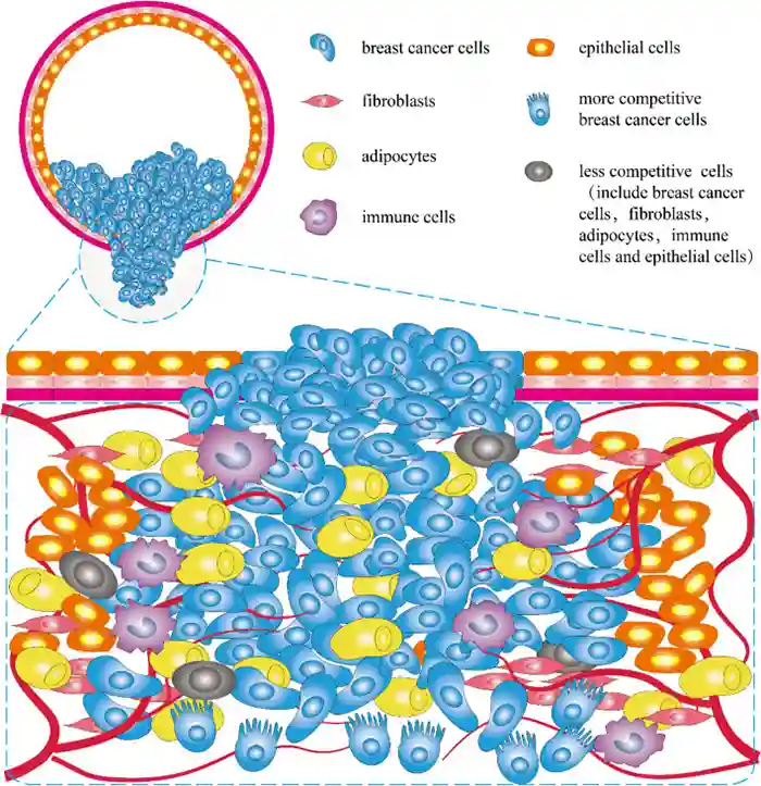
Figure 3. Cellular interactions within the breast cancer microenvironment (DOI: 10.1186/s40164‑022‑00363‑1)
Breast cancer cells reside within a local milieu comprising stromal cells, immune cells, vasculature‑associated cells, and the extracellular matrix. The microenvironment is not a passive background formed merely after tumor growth; through intercellular signaling and tissue structural changes, it actively influences tumor cell proliferation, survival, and stress responses. Simultaneously, tumor cells actively remodel their surrounding environment by secreting signaling molecules, modifying the matrix, and modulating immune cell functions.
Cancer‑associated fibroblasts are key constituents of this bidirectional relationship. They modify extracellular matrix composition and architecture while releasing diverse growth factors, cytokines, and signaling molecules. These alterations impact local signals received by tumor cells and modify cell‑matrix interactions. Because CAFs themselves exhibit functional heterogeneity, their impact on breast cancer cells is context‑dependent, varying with their specific cellular states and tissue microenvironments.
Crosstalk between immune cells and tumor cells further underscores the microenvironment's role in treatment adaptation. In certain estrogen receptor (ER)‑positive breast cancer research models, inflammatory signaling involving tumor‑associated macrophages (TAMs) accompanies the activation of NF‑κB, STAT3, and ERK pathways in tumor cells, thereby modulating ERα activity. This indicates that tumor cell responses to hormonal signals are not determined solely by receptor expression; inflammatory signals generated by neighboring cells can alter regulatory networks and affect endocrine therapy resistance. Nevertheless, such effects are model‑ and context‑specific and should not be generalized to assume all TAMs promote breast cancer resistance.
The influence of the microenvironment on tumor evolution extends to metastasis. Upon disseminating from the primary tumor into distant tissues, breast cancer cells encounter distinct matrix architectures, cell compositions, and immune milieus. Cells possessing survival advantages in the primary tumor may not necessarily sustain growth in distant organs; conversely, novel tissue environments further select for cell populations capable of adapting to local conditions. Consequently, metastasis involves not only intrinsic alterations in tumor cells but also adaptation between tumor cells and distant organ environments.
Integrating these processes reveals that the microenvironment is not a tertiary factor independent of tumor heterogeneity and treatment adaptation. Distinct internal cell populations may respond differently to identical local signals, while microenvironmental shifts can alter the relative survival advantage of these cells. Conversely, surviving and expanding tumor cells can further modify matrix structure, inflammatory signaling, and immune cell states. These three components form an interdependent dynamic process, though specific relationships vary across breast cancer subtypes, disease stages, and therapeutic contexts, precluding a universal evolutionary pathway applicable to all tumors.
Conclusion: Shifting from Single Molecular Markers to Dynamic Tumor Biology
Comprehending breast cancer requires connecting changes across different time points, cell populations, and tissue environments. Tumor heterogeneity unmasks pre‑existing cellular differences, therapeutic pressure alters the relative importance of these variations, and the microenvironment further modulates cell states and signaling networks. While interrelated, these elements do not form a rigid, invariant evolutionary trajectory.
For breast cancer mechanistic research, this perspective facilitates precise questioning: Does an observed molecular change reflect shifts in cellular subpopulation proportions, or a state transition within the same cell type? Is an alteration in a signaling pathway driven by intrinsic tumor cell regulation or influenced by adjacent stromal and immune cells? Are these changes time‑dependent and spatially specific? Step‑by‑step resolution of these questions will yield a more accurate understanding of the links between breast cancer progression, treatment adaptation, and metastasis, pointing the way forward for future mechanistic studies.
abinScience Breast Cancer Research Tools
Focusing on molecular mechanisms and tumor microenvironment research in breast cancer, abinScience offers a comprehensive portfolio of research‑grade antibodies and proteins covering diverse targets and application directions.
Proteins
| Catalog No. |
Product Name |
| HY286021 |
Human ErbB2/Her2 Recombinant Protein (C‑Fc) |
| HY286011 |
Human ErbB2/Her2 Recombinant Protein (C‑His) |
| HB862012 |
Human N‑Cadherin Recombinant Protein (N‑His) |
| HB862011 |
Human N‑Cadherin Recombinant Protein (C‑Fc) |
| HB241022 |
Human Cyclin D1 Recombinant Protein (N‑His) |
| HY335012 |
Human ESR1/ER‑alpha Recombinant Protein (N‑His) |
Antibodies
| Catalog No. |
Product Name |
| HV985013 |
Anti‑Human Nectin‑4 Recombinant Antibody (P1E7) |
| MV985013 |
Anti‑Mouse Nectin‑4 Recombinant Antibody (R4P1) |
| HY286013 |
Anti‑Human ErbB2/Her2 Recombinant Nanobody (SAA0836) |
| HY286023 |
Anti‑Human ErbB2/Her2 Recombinant Nanobody (SAA1045) |
| HY286033 |
Anti‑Human ErbB2/Her2 Recombinant Nanobody (SAA1170) |
| HB116013 |
Anti‑Human ErbB3/Her3 Recombinant Nanobody (SAA1309) |
| HB862014 |
Anti‑Human N‑Cadherin Polyclonal Antibody |
| HB862107 |
Anti‑Human N‑Cadherin Recombinant Antibody (1H7) |
| HB241024 |
Anti‑Human Cyclin D1 Polyclonal Antibody |
| HY335014 |
Anti‑ESR1/ER‑alpha Polyclonal Antibody |
| HY335013 |
Anti‑Human ESR1/ER‑alpha Recombinant Antibody (RB125) |
| HB610043 |
Anti‑Phospho‑AKT (pSer473) Recombinant Antibody (SRR0405) |
| HB659013 |
Anti‑Phospho‑ERK1 (T202/Y204)/ERK2 (T185/Y187) Recombinant Antibody (SRR0206) |
References
- [1] Guo, L., Kong, D., Liu, J., Zhan, L., Luo, L., Zheng, W., Zheng, Q., Chen, C., & Sun, S. (2023). Breast cancer heterogeneity and its implication in personalized precision therapy. Experimental Hematology & Oncology, 12(1), 3. https://doi.org/10.1186/s40164‑022‑00363‑1
- [2] Mavrommati, I., Johnson, F., Echeverria, G. V., & Natrajan, R. (2021). Subclonal heterogeneity and evolution in breast cancer. NPJ Breast Cancer, 7(1), 155. https://doi.org/10.1038/s41523‑021‑00363‑0
- [3] Herrera‑Abreu, M. T., Palafox, M., Asghar, U., Rivas, M. A., Cutts, R. J., Garcia‑Murillas, I., Pearson, A., Guzman, M., Rodriguez, O., Grueso, J., Bellet, M., Cortés, J., Elliott, R., Pancholi, S., Baselga, J., Dowsett, M., Martin, L. A., Turner, N. C., & Serra, V. (2016). Early Adaptation and Acquired Resistance to CDK4/6 Inhibition in Estrogen Receptor‑Positive Breast Cancer. Cancer Research, 76(8), 2301–2313. https://doi.org/10.1158/0008‑5472.CAN‑15‑0728
- [4] Kumarasamy, V., Vail, P., Nambiar, R., Witkiewicz, A. K., & Knudsen, E. S. (2021). Functional Determinants of Cell Cycle Plasticity and Sensitivity to CDK4/6 Inhibition. Cancer Research, 81(5), 1347–1360. https://doi.org/10.1158/0008‑5472.CAN‑20‑2275
- [5] Li, Z., Metzger Filho, O., Viale, G., dell'Orto, P., Russo, L., Goyette, M. A., Kamat, A., Yardley, D. A., Gupta Abramson, V., Arteaga, C. L., Spring, L. M., Chiotti, K., Halsey, C., Waks, A. G., King, T. A., Lester, S. C., Bellon, J. R., Winer, E. P., Spellman, P. T., Krop, I. E., … Polyak, K. (2024). HER2 heterogeneity and treatment response‑associated profiles in HER2‑positive breast cancer in the NCT02326974 clinical trial. The Journal of Clinical Investigation, 134(7), e176454. https://doi.org/10.1172/JCI176454
- [6] Castellaro, A. M., Rodriguez‑Baili, M. C., Di Tada, C. E., & Gil, G. A. (2019). Tumor‑Associated Macrophages Induce Endocrine Therapy Resistance in ER+ Breast Cancer Cells. Cancers, 11(2), 189. https://doi.org/10.3390/cancers11020189


 中文
 English
 한국어
 日本語
 Español
 Français
 Русский


