In flow cytometry experiments, staining buffers are widely used in steps such as cell resuspension, antibody incubation, and washing. Different experimental protocols employ varying buffer formulations: some use PBS or HBSS directly, while others incorporate components such as bovine serum albumin (BSA), foetal bovine serum (FBS), EDTA, or sodium azide into the base buffer.
These formulations do not follow a single universal standard applicable to all experiments. For routine cell surface antigen detection, PBS or HBSS alone can satisfy basic requirements; however, in scenarios prone to cell aggregation, non‑specific adsorption, or where specific cellular states must be maintained, the addition of other components may be more appropriate.
What exact roles do different buffer components play? Does the addition of extra components invariably improve staining performance? How should an appropriate buffer system be selected according to detection targets?
Phosphate‑buffered saline (PBS) and Hanks' balanced salt solution (HBSS) are common base buffer systems in flow cytometry experiments. Both can be used directly in certain experiments or serve as the formulation base for additive‑containing staining buffers.
PBS maintains a relatively stable pH via a phosphate buffer system and provides a suitable ionic environment. In many routine flow cytometry experiments, PBS can be used directly for cell washing, antibody incubation, and cell resuspension following staining. It is particularly suitable for procedures with relatively short processing times, stable cell states, and no significant cell aggregation or non‑specific adsorption issues.
HBSS can likewise be used directly in certain flow cytometry experiments. Compared with PBS, HBSS typically contains a greater variety of inorganic salts, with some formulations also incorporating glucose or bicarbonate, and is frequently employed in cell processing and experiments requiring the maintenance of a specific physiological environment.
It should be noted that both PBS and HBSS exist in various formulations, and particular attention should be paid to whether they contain Ca2+ and Mg2+. These two divalent cations participate in various cell adhesion and protein interaction processes, and their presence or absence may influence the cellular dispersion state and the recognition of certain antigens.
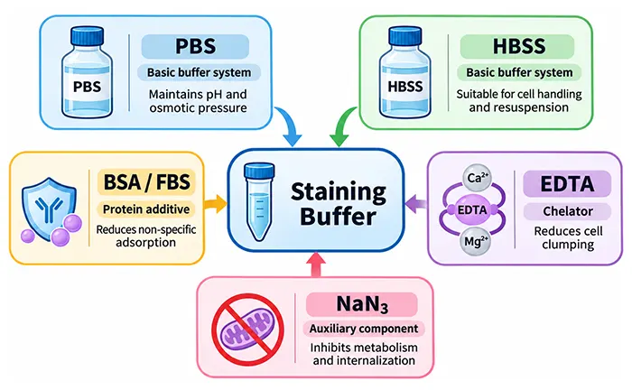
Figure 1. Principal composition and functions of flow cytometry staining buffers.
The direct use of PBS or HBSS does not imply inferior staining performance, nor does the addition of components such as BSA or FBS necessarily enhance staining efficacy. For routine detection, if the base buffer already maintains good cell viability, low non‑specific signals, and stable target antigen recognition, altering the formulation merely to increase component count is unnecessary.
When base buffers cannot fully meet experimental requirements, protein additives, chelating agents, or other auxiliary components can be incorporated according to specific needs.
| Common Components | Main Function | Points for Attention |
|---|---|---|
| BSA | Reduces certain types of non‑specific adsorption; provides a protein environment | Concentration, purity, and experimental compatibility |
| FBS | Provides serum proteins; reduces certain types of non‑specific adsorption | Lot‑to‑lot variability and additional biological components |
| EDTA | Chelates divalent cations; reduces intercellular adhesion | May affect divalent cation‑dependent epitopes |
| Sodium Azide (NaN3) | Inhibits microbial growth and certain cellular metabolic activities | Unsuitable for experiments requiring the maintenance of cell function |
| HEPES | Assists in maintaining pH stability under specific conditions | Not required for all routine staining procedures |
These additive components exert distinct functions, and their selection should depend on sample status, target antigen characteristics, and downstream experimental requirements, rather than incorporating all components simultaneously into the staining buffer.
In routine flow cytometry staining, both BSA and FBS can be used to reduce non‑specific adsorption between antibodies and surfaces such as cells and experimental containers. Although both serve similar purposes, their compositions differ.
BSA is a relatively well‑defined protein component, whereas FBS contains various serum proteins and multiple biological components in addition to albumin. Consequently, BSA typically facilitates better control over system composition, whereas FBS is frequently used in established routine cell staining protocols.
For general cell surface antibody staining, both can serve as protein additives, but they cannot be assumed to be unconditionally interchangeable across all experimental systems.
A classic example is the biotin‑avidin detection system. When biotinylated antibodies are used with fluorophore‑labelled avidin, free biotin in serum may interfere with detection. A staining buffer without free biotin may therefore be preferable.
Furthermore, protein additives are not equivalent to Fc receptor blocking. The former primarily reduces certain forms of non‑specific adsorption and cannot substitute for dedicated blocking treatments targeting Fc receptor‑mediated binding.
Therefore, for experiments where satisfactory staining results can be achieved directly using PBS or HBSS, protein addition is not strictly necessary. When significant non‑specific adsorption, cell loss, or system stability issues arise, the addition of BSA or serum can be considered on a case‑by‑case basis.
EDTA is a common metal ion chelating agent capable of binding divalent cations such as Ca2+ and Mg2+. Because the function of certain cell adhesion molecules relies on these ions, EDTA is frequently utilised to reduce intercellular adhesion and improve single‑cell suspension quality.
However, divalent cations not only participate in cell adhesion but may also influence the spatial conformation of certain proteins. When an antibody recognises an epitope that depends on a specific conformation, EDTA may indirectly affect antibody binding.
For example, when different anti‑human CD8 antibody clones were used to stain peripheral blood mononuclear cells (PBMCs), EDTA markedly reduced the staining intensity of certain anti‑CD8α antibodies. With the RPA‑T8 clone, 1 mmol/L EDTA can reduce fluorescence intensity by approximately 95%. This alteration is not caused by CD8 internalisation; upon supplementation with calcium ions, the staining signal can be restored. Other anti‑CD8 clones, such as OKT8, do not exhibit the same degree of EDTA sensitivity. This demonstrates that the effect of EDTA on flow cytometry staining is antibody clone‑dependent. Even when detecting the same marker, different clones may exhibit distinct responses to the ionic environment owing to differences in recognised epitopes.
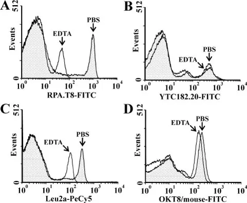
Figure 2. Effect of EDTA on different anti‑CD8 antibody clones (DOI: 10.1002/cyto.a.20084).
Therefore, when processing samples prone to cell clumping, the addition of EDTA may be considered; however, for detection targets possessing divalent cation‑dependent epitopes, preferential confirmation should be sought regarding whether the relevant antibody clone is compatible with EDTA‑containing staining systems.
Sodium azide acts both as a preservative and as an inhibitor of cellular energy metabolism, thereby affecting certain active biological processes.
For certain cell surface receptors, antibody binding may induce receptor redistribution, internalisation, or other dynamic alterations. By inhibiting cellular metabolism, sodium azide can mitigate surface signal loss caused by these changes to a certain extent. Some commercial flow cytometry staining buffers contain no more than 0.09% sodium azide and are primarily used for routine cell surface immunofluorescence staining and subsequent detection.
If stained cells require continued culture, functional assays, or live cell sorting, the metabolic inhibition caused by sodium azide may compromise downstream results. In such cases, buffer systems lacking sodium azide and suitable for maintaining cellular physiological functions should be selected.
Having understood the roles of individual components, the selection of staining buffers can be distilled into three considerations: whether the base buffer meets experimental requirements, whether target antigens are sensitive to specific components, and whether cells must retain normal physiological function following staining.
For routine cell surface antigen detection, direct antibody incubation and washing using PBS or HBSS are feasible, and are particularly suited to experiments involving relatively short sample processing times, stable cell viability, and minimal non‑specific adsorption. If stable and reproducible staining results are attainable using the base buffer, the additional inclusion of BSA, FBS, or other components is unnecessary.
When cells tend to adhere to experimental containers, non‑specific adsorption is pronounced, or cell suspension quality requires improvement, buffers containing appropriate protein additives should be considered.
Accordingly, the selection of routine staining buffers should not be viewed as a simple choice of superiority between PBS and additive‑containing buffers, but rather evaluated based on actual detection performance.
For certain primary cells or adherent cultured cells prone to clumping, buffer systems devoid of Ca2+ and Mg2+ can be considered, supplemented with an appropriate amount of EDTA as required to improve cell dispersion. Nevertheless, when cell clumping is primarily driven by free DNA released from dead cells, EDTA may not resolve the issue. Such samples may require alternative handling methods rather than an indiscriminate increase in EDTA concentration.
When the spatial conformation of a target antigen or antibody binding is dependent upon Ca2+ and Mg2+, the ionic conditions of the buffer warrant careful attention.
For example, when utilising certain clones of CD8, potential reductions in staining signal caused by EDTA must be accounted for.If the antibody has not been evaluated in an EDTA‑containing buffer, parallel comparisons can help determine whether EDTA affects staining, rather than directly attributing fluorescence changes to alterations in antigen expression levels.
If cells are intended for subsequent culture, functional assays, or other live cell procedures following flow cytometric analysis, buffers lacking sodium azide must be prioritised. Furthermore, cell sorting procedures may involve extended sample processing times, pH stability maintenance, and cell dispersion requirements; hence, certain sorting protocols incorporate HEPES and protein additives into appropriate base salt solutions.
| Experimental Requirement | Buffer Selection Focus |
|---|---|
| Routine surface antigen staining | PBS or HBSS can be used directly; incorporate protein or other components only if necessary |
| Pronounced non‑specific adsorption | Consider adding BSA or serum |
| Tendency to form cell aggregates | Consider calcium/magnesium‑free buffer systems and EDTA, while verifying antigen compatibility |
| Divalent cation‑dependent antigens | Pay attention to EDTA and Ca2+/Mg2+ conditions; compare different formulations when necessary |
| Post‑staining culture or functional analysis | Avoid sodium azide; maintain appropriate cellular physiological environments |
| Live cell sorting | Comprehensively evaluate cell viability, single‑cell state, and buffering capacity |
| Biotin‑avidin detection | Account for potential interference from free biotin in serum; prioritise compatible formulations |
It must be emphasized that these principles serve to guide the evaluation of formulation suitability and do not imply that a single optimal buffer exists for every experimental scenario. For established and validated staining protocols, consistency with original conditions should generally be maintained.
Although staining buffers are classified as basic reagents, altering their formulation may modify protein concentrations, ionic environments, or cellular metabolic states within the experimental system. Consequently, substitutions between different buffers should not be judged solely by their names.
For example, replacing PBS with a staining buffer containing BSA and EDTA, or replacing a commercial staining buffer with PBS alone, changes the staining environment even when the base buffer remains similar. Such differences may exert negligible effects on routine markers, yet prove critical for certain specialised antigens or detection systems.
If staining results change after switching buffers, parallel comparisons can be performed using the same cell sample, antibody clone, amount of antibody used for staining, incubation conditions and instrument acquisition settings. Alongside evaluations of positivity rates and fluorescence intensity shifts, cell viability and aggregation ratios must also be monitored to avoid conflating sample state changes with alterations in antigen recognition.
For studies that compare results across different batches or treatment groups over time, maintaining a consistent staining buffer formulation helps minimise unnecessary technical variability. If formulation changes are unavoidable, the necessity for re‑validation should be determined according to the significance and potential sensitivity of the detection targets.
The selection of flow cytometry staining buffers is straightforward, provided the practical functions of individual components are understood. While PBS or HBSS can directly satisfy the demands of various routine staining experiments, components such as BSA, FBS, EDTA, and sodium azide can be selectively added based on sample characteristics and detection targets.
A suitable staining buffer is not necessarily the one with the most components, but one that maintains appropriate cellular conditions while supporting reliable antibody recognition and detection.
abinScience was founded in 2023 as a strategic venture of AtaGenix (established 2011), dedicated to delivering premium life science reagents that accelerate discovery.
abinScience flow cytometry antibody products cover commonly used detection markers, with a wide variety to meet the research needs of multiple species(Human, Mouse, Rat, Dog, Hamster, Monkey, etc.)We provide stable and reliable support for scientific research.

お問い合わせ
+86-027-65523339
中国武漢市深敦寺路666号C棟、武漢、430206


 中文
 English
 한국어
 日本語
 Español
 Français
 Русский