
 中文
 English
 한국어
 日本語
 Español
 Français
 Русский
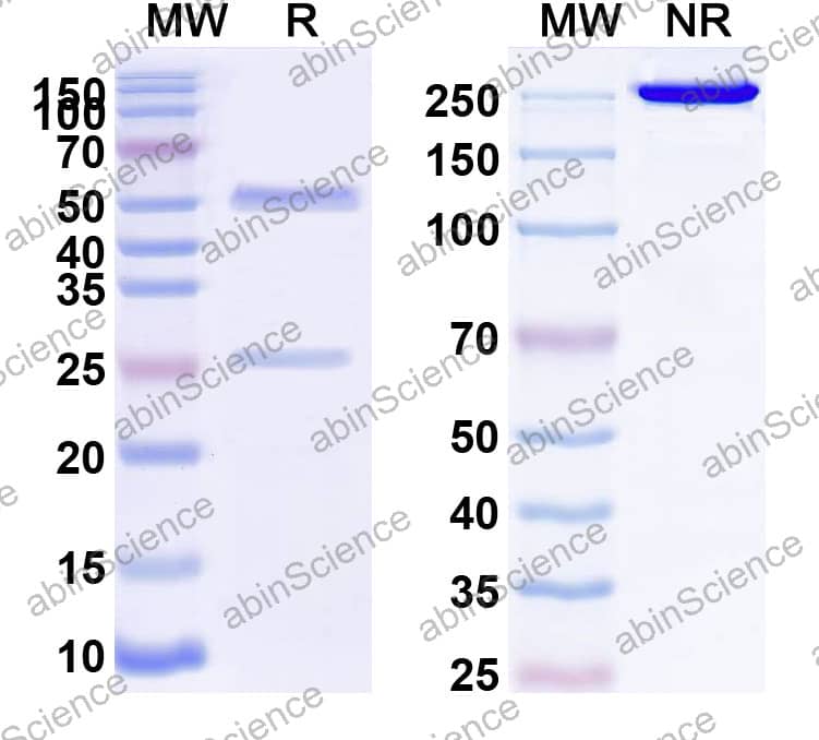
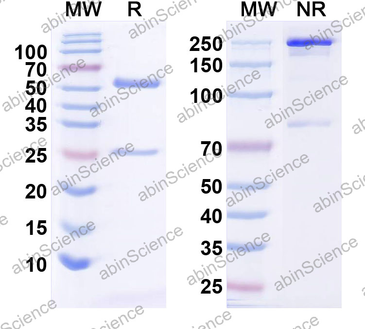
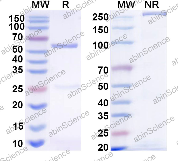
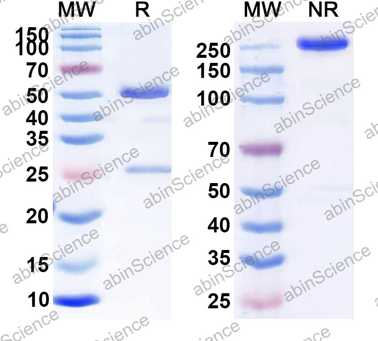
| カタログ番号 | HV974133 |
|---|---|
| 説明 |
Anti-Human PD-L1 Recombinant Antibody (SRR0417) (HV974133) is a rabbit monoclonal antibody detecting PD-L1 in IF, FCM, WB. Suitable for Human.
Highlights
|
| 種反応性 | Human |
| アプリケーション | IF, FCM, WB |
| 宿主種 | Rabbit |
| クローン性 | Monoclonal |
| アイソタイプ | IgG |
| クローンID | SRR0417 |
| テスト済みアプリケーション | IF, FCM, WB |
| ターゲット | B7 homolog 1, B7-H1, B7H1, CD274, PD-L1, PDCD1 ligand 1, PDCD1L1, PDCD1LG1, PDL1, Programmed cell death 1 ligand 1, Programmed death ligand 1, hPD-L1 |
| エンドトキシンレベル | < 10 EU/mg as determined by LAL method. |
| 純度 | >95% as determined by SDS-PAGE. |
| 精製 | Protein A/G purified from cell culture supernatant. |
| アクセッション番号 | Q9NZQ7 |
| 形態 | Liquid |
| 保存バッファー | 0.01M PBS pH 7.4. データシートのハードコピーまたはロット固有のCOAに記載された具体的なバッファー情報を参照してください。 |
| 再構成 | Reconstitute in sterile water for a stock solution. A copy of datasheet will be provided with the products, please refer to it for details. |
| 安定性と保存 | Use a manual defrost freezer and avoid repeated freeze thaw cycles. Store at 2 to 8°C for frequent use. Store at -20 to -80°C for twelve months from the date of receipt. |
| 背景 | Programmed cell death 1 ligand 1 (CD274/PD-L1) is a ~33 kDa protein. Plays a critical role in induction and maintenance of immune tolerance to self. As a ligand for the inhibitory receptor PDCD1/PD-1, modulates the activation threshold of T-cells and limits T-cell effector response. Through a yet unknown activating receptor, may costimulate T-cell subsets that predominantly produce interleukin-10 (IL10). Can also act as a transcription coactivator: in response to hypoxia, translocates into the nucleus via its interaction with phosphorylated STAT3 and promotes transcription of GSDMC, leading to pyroptosis. CD274 is the therapeutic target of atezolizumab (Tecentriq) and durvalumab (Imfinzi). 1. Freeman, GJ. et al. (2000) The Journal of experimental medicine 192, 1027-34. PMID: 11015443 2. Mezzadra, R. et al. (2017) Nature 549, 106-110. PMID: 28813410 3. Burr, ML. et al. (2017) Nature 549, 101-105. PMID: 28813417 4. Huang, X. et al. (2019) Cancer immunology research 7, 1580-1590. PMID: 31399419 5. Kennedy, A. et al. (2023) The EMBO journal 42, e111556. PMID: 36727298 6. Dong, H. et al. (1999) Nature medicine 5, 1365-9. PMID: 10581077 7. Hou, J. et al. (2020) Nature cell biology 22, 1264-1275. PMID: 32929201 |
| 注意事項 | For research use only. |
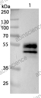
Western blot analysis was performed using anti-PD-L1 monoclonal antibody at 1 ug/mL on various samples.
Lane 1: PD-L1-overexpressing HEK293T cell lysate
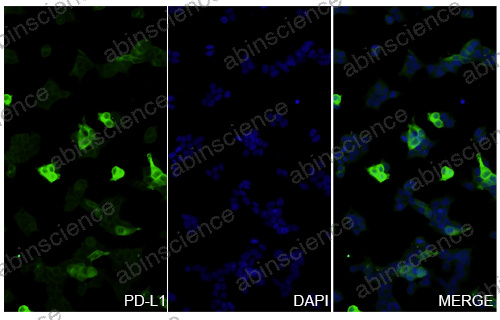
293T cells overexpressing PD-L1 were fixed with 4% paraformaldehyde for 20 minutes, followed by blocking with 5% goat serum for 1 hour. The cells were then incubated with a primary antibody targeting PD-L1 (HV974133) at a concentration of 20 ug/ml overnight at 4°C. Afterward, the cells were incubated with a secondary antibody, Goat Anti-Rabbit IgG (Alexa Fluor-488) at a concentration of 6.5 ug/ml for 50 minutes at room temperature. The localization of PD-L1 was visualized (shown in green), and nuclear DNA was stained with DAPI (shown in blue).
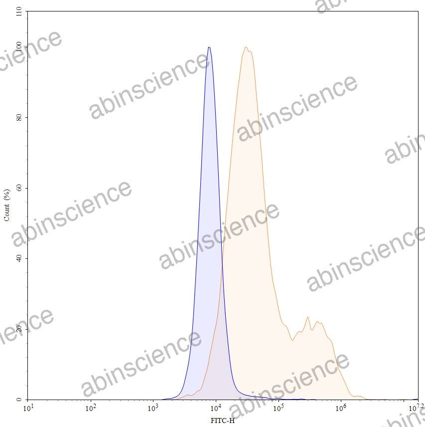
Flow-cytometry using anti-Human PD-L1 antibody. Overexpression cells and Untransfected cells were stained with an anti-Human CD274 monoclonal antibody (Catalog HV974133, Yellow Histogram) at a concentration of 5 µg/ml for 30 mins at RT. After washing, bound antibody was detected using a Goat Anti-Rabbit IgG H&L Polyclonal Antibody, FITC (abinScience:TF690414) and cells analysed on a NovoCyte Flow Cytometer.
お問い合わせ
+86-027-65523339
中国武漢市深敦寺路666号C棟、武漢、430206


