
 中文
中文 English
 한국어
한국어 日本語
日本語 Español
 Français
 Русский

| Numéro de catalogue | HY546307 |
|---|---|
| Reactivité des espèces | Human |
| Applications | FCM |
| Espèce hôte | Human |
| Clonalité | Monoclonal |
| Isotype | IgG1, kappa |
| ID de clone | RG7813 |
| Applications testées | FCM |
| Cible | CD66e, CEA, CEA cell adhesion molecule 5, CEACAM5, Carcinoembryonic antigen, Carcinoembryonic antigen-related cell adhesion molecule 5, Cell adhesion molecule CEACAM5, Meconium antigen 100 |
| Niveau d'endotoxines | < 10 EU/mg |
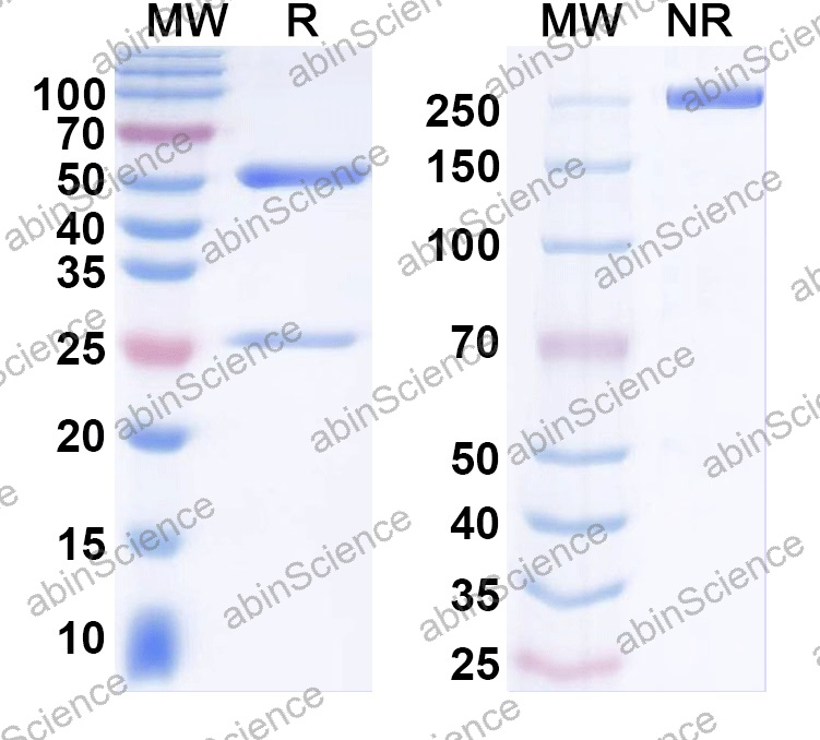
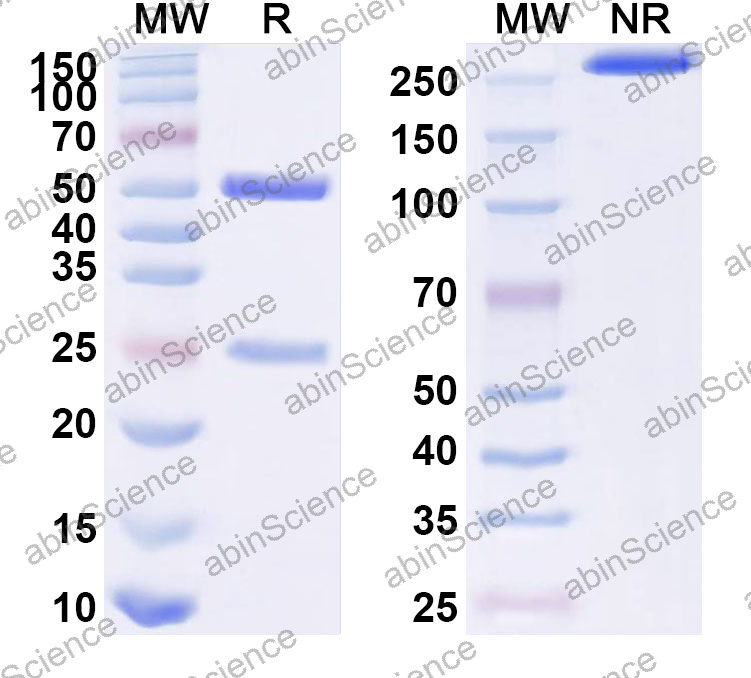
| Pureté | >95% as determined by SDS-PAGE. |
| Purification | Protein A/G purified from cell culture supernatant. |
| Numéro d'accès | P06731 |
| Forme | Liquid |
| Tampon de stockage | 0.01M PBS pH 7.4. Reférez-vous aux informations spécifiques sur le tampon dans la copie papier du datasheet ou dans le COA spécifique au lot. |
| Stabilité et stockage | Use a manual defrost freezer and avoid repeated freeze thaw cycles. Store at 2 to 8°C for frequent use. Store at -20 to -80°C for twelve months from the date of receipt. |
| Contexte | Cell adhesion molecule CEACAM5 (CD66e) is a ~76 kDa protein. Cell surface glycoprotein that plays a role in cell adhesion, intracellular signaling and tumor progression. Mediates homophilic and heterophilic cell adhesion with other carcinoembryonic antigen-related cell adhesion molecules, such as CEACAM6. Plays a role as an oncogene by promoting tumor progression; induces resistance to anoikis of colorectal carcinoma cells. 1. Taheri, M. et al. (2000) The Journal of biological chemistry 275, 26935-43. PMID: 10864933 2. Ordoñez, C. et al. (2000) Cancer research 60, 3419-24. PMID: 10910050 3. Oikawa, S. et al. (1989) Biochemical and biophysical research communications 164, 39-45. PMID: 2803308 |
| Note | For research use only. |
SEC-HPLC detection for Anti-Human CD66e/CEA/CEACAM5 Antibody (RG7813).
Flow-cytometry using anti-human CD66e antibody.CD66e Transfected CHO cells were stained with an irrelevant antibody (Blue Histogram) or an anti-human CD66e antibody monoclonal antibody (Catalog # HY546307 ,Green Histogram) at a concentration of 5 µg/ml for 30 mins at RT. After washing, bound antibody was detected using a FITC conjugated goat anti-human antibody (Catalog # HF690414) and cells analysed on a NovoCyte Flow Cytometer.
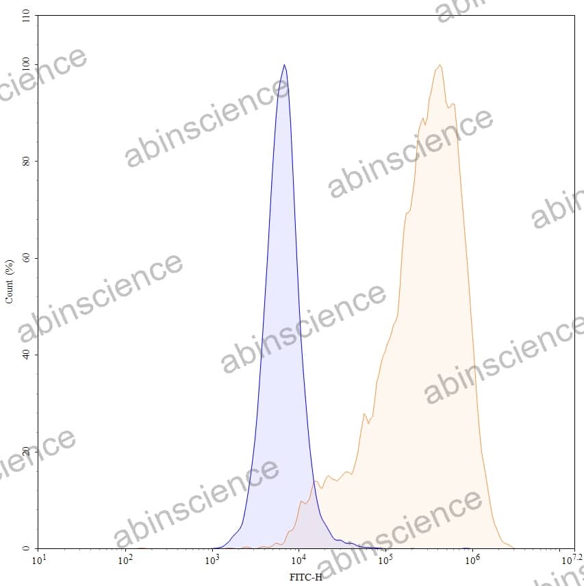
Flow-cytometry using anti-human CD66e antibody. HPAC cells were stained with an irrelevant antibody (Blue Histogram) or an anti-human CD66e monoclonal antibody (Catalog HY546307, Yellow Histogram) at a concentration of 5 µg/ml for 30 mins at RT. After washing, bound antibody was detected using a a Goat Anti-Human IgG H&L Polyclonal Antibody, FITC (abinScience: HF690414) and cells analysed on a NovoCyte Flow Cytometer.
Contactez-nous pour les devis personnalisés, les demandes en gros et tout autre problème.
Courrier : [email protected]
+86-027-65523339
Bâtiment C, No. 666, Rue Shendunsi, Wuhan, 430206, Chine


