
 中文
中文 English
 한국어
한국어 日本語
日本語 Español
 Français
 Русский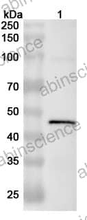


| カタログ番号 | VK564054 | ||||||||
|---|---|---|---|---|---|---|---|---|---|
| 説明 |
Anti-PEDV S1/Spike glycoprotein 1 Polyclonal Antibody (VK564054) is a rabbit polyclonal antibody detecting Spike glycoprotein in ELISA, IHC, WB.
Highlights
|
||||||||
| 種反応性 | Porcine epidemic diarrhea virus (PEDV) | ||||||||
| アプリケーション | ELISA, IHC, WB | ||||||||
| 宿主種 | Rabbit | ||||||||
| クローン性 | Polyclonal | ||||||||
| アイソタイプ | IgG | ||||||||
| 免疫原 | E. coli - derived recombinant PEDV S1/Spike glycoprotein 1 (Gln21-Glu496). | ||||||||
| テスト済みアプリケーション | WB | ||||||||
| ターゲット | Spike glycoprotein, S glycoprotein, E2, Peplomer protein, S, Porcine epidemic diarrhea virus, PEDV | ||||||||
| 精製 | Purified by antigen affinity column. | ||||||||
| アクセッション番号 | S0AP48 | ||||||||
| 形態 | Liquid | ||||||||
| 保存バッファー | 0.01M PBS, pH 7.4, 50% Glycerol, 0.05% Proclin 300. データシートのハードコピーまたはロット固有のCOAに記載された具体的なバッファー情報を参照してください。 |
||||||||
| 製品使用情報 |
| ||||||||
| 安定性と保存 | Use a manual defrost freezer and avoid repeated freeze thaw cycles. Store at 2 to 8°C for frequent use. Store at -20 to -80°C for twelve months from the date of receipt. | ||||||||
| 背景 | Spike glycoprotein (S1/Spike glycoprotein 1) is a ~151 kDa protein. S1 region attaches the virion to the cell membrane by interacting with the host receptor, initiating the infection. Binding to the receptor probably induces conformational changes in the S glycoprotein unmasking the fusion peptide of S2 region and activating membranes fusion. S2 region belongs to the class I viral fusion protein. Under the current model, the protein has at least 3 conformational states: pre-fusion native state, pre-hairpin intermediate state, and post-fusion hairpin state. During viral and target cell membrane fusion, the coiled coil regions (heptad repeats) regions assume a trimer-of-hairpins structure, positioning the fusion peptide in close proximity to the C-terminal region of the ectodomain. | ||||||||
| 注意事項 | For research use only. |
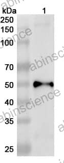
Western blot analysis was performed using anti-S1 polyclonal antibody at 1ug/mL on various samples.
Lane 1: recombinant PEDV S1/Spike glycoprotein 1 (Catalog No: VK564012)
+86-027-65523339
中国武漢市深敦寺路666号C棟、武漢、430206


