A common challenge in immuno‑biology research using animal models is determining whether an observed biological effect is directly driven by a specific immune activation pathway. While preliminary data may suggest immune cell dysfunction or demonstrate phenotypic shifts following global immune enhancement, these findings rarely isolate which specific activating signal plays the key functional role.
Utilizing agonist or stimulatory invivo functional antibodies provides a targeted approach to answer this question. By selectively amplifying signals through specific receptors or co‑stimulatory pathways, researchers can actively modulate immune responses to evaluate downstream functional consequences and directly test pathway necessity.
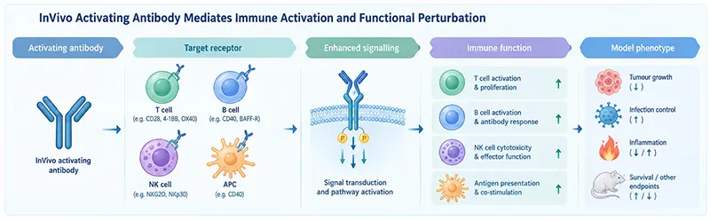
Figure 1. Schematic representation of invivo functional intervention using activating antibodies.
Immune cell activation and effector functions are regulated by multi‑receptor signaling networks, making it difficult to attribute a specific phenotype to a single activation pathway. When correlative evidence links an immune response to a target phenotype, the critical next step is demonstrating whether selectively enhancing a candidate pathway directly drives expected biological functions.
In oncology models, for instance, effector T cells may be present but functionally constrained. Rather than aiming for non‑specific immune cell expansion or general hyper‑activation, researchers can use agonist invivo antibodies to boost candidate co‑stimulatory signals and evaluate whether this targeted enhancement delivers expected functional shifts that alter tumor progression. This experimental logic extends to infection, auto‑immunity, and chronic inflammation models.
Targeted functional intervention elevates candidate pathways from correlative observations into testable hypotheses. Demonstrating concomitant enhancements in cellular function and model phenotype provides direct evidence of a pathway's functional contribution.
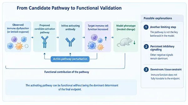
Figure 2. Experimental logic for validating candidate immune activation pathways using activating invivo antibodies.
When evaluating agonist invivo antibodies, the focus must extend beyond terminal phenotypic changes to include granular functional readouts.
First, confirm that the intervention induces specific functional activation in target cell populations (e.g., T cells, B cells, or natural killer [NK] cells) consistent with the target pathway. Second, determine whether these cellular shifts translate into meaningful systemic or tissue‑level outputs. The primary research question shifts from "did the model phenotype change?" to "which specific immune mechanism was altered by amplifying this signal?".
Functional readouts should be tailored to the cell type and receptor axis:
Aligning cell‑specific functional metrics with overall endpoints provides clear evidence of pathway involvement.
Target selection should not focus on identifying the "strongest" immune activator, but rather on pinpointing the specific functional bottleneck in the system—whether that is insufficient T‑cell co‑stimulation, restricted B‑cell activation, blunted NK cytotoxicity, or impaired antigen presentation.
| Research Focus | Representative Activation / Co‑stimulatory Nodes | Primary Readouts & Functional Focus |
|---|---|---|
| T‑Cell Activation & Co‑stimulation | CD28, ICOS, CD27, 4‑1BB, OX40, GITR, CD2 | T‑cell priming, expansion, effector function, and persistence |
| B‑Cell Activation & Survival | CD40, CD27, BAFF‑R, TACI, BCMA | B‑cell activation, survival, differentiation, and antibody responses |
| NK‑Cell Activation | NKG2D, CD16, NKp30, NKp44, NKp46, DNAM‑1 | NK recognition, activation, cytotoxicity, and effector function |
| Dendritic Cell / APC Regulation | CD40 and related co‑stimulatory axes | Antigen presentation, co‑stimulatory capacity, and T‑cell priming |
| Innate Immune Activation | TLRs, STING, RIG‑I | Pathogen recognition, innate immune priming, and inflammatory signaling |
| Regulatory Immune Modulation | GITR, OX40, CD27, 4‑1BB | Balance between effector and regulatory immune functions |
| Cross‑Cellular Immune Regulation | CD40, CD27, 4‑1BB | Intercellular signal transduction and functional amplification |
A critical, highly informative scenario in agonist antibody studies occurs when target cellular function is successfully enhanced, but terminal disease endpoints show minimal change.
For example, an agonist antibody may drive robust effector cell activation and expansion without significantly altering tumor burden or pathology. This discrepancy highlights essential regulatory insights:
Evaluating experimental outcomes across distinct functional tiers provides clear mechanistic direction:
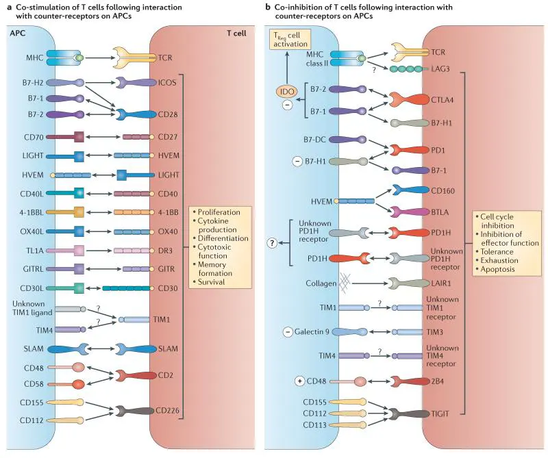
Figure 3. Co‑stimulatory and co‑inhibitory signal integration in T cells (DOI: 10.1038/nri3405).
When modulating a single positive pathway is insufficient to alter disease outcomes, investigating the balance between co‑stimulatory and co‑inhibitory pathways becomes essential.
A powerful approach involves comparing three conditions: Pathway Activation, Checkpoint Blockade, and Combined Activation + Blockade. Rather than assuming combination therapy will automatically yield superior results, this framework determines which regulatory axis limits the system:
In immuno‑oncology, combining agonist co‑stimulatory antibodies with immune checkpoint blockers leverages these complementary pathways to overcome multi‑layered immunosuppression.
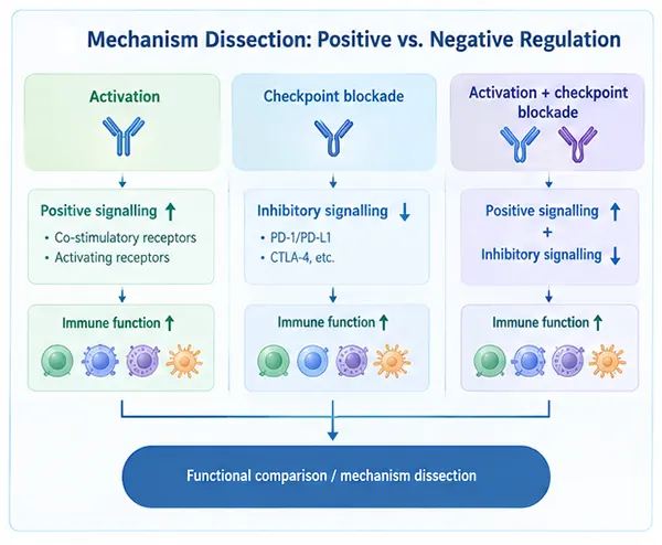
Figure 4. Combinatorial strategies integrating positive activation and checkpoint blockade for mechanistic resolution.
Agonist invivo antibodies serve as precise functional tools to perturb targeted signaling nodes and test their causal role in immune responses. This methodology applies across T cells, B cells, NK cells, antigen‑presenting cells (APCs), and innate signaling networks.
Concomitant shifts in cell function and endpoint phenotype confirm pathway necessity. Enhanced cellular responses paired with static endpoints highlight downstream rate‑limiting steps. Combining activation signals with checkpoint blockade resolves multi‑node regulatory networks.
Ultimately, stimulatory invivo antibodies move beyond simple "immune enhancers"—they provide the active perturbation tools required to convert correlative activation pathways into validated invivo mechanisms.
abinScience offers a comprehensive portfolio of high‑grade invivo functional antibodies targeting key co‑stimulatory and activation nodes across T cells, B cells, NK cells, and innate immune pathways. These tools support pathway validation, functional characterization, and combination immunotherapy studies.
| Catalog No. | Product Name |
|---|---|
| HW571010 | InVivoMAb Anti‑Human 4‑1BB/TNFRSF9/CD137 Antibody (Iv0139) |
| HB782010 | InVivoMAb Anti‑Human CD40/TNFRSF5 Antibody (Iv0136) |
| HX011010 | InVivoMAb Anti‑Human IL‑2 Antibody (Iv0020) |
| HF813010 | InVivoMAb Anti‑Human IFN‑gamma Antibody (Iv0049) |
| HB769010 | InVivoMAb Anti‑Human IL‑12/IL‑23 p40 Antibody (Iv0026) |
| HW688010 | InVivoMAb Anti‑Human Flt‑3 Ligand/FLT3L Antibody (Iv0057) |
| HF827010 | InVivoMAb Anti‑Human IFN‑alpha 2/IFNA2 Antibody (Iv0001) |
| HF004030 | InVivoMAb Anti‑Human EGFR & Mouse CD3 epsilon Antibody (Iv0228) |
| HV599010 | InVivoMAb Anti‑Human BAFFR/TNFRSF13C Antibody (CB3s) |
| HF858010 | InVivoMAb Anti‑Human NKp46/NCR1 Antibody (Iv0208) |
| MC336209 | InVivo Plus Anti‑Mouse 4‑1BB/TNFRSF9/CD137 Antibody(3H3) |
| MY422019 | InVivo Plus Anti‑Mouse CD28 Antibody (PV‑1) |
| MB782109 | InVivo Plus Anti‑Mouse CD40/TNFRSF5 Antibody(FGK45) |
| MB123019 | InVivo Plus Anti‑Mouse NKG2D/CD314 Antibody(CX5) |
Click here to explore our complete portfolio of InVivo antibody products and InVivo Plus antibody products
+86-027-65523339
中国武漢市深敦寺路666号C棟、武漢、430206


 中文
中文 English
 한국어
한국어 日本語
日本語 Español
 Français
 Русский