From Infection to Malignancy: How HPV Rewrites Cellular Fate
Дата выпуска:
2026-08-21 Количество просмотров: 35
Among female malignancies globally, cervical cancer has long ranked near the top in both incidence and mortality. Over 99% of cervical cancer cases are directly linked to persistent infection with high-risk human papillomaviruses (HPV), establishing HPV as an enduring research focus in virology and oncology.
HPV infection is extremely common in the general population, yet the vast majority of infections are transient—cleared spontaneously by the host immune system within several months to two years without causing overt pathological lesions. Fewer than 1% of high-risk HPV infections persist, progressively inducing aberrant cellular proliferation and ultimately driving progression to precancerous lesions and invasive malignancies.
This highlights a fundamental question in HPV research: given that all represent HPV infections, why are some completely eliminated by the immune system, while certain high-risk strains persist long-term intracellularly, ultimately driving normal cells toward malignant transformation?
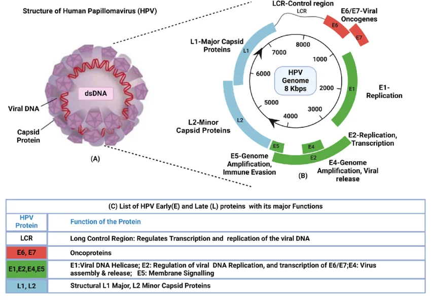
Figure 1. HPV structure, genomic organization, and protein functions (DOI: 10.1007/s12038-025-00493-8)
I. Infection ≠ Carcinogenesis: How the HPV Life Cycle Dictates Disease Outcome
1.1 Understanding HPV: Classification and Pathogenicity of Double-Stranded DNA Viruses
HPV comprises a group of non-enveloped, double-stranded circular DNA viruses exhibiting strict epitheliotropism, specifically infecting human cutaneous or mucosal epithelial tissues. Over 200 HPV genotypes have been identified to date, categorized into low-risk and high-risk types based on pathogenic potential and oncogenic risk.
Low-risk types, typified by HPV-6 and HPV-11, usually cause only benign proliferative lesions of the skin or mucosa (such as common warts, plantar warts, and condyloma acuminata). Their viral genomes remain extrachromosomal without integrating into host chromosomes, posing virtually no risk of malignant transformation.
High-risk types (approximately 15 established genotypes) are strongly correlated with malignancies, including cervical, anal, oropharyngeal, and vaginal cancers. Among these, HPV-16 and HPV-18 exhibit the highest oncogenicity, accounting for approximately 70% of cervical cancer cases worldwide.
1.2 The Infection Cascade: How HPV Replicates Within Epithelial Cells
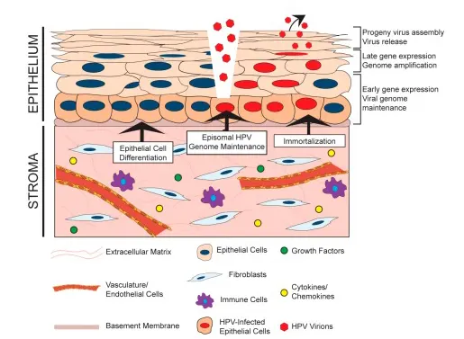
The HPV life cycle relies entirely on host epithelial cell differentiation. Viral infection and replication are tightly regulated in a spatiotemporal manner, proceeding without inducing cell lysis or systemic virion dissemination.
The primary target of infection is proliferating stem cells in the basal layer of epithelial tissue. Accessing deeper tissues through micro-abrasions in the skin or mucosa, the virus binds to cell-surface receptors on basal stem cells, enters via endocytosis, and releases its viral genome into the nucleus. During early infection, the viral genome exists as an episomal circular DNA plasmid, expressing low levels of early proteins such as E1, E2, E6, and E7 to maintain stable, low-copy-number viral genome replication, without inducing obvious morphological alterations in host cells.
As basal cells divide, migrate toward the epithelial surface, and progressively differentiate, viral gene expression undergoes stage-specific transitions: early proteins continuously regulate the cell cycle, delaying terminal differentiation to supply host machinery and substrates for robust viral DNA amplification. Upon reaching the superficial epithelial layers and entering terminal differentiation, late proteins L1 and L2 are expressed abundantly, self-assembling to form the viral capsid that encapsidates progeny viral genomes. Mature infectious virions are then shed into the environment along with the natural desquamation of superficial epithelial cells, initiating a new round of infection.
Figure 2. The HPV life cycle (DOI: 10.3390/v9080219)
II. Structural Proteins of HPV: How L1 and L2 Drive Virion Assembly and Entry
2.1 Core Structural Proteins: L1 and L2
Mature HPV virions consist of an icosahedral protein capsid encapsidating an internal double-stranded circular DNA genome. The capsid is assembled from two structural proteins, L1 and L2, which constitute the structural framework for the viral life cycle.
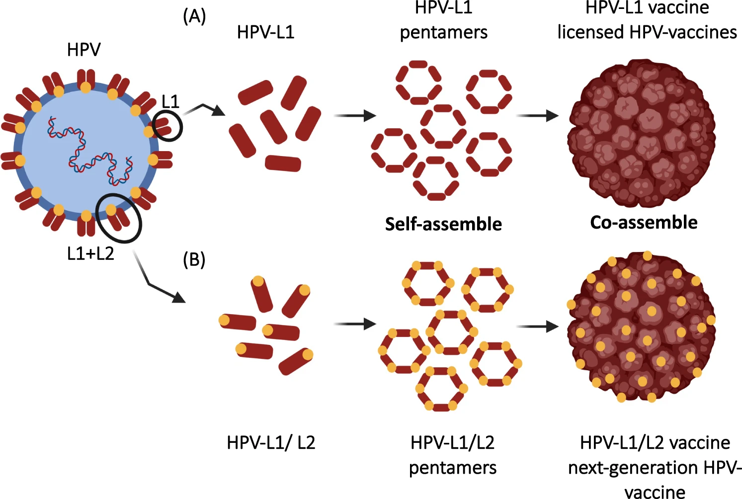
L1 is the major capsid protein, accounting for over 80% of total capsid protein mass. Featuring a highly conserved amino acid sequence, it forms the structural backbone of the capsid. Individual L1 monomers form pentameric capsomers, the basic building blocks of assembly. Recombinantly expressed L1 can self-assemble into virus-like particles (VLPs) that mirror authentic virions in morphology and spatial conformation. Lacking viral nucleic acids, VLPs are non-infectious yet preserve native antigenic epitopes, making them critical tools in HPV structural biology and vaccinology/immunology research.
L2 is the minor capsid protein. Mostly buried within the capsid shell with only a minor N-terminal peptide exposed on the virion surface, L2 shows lower sequence conservation than L1 but plays indispensable roles in several key stages of infection. Furthermore, L2 interacts with the viral genome via its nucleic acid-binding domain, guiding accurate genome packaging during assembly and ensuring functional integrity of progeny virions.
Figure 3. HPV L1 and L2 capsid proteins. A: VLPs assembled from L1 protein; B: An HPV vaccine expressing both L1 and L2 capsid proteins (DOI: 10.1038/s41423-022-00897-8)
2.2 Synergistic Roles of L1 and L2 in Virion Assembly and Entry
The functions of L1 and L2 span both ends of the HPV life cycle, orchestrating virion assembly and maturation as well as mediating host cell attachment and entry—providing the molecular foundation for viral transmission.
Assembly Phase: As infected host cells migrate to the superficial epithelial layer and enter terminal differentiation, the viral late promoter is activated, triggering high-level expression and nuclear translocation of L1 and L2 proteins. L1 pentamers spontaneously assemble into an icosahedral capsid frame, while L2 binds to viral genomic DNA, facilitating its packaging into the capsid interior while stabilizing overall capsid architecture via protein-protein interactions. This yields structurally complete, infectious progeny virions that are shed alongside desquamating epithelial cells.
Infection Initiation Phase: L1 mediates primary cell attachment: the virus targets heparan sulfate proteoglycans (HSPGs) and other cell-surface receptors on basal epithelial cells via L1, triggering receptor-mediated endocytosis into endosomes. Once internalized, L2 exerts crucial membrane-penetration and trafficking functions: specific L2 peptides mediate endosomal membrane disruption/perforation, aiding nuclear escape of the viral genome, and interact with host transport factors to precisely target viral DNA into the cell nucleus. This establishes primary infection, paving the way for genome replication and early gene expression.
III. Oncoproteins E6 and E7: Core Effectors Driving Malignant Transformation
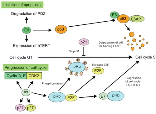
Figure 4. Roles of E6 and E7 proteins in HPV-associated carcinogenesis (DOI: 10.4081/oncol.2012.e17)
3.1 Mechanisms of Persistence and Carcinogenesis: Breakdown of Tumor Suppressor Defenses
Normal cellular proliferation, differentiation, and survival are controlled by intricate networks: (1) cell cycle checkpoints stringently regulate division pace and limit; (2) DNA damage response systems safeguard genomic integrity; and (3) apoptotic pathways selectively eliminate irreversibly damaged or aberrantly proliferating cells. Together, these form a robust tumor suppressor barrier preventing transformation.
In low-risk HPV infections, E6/E7 proteins exert minimal disruption on host regulatory networks, and the viral life cycle terminates naturally as epithelial cells shed, resulting in transient, non-oncogenic infections. In contrast, persistent high-risk HPV infection can induce pivotal genomic alterations: viral episomal circular DNA undergoes double-strand breaks and randomly integrates into the host cell chromosome.
Upon viral integration, local genomic regulation is disrupted, leading to dysregulated, constitutive overexpression of the E6 and E7 early proteins. This deregulates the host cell's tumor suppressor network, allowing cells to escape normal growth controls. Over prolonged periods of infection, cumulative somatic mutations accumulate, culminating in malignant transformation.
3.2 E6 Protein: Targeted Degradation of p53 to Dismantle the "Guardian of the Genome"
Within the cell's tumor-suppressive machinery, p53 serves as the central "guardian of the genome." Under cellular stress—such as DNA damage or oncogene activation—p53 is rapidly activated to arrest cell cycle progression (enabling DNA repair) or initiate apoptosis if damage is irreparable, purging mutated cells at the source.
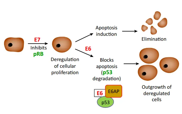
High-risk HPV E6 is the key effector disarming this host defense. E6 recruits host E6-associated protein (E6AP, an E3 ubiquitin ligase) to form an E6–E6AP complex, which specifically binds p53, targeting it for polyubiquitination and subsequent proteasomal degradation. Chronic depletion of p53 deprives the cell of genome surveillance and apoptotic induction; consequently, unrepaired DNA damage accumulates through successive cell divisions, causing pronounced chromosomal instability and providing a mutagenic foundation for malignant conversion.
Additionally, E6 prolongs infected cell lifespan and evades immune clearance by activating telomerase (hTERT), disrupting cellular adhesion, and suppressing immune recognition pathways. Conversely, low-risk HPV E6 exhibits negligible binding affinity for p53, explaining its lack of oncogenic drive.
3.3 E7 Protein: Disruption of the Rb Pathway to Release Cellular Proliferation Brakes
While E6 dismantles the cell's genomic security network, E7 directly releases the molecular brakes on cell proliferation by targeting the Retinoblastoma protein (Rb).
Rb is a master regulator of the G1/S cell cycle transition. In quiescent cells, unphosphorylated Rb sequesters E2F family transcription factors, repressing E2F activity and blocking entry into S phase. When physiological division is required, cyclin-dependent kinases (CDKs) phosphorylate Rb, liberating E2F to advance the cell cycle in a regulated manner.
High-risk HPV E7 binds directly to Rb via its conserved CR2 domain, disrupting the Rb–E2F complex. This results in constitutive E2F activation, driving uncontrolled transition from G1 to S phase. E7 also targets cyclin-dependent kinase inhibitors such as p21 and p27, further dismantling negative growth regulation and locking host cells in a state of hyperproliferation.
Similar to E6, low-risk HPV E7 binds Rb with significantly lower affinity, causing minimal cell cycle perturbation and failing to induce persistent dysregulated proliferation.
3.4 Synergistic Transformation: Coordinated Action of E6 and E7
HPV-driven carcinogenesis is a multi-step, multi-factorial long-term cascade, with E6–E7 synergy acting as the primary driver:
Figure 5. Synergistic action of HPV E6 and E7 proteins in malignant cell transformation (DOI: 10.1016/j.tim.2017.07.007)
IV. From Protein Function to Disease Pathogenesis: Key Viral Proteins as Core Targets in HPV Research
As HPV research advances, focus in the field has shifted from diagnostic presence ("Is HPV present?") to functional pathology ("How does the virus reprogram host cells to drive disease progression?"), moving from simple detection to detailed molecular mechanistic inquiry.
Early research and clinical diagnostics focused primarily on HPV DNA detection and genotyping to evaluate infection status and assess epidemiological risk.
Current research focuses heavily on the precise functional mechanisms of key viral proteins:
• Elucidating structural and functional differences across genotypes: Clarifying why distinct HPV types exhibit marked differences in oncogenic potential, providing a theoretical foundation for regional prevention and control strategies.
• Correlating viral protein expression with disease progression: Quantifying E6/E7 mRNA or protein levels directly reflects viral activity, offering higher scientific and predictive value for monitoring lesion progression.
• Targeting viral proteins for therapeutic intervention: Beyond L1-based prophylactic vaccines, current frontiers include therapeutic vaccines against E6/E7, small-molecule inhibitors, and nucleic acid-based therapeutics (e.g., RNAi and CRISPR system delivery).
Conclusion
The continuum from HPV infection to malignant transformation represents a prolonged molecular tug-of-war between viral proteins and host cell regulatory networks. Structural capsid proteins L1 and L2 mediate infectious entry and form the primary targets for neutralizing immunity and vaccine design. Meanwhile, early oncoproteins E6 and E7 act as the master drivers of malignant transformation by degrading key tumor suppressors and dismantling cell cycle checkpoints.
Deciphering the precise molecular mechanisms of L1/L2 and E6/E7 not only answers fundamental questions about why specific HPV infections progress to cancer, but also provides a robust scientific foundation for developing next-generation preventive, diagnostic, and therapeutic strategies.
abinScience HPV Research Tools
To support research on HPV structural biology, infection mechanisms, and protein expression, abinScience offers high-quality recombinant proteins and antibodies targeting key viral proteins across multiple HPV genotypes. Covering essential targets such as L1, L2, E6, and E7, our portfolio supports diverse research applications including ELISA, Western blotting, and immunohistochemistry (IHC).
[1] Doorbar J, Egawa N, Griffin H, Kranjec C, Murakami I. Human papillomavirus molecular biology and disease association. Rev Med Virol. 2015 Mar;25 Suppl 1(Suppl Suppl 1):2-23. doi: 10.1002/rmv.1822. PMID: 25752814; PMCID: PMC5024016.
[2] Moody CA, Laimins LA. Human papillomavirus oncoproteins: pathways to transformation. Nat Rev Cancer. 2010 Aug;10(8):550-60. doi: 10.1038/nrc2886. Epub 2010 Jul 1. PMID: 20592731.
[3] Schiller JT, Lowy DR. Understanding and learning from the success of prophylactic human papillomavirus vaccines. Nat Rev Microbiol. 2012 Oct;10(10):681-92. doi: 10.1038/nrmicro2872. Epub 2012 Sep 10. PMID: 22961341; PMCID: PMC6309166.
[4] Gupta C, Dolma KG, Sherpa ML, Bag A, Byahut A. Human papillomavirus vaccine: Success and challenges. J Biosci. 2025;50:51. PMID: 40619783.
[5] Spurgeon ME, Lambert PF. Human Papillomavirus and the Stroma: Bidirectional Crosstalk during the Virus Life Cycle and Carcinogenesis. Viruses. 2017 Aug 9;9(8):219. doi: 10.3390/v9080219. PMID: 28792475; PMCID: PMC5580476.
[6] Mohsen MO, Bachmann MF. Virus-like particle vaccinology, from bench to bedside. Cell Mol Immunol. 2022 Sep;19(9):993-1011. doi: 10.1038/s41423-022-00897-8. Epub 2022 Aug 12. PMID: 35962190; PMCID: PMC9371956.
[7] Hoppe-Seyler K, Bossler F, Braun JA, Herrmann AL, Hoppe-Seyler F. The HPV E6/E7 Oncogenes: Key Factors for Viral Carcinogenesis and Therapeutic Targets. Trends Microbiol. 2018 Feb;26(2):158-168. doi: 10.1016/j.tim.2017.07.007. Epub 2017 Aug 17. PMID: 28823569.


 中文
中文 English
 한국어
한국어 日本語
日本語 Español
 Français
 Русский