Journal of Nanobiotechnology · Male Reproductive Health Research Digest
Non-obstructive azoospermia (NOA) is the most severe form of male infertility, accounting for the majority of azoospermia cases. Its etiology is highly heterogeneous, encompassing known causes such as Klinefelter syndrome (KS), Y-chromosome microdeletions (YCMD), and mumps orchitis (MO), while a substantial proportion of cases remain unexplained and are classified as idiopathic NOA (iNOA). Current evaluation of NOA still relies heavily on invasive procedures, including hormone testing, testicular volume measurement, chromosomal and genetic testing, and testicular biopsy—the diagnostic gold standard. A reliable non-invasive alternative that could replace or triage biopsy is still lacking.
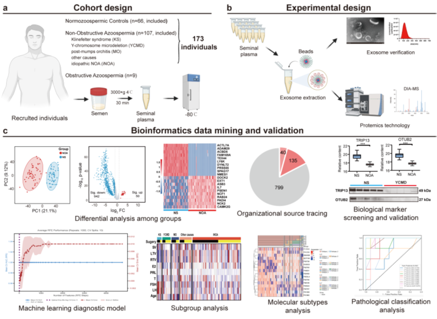
Seminal plasma extracellular vesicles (SPEVs), secreted by epithelial cells and germ cells of the male reproductive tract, carry proteins that are relatively stable and are considered promising candidates for liquid biopsy. Earlier studies have attempted to use SPEV proteins to distinguish NOA from obstructive azoospermia, but were limited by small sample sizes and the lack of a scalable enrichment method suited to in-depth proteomics. This study, conducted by a team at Peking University First Hospital and published in Journal of Nanobiotechnology, analyzed a large cohort of seminal plasma samples to systematically evaluate the value of the SPEV proteome in distinguishing NOA from healthy controls, as well as in molecular subtyping within NOA.
Large Seminal Plasma CohortSystematic evaluation of SPEV proteomics for distinguishing NOA from healthy controls
Two Molecular Subtypes of iNOAUnsupervised clustering reveals a stratification dimension independent of clinical indicators
CLPSL1 Validated by Western BlotabinScience antibody (HB388014) confirms downregulation in the iNOA group
01Isolation and Characterization of SPEVs
The researchers used a functionalized magnetic bead-based capture method to isolate SPEVs—a fast, straightforward enrichment process that avoids the cumbersome steps of traditional approaches such as ultracentrifugation. Electron microscopy, particle size analysis, and detection of marker proteins together confirmed that the captured vesicles exhibited typical morphology and purity, laying the groundwork for downstream proteomic analysis. Building on this workflow, the team used high-resolution mass spectrometry to achieve deep-coverage detection of seminal plasma vesicle proteins.
02Proteomic Comparison Between NOA and Normal Subjects (NS)
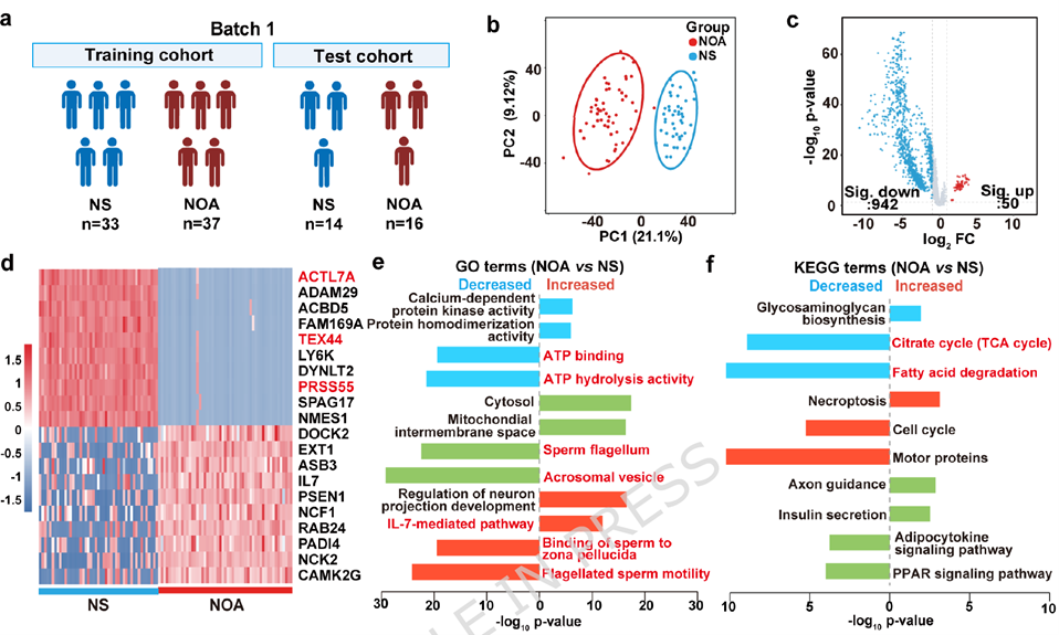
The researchers first compared the SPEV protein profiles of NOA patients with those of normal subjects (NS). The two groups were clearly distinguishable, with NOA patients showing widespread changes in protein expression—predominantly downregulation—affecting core reproductive pathways such as spermatogenesis and sperm flagellar structure and motility. The activation of certain immune-related pathways and broad suppression of energy metabolism pathways were also observed.
This pattern of differences was reproduced in an independent sample set, indicating that the detection workflow is highly reproducible.
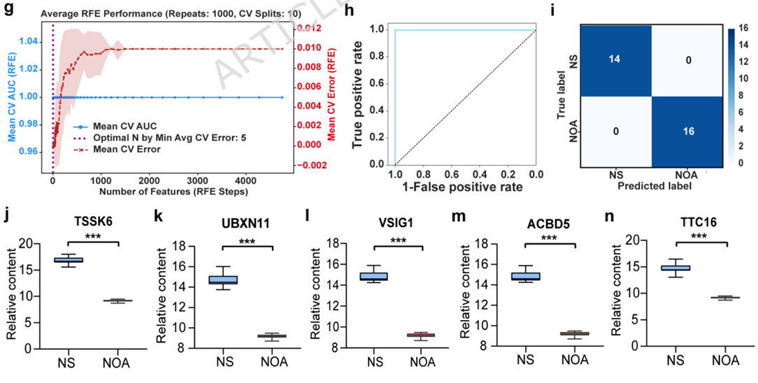
Building on these findings, the researchers identified a core set of proteins that effectively distinguishes NOA from normal subjects. The resulting classification model achieved strong discriminative performance in the test set and maintained good classification accuracy in a fully independent second batch of samples, suggesting these proteins hold promise as non-invasive diagnostic markers.
03Comparison Across NOA Etiological Subgroups
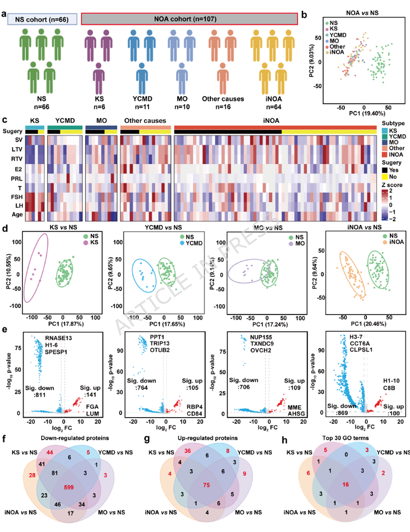
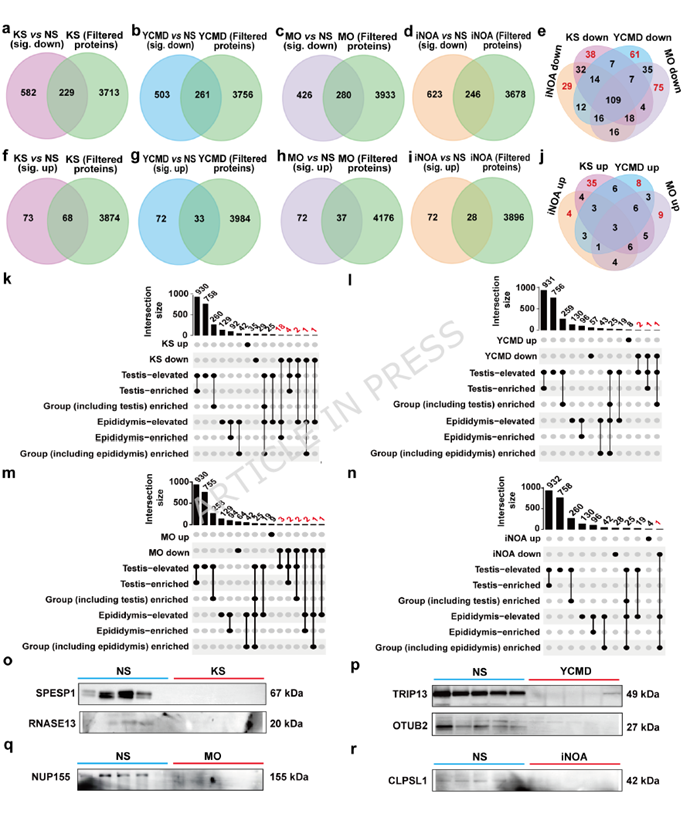
NOA can be divided into several subgroups by etiology. The researchers found that, regardless of underlying cause, each subgroup showed a similar set of core molecular changes relative to normal subjects, centered on impaired spermatogenesis and disrupted energy metabolism—suggesting that different etiologies of NOA may converge on common downstream pathological mechanisms. At the same time, each etiological subgroup also carried its own subset of specific protein changes. For example, protein changes associated with Klinefelter syndrome were linked to complement immune activation; those associated with Y-chromosome microdeletion were more related to nuclear structure and transport functions; changes associated with mumps orchitis were concentrated in ciliary structures; and idiopathic NOA showed more changes in protein folding and microtubule-related pathways.
Further analysis using tissue-specificity databases confirmed that these differentially expressed proteins were primarily derived from testicular tissue. Several representative proteins were selected and validated by Western blot in an independent sample set, and the results were consistent with the proteomic screening conclusions.
04Molecular Subtyping of iNOA
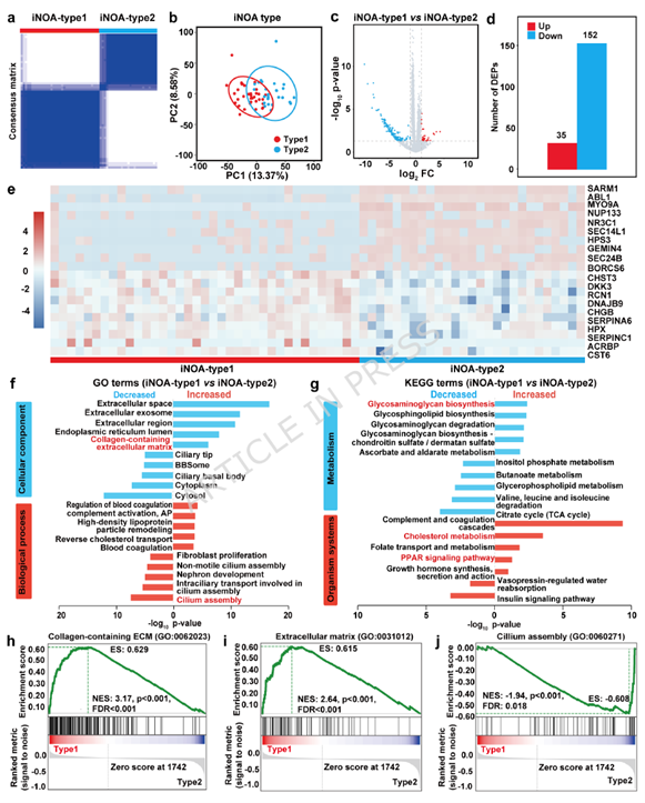
For the currently unexplained cases of idiopathic NOA, the researchers performed unsupervised clustering based on proteomic expression profiles, which further divided these patients into two subgroups with distinct molecular features. Interestingly, this proteome-based classification showed no clear association with existing clinical parameters, indicating that it captures a new dimension independent of conventional clinical assessment. Functionally, the two subtypes were respectively associated with the activity of pathways related to extracellular matrix metabolism and ciliary structure assembly. The authors also noted that these pathway-level differences do not directly correspond to differences in sperm retrieval capability, but rather reflect distinct underlying directions of functional impairment.
05Exploratory Prediction: Testicular Histopathological Classification
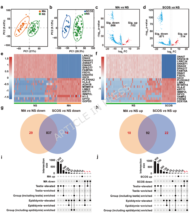
Beyond etiology, the researchers also examined another dimension—testicular histopathological type, specifically the two clinically common categories of Sertoli cell-only syndrome (SCOS) and maturation arrest (MA). These two pathological types reflect different degrees and natures of impaired spermatogenic function within the testis, and can currently only be confirmed by biopsy. The researchers first compared SPEV protein differences between these two patient groups and normal subjects, finding that both groups were clearly distinguishable from normal subjects, each showing a set of characteristic protein changes; tissue-specificity database analysis further confirmed the testicular origin of these proteins. Building on this, the researchers attempted to directly compare protein differences between the SCOS and MA groups and built a classification model to test whether SPEV proteins could distinguish between these two pathological types. Under a conventional train-test split, the model showed relatively high discriminative performance, but due to the small sample size in the SCOS group, its ability to identify this category was unstable. When the researchers re-evaluated the model using a more rigorous nested cross-validation approach, overall performance declined markedly. The authors concluded that the SPEV proteome is not yet sufficient to support non-invasive classification of testicular pathology, and that this line of investigation remains at an early stage.
06Exploratory Prediction: Surgical Sperm Retrieval Outcome
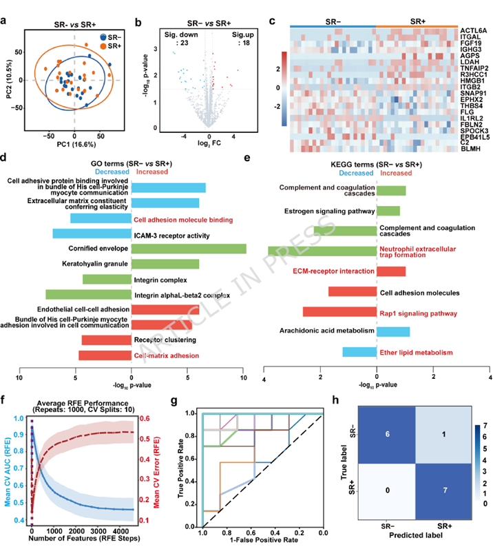
Another question of clinical relevance is whether seminal plasma proteins measured before surgery can predict whether microdissection testicular sperm extraction will successfully retrieve sperm. This has practical value for guiding treatment choices and assessing the necessity of surgery. The researchers compared SPEV protein profiles between patients with successful and failed sperm retrieval and found that overall differences between the two groups were not particularly pronounced; only after relaxing the statistical threshold did a subset of differentially expressed proteins emerge, involving functions such as cell adhesion and extracellular matrix interactions. Based on this, the researchers built a combined predictive model incorporating select protein features and clinical information (age). Similarly, the model demonstrated good discriminative ability under conventional validation, but its performance dropped noticeably when re-assessed using more rigorous validation methods, and its advantage over a model built solely on clinical indicators was not substantial.
In both of these exploratory analyses, the researchers adopted a cautious stance: on the one hand, the preliminary results suggest meaningful potential applications; on the other, under more stringent statistical validation, the stability and generalizability of the models were constrained by limited sample size. As such, the value of the SPEV proteome for testicular pathology classification and prediction of sperm retrieval outcomes remains at the hypothesis-generating stage, and will require larger, multi-center, independent cohorts for further verification.
abinScience Experimental Support
Notably, in the Western blot validation of etiology-subgroup-specific candidate proteins, the researchers used an abinScience CLPSL1 primary antibody (Cat. No. HB388014) to confirm the downregulation trend of CLPSL1 in the idiopathic NOA (iNOA) group relative to normal subjects. This result was consistent with the earlier proteomic screening findings, providing independent validation support for the proposal of CLPSL1 as a potential biomarker for iNOA.


 中文
中文 English
 한국어
한국어 日本語
日本語 Español
 Français
 Русский