Non-Human Primate Models: Platforms for Translational B Cell and Humoral Immunity Research
Non-human primates (NHPs), including rhesus macaques and African green monkeys, possess immune systems highly homologous to humans, making them essential models for preclinical vaccine evaluation, infectious immunity studies, and autoimmune disease research. Whether analyzing protective humoral responses induced by vaccination or clarifying the functional roles and differentiation trajectories of B and plasma cells during disease progression, these NHP models provide irreplaceable biological data and theoretical support for clinical translational studies.
Limitations of Conventional Approaches
Previous NHP B cell studies largely relied on flow cytometry panels customized for specific scientific questions, often adapted from human B cell detection systems. Limitations include insufficient validation of cross-reactivity between anti-human monoclonal antibodies and NHP antigens, lack of systematic testing across multiple tissues and sample types, and inconsistent definitions of subsets and gating strategies. These factors pose challenges for cross-study data comparison and result validation.
Multidimensional, Broadly Applicable Panels for B/Plasma Cell Analysis
OMIP-026 was specifically developed as a 10-color B cell panel and an 11-color plasma cell panel, covering “lineage – differentiation – activation – homing” axes to enable full-dimensional analysis of target cells in a single staining. Notably, these panels achieve triple adaptability across species, sample types, and tissue sources, and through careful antibody clone validation and fluorochrome optimization, they overcome longstanding technical bottlenecks in NHP immunophenotyping, providing a critical tool applicable across disease model studies.
As the first standardized multicolor flow cytometry panel for rhesus macaque B and plasma cells, OMIP-026 fills the gap in systematic, reproducible tools for NHP B cell phenotyping and provides a unified methodological reference for human immune disease research modeled in rhesus macaques, including HIV, autoimmune diseases, and vaccine development.
1. OMIP-026 Panel
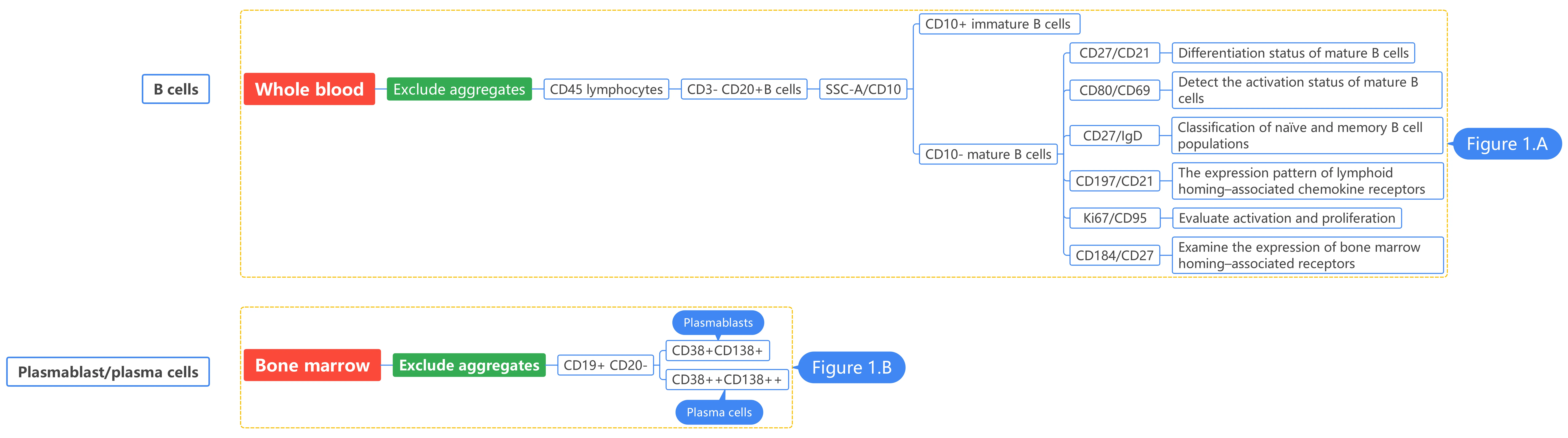
2. Gating Logic
3. Experimental Results
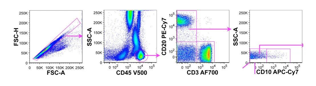
Figure A: Whole blood sample, B-cell comprehensive analysis.
B cells are identified as CD45+ SSClow CD3− CD20+ populations following exclusion of aggregates. Immature and mature B cells are distinguished based on CD10 expression.
Within mature B cells, multidimensional analysis is performed using combinations of CD21/CD27, CD27/IgD, CD80/CD69, CD21/CD197, CD95/Ki67, and CD27/CD184 to resolve differentiation states, activation profiles, and functional heterogeneity.
CD21 / CD27
Illustrates the differentiation status of mature B cells, separating naïve and memory subsets and showing their relative distribution.
CD80 / CD69
Reflects the activation status of mature B cells and highlights functionally activated populations.
CD27 / IgD
Provides a more precise classification of naïve and memory B cell populations, allowing discrimination of different memory compartments.
CD21 / CD197
Shows the expression pattern of lymphoid homing–associated chemokine receptors, indicating potential migration toward secondary lymphoid tissues.
CD95 / Ki67
Simultaneously reflects activation and proliferation, revealing functionally active and cycling B cell subsets.
CD27 / CD184
Demonstrates the expression of receptors associated with bone marrow homing, indicating a potential tendency toward terminal differentiation or plasma cell–related niches.
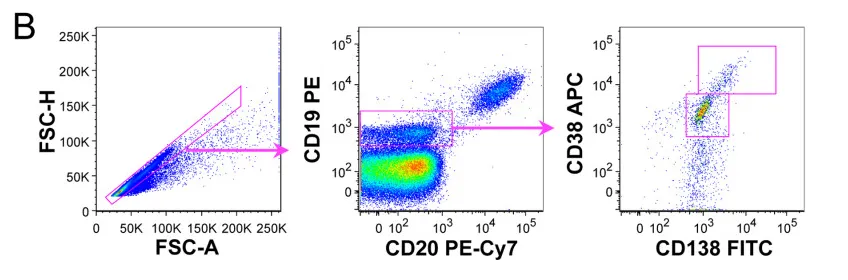
Figure B: Bone marrow sample, analysis of plasma cell lineage
Plasmablast and plasma cell gating strategy. Plasmablasts were defined as CD19+ CD20− CD38+ CD138+ and plasma cells as CD19+ CD20− CD38++ CD138++.
4. Panel Interpretation
4.1 Multidimensional Phenotyping for B/Plasma Cells
The 10-color B cell and 11-color plasma cell panels allow single-staining analysis covering lineage, differentiation, activation, proliferation, and homing. CD3/CD19/CD20/CD45 defines target cells. CD10, IgD/CD21/CD27, and CD38/CD138 capture full differentiation from naive B cells to terminal plasma cells. CD69/CD80/CD95 and Ki67 quantify activation and proliferation levels, while CD197 and CD184 reveal migration and homing properties. This integrated design substantially improves detection efficiency and data completeness.
4.2 Triple Compatibility: Species, Sample Type, and Tissue Source
The panels are applicable across species (rhesus macaques, African green monkeys), sample types (fresh whole blood, PBMCs, thawed bone marrow), and tissues (peripheral blood, bone marrow, lymph nodes, spleen, tonsils), enabling comprehensive cross-context phenotypic comparisons without additional optimization.
4.3 Foundational and Translational Value
OMIP-026 establishes a full research workflow: methodological development → reference dataset construction → practical application. It provides baseline phenotypic references in healthy rhesus macaques and has been applied in SIV infection and autoimmune disease models. The panels also support vaccine development and drug efficacy assessment, bridging animal models and translational clinical applications.
5. Applications
Vaccine development Drug efficacy evaluation Anti-infective immune response studie Autoimmune disease research Humoral immunity analysis B cell development and differentiation studies
6. Conclusion
OMIP-026 provides a standardized and highly versatile solution for multidimensional characterization of B and plasma cells in non-human primates. By integrating comprehensive phenotypic markers with broad applicability across species, sample types, and tissues, it significantly enhances data comparability and reproducibility. This panel not only advances fundamental immunological research but also supports translational efforts bridging animal models and clinical applications.
Get More Flow Cytometry Antibodies
abinScience offers high-quality Flow Cytometry Antibodies for B cell and plasma cell immunophenotyping, supporting research in non-human primates and other model systems.
References
[1] Neumann B, Sopper S, Stahl-Hennig C. OMIP-026: Phenotypic analysis of B and plasma cells in rhesus macaques. Cytometry A. 2015 Sep;87(9):800-2. doi: 10.1002/cyto.a.22712. Epub 2015 Jun 26. PMID: 26115002.

About Us
As a strategic venture of AtaGenix (established 2011), abinScience was founded in 2023 to deliver premium life science reagents that accelerate discovery. Our flow cytometry antibody products cover commonly used detection markers, with a wide variety to meet the research needs of multiple species (Human/Mouse/Rat/Dog/Hamster/Monkey, etc.). We provide stable and reliable support for scientific research.
Explore abinScience Flow Cytometry Antibodies


 中文
中文 English
 한국어
한국어 日本語
日本語 Español
 Français
 Русский