On May 16, the WHO Director-General declared that the Bundibugyo virus disease (BVD) outbreak affecting the Democratic Republic of the Congo (DRC) and Uganda constitutes a Public Health Emergency of International Concern (PHEIC). This marks the DRC's 17th Ebola outbreak, with 500 suspected cases and 130 deaths (including confirmed fatalities) reported to date, alongside imported cases identified in Uganda. Unlike Zaire ebolavirus (EBOV), no approved vaccines or specific therapeutics currently exist for BDBV, and diagnostic capacity remains largely confined to specialized reference laboratories — severely limiting the ability to assess the true scale of the outbreak.
Bundibugyo virus (BDBV) is a member of the genus Ebolavirus within the family Filoviridae. First isolated in Bundibugyo District, Uganda in 2007, it is the third known ebolavirus species to cause large-scale human disease, following the discovery of Zaire ebolavirus (EBOV) and Sudan virus (SUDV) in 1976. At the genomic level, BDBV diverges substantially from EBOV, sharing only approximately 58–61% nucleotide sequence identity. Historical epidemiological data from the two major BDBV outbreaks — Uganda in 2007 and the DRC in 2012 — recorded case fatality rates ranging from 30% to 50%.
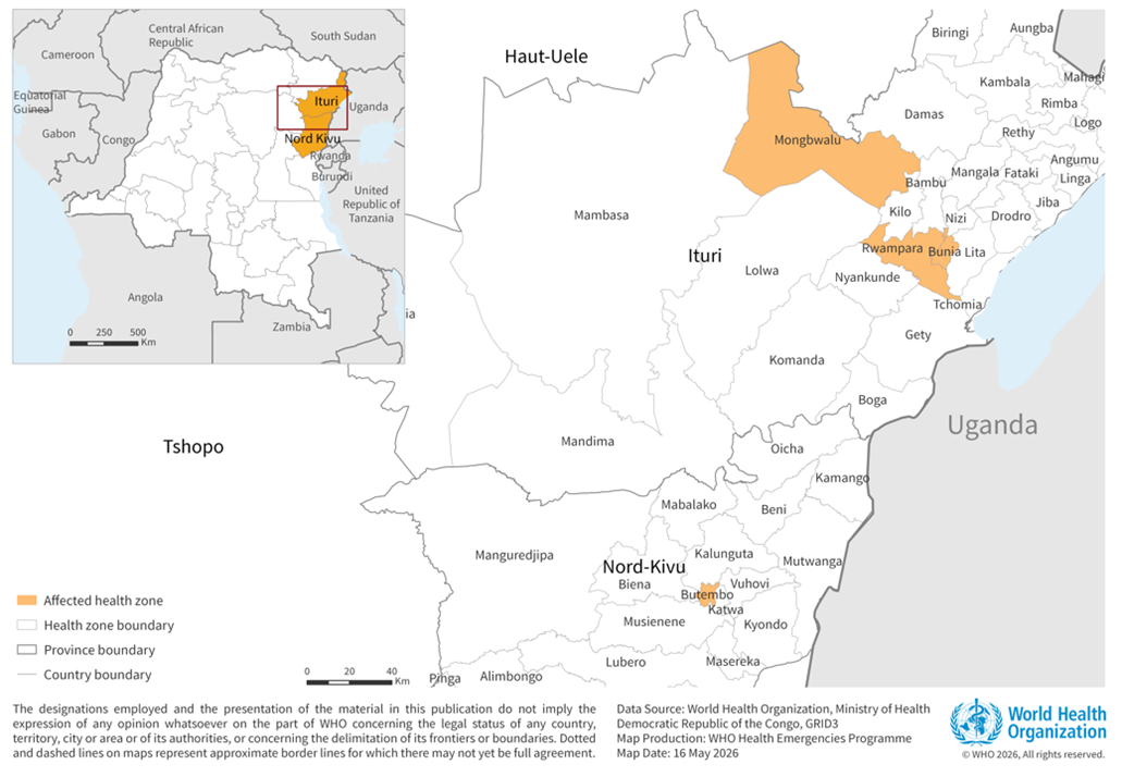
Figure 1. Health zones in the Democratic Republic of the Congo affected by Bundibugyo virus disease as of May 16, 2026.
(Source: WHO official website)
The BDBV genome is a single-stranded, negative-sense RNA (ssRNA⁻) approximately 18,940 nucleotides in length — consistent with the typical 18.9–19 kb range of the genus. It encodes seven major structural proteins in the following order: NP, VP35, VP40, GP, VP30, VP24, and L. These proteins act in concert to drive viral replication, particle assembly, and evasion of host immune defenses. Viral particles display the characteristic filamentous or thread-like morphology of filoviruses, with a uniform diameter of approximately 80 nm and lengths reaching up to 14 μm. Particles can adopt branched, U-shaped, "6"-shaped, or circular conformations, and are enclosed by a lipid envelope studded with GP glycoprotein spikes.
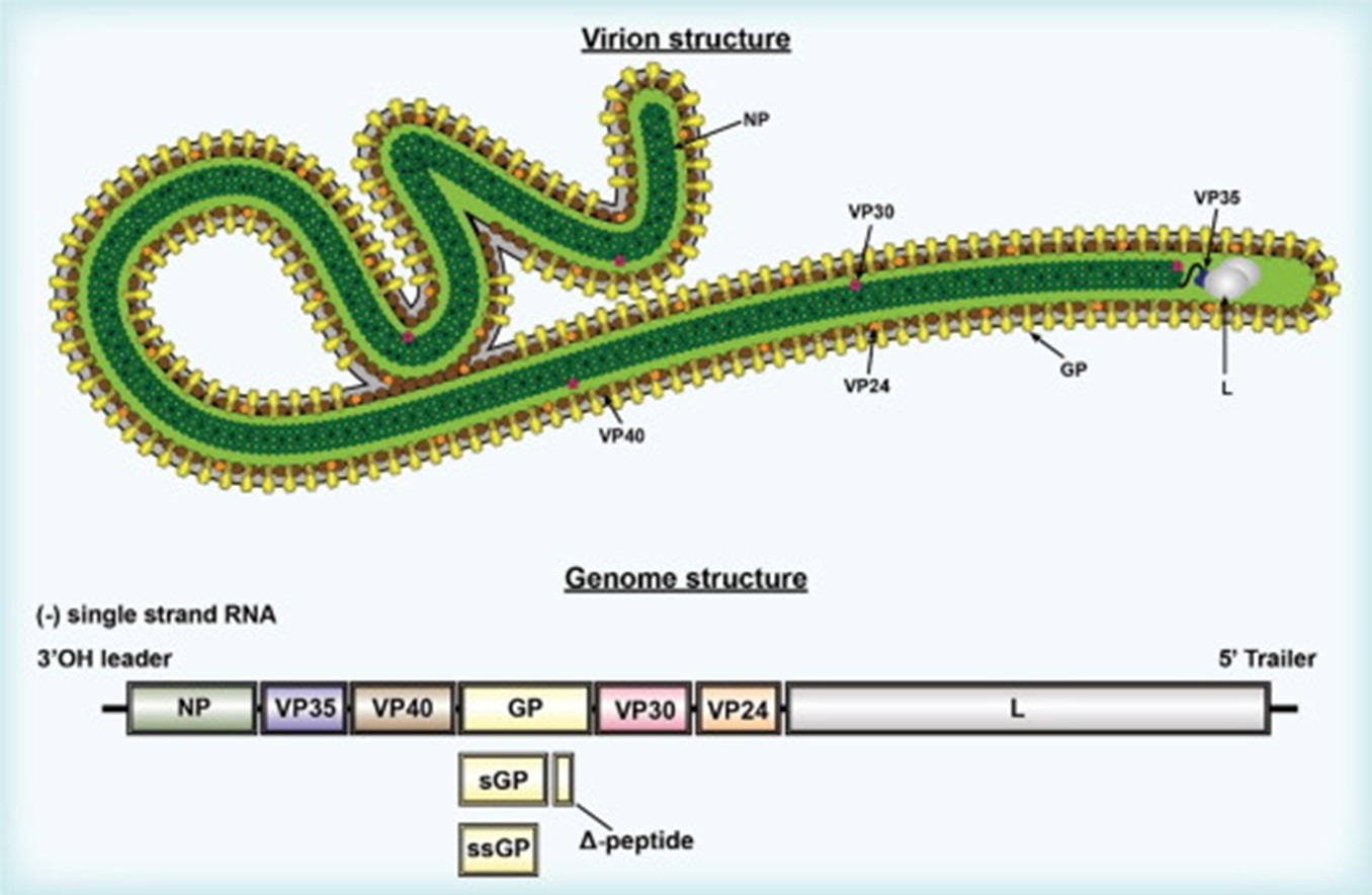
Figure 2. Virion and genome structure of EBOV.
(Source: https://doi.org/10.1016/j.jobb.2023.05.001)
| Protein | Full Name | Primary Function | Key Features |
|---|---|---|---|
| GP | Glycoprotein | Mediates viral attachment and entry into host cells | Surface spike protein; primary immunological target and central focus of vaccine development. BDBV GP harbors distinct epitopes; high-resolution crystal structures have been resolved. |
| NP | Nucleoprotein | Encapsidates the RNA genome to form the nucleocapsid | Highly conserved across ebolaviruses; essential for viral RNA packaging. |
| VP35 | VP35 protein | Polymerase cofactor | Potent antagonist of host interferon responses, facilitating immune evasion. |
| VP40 | VP40 matrix protein | Drives viral assembly and budding | Among the most variable genes in the genome; the principal driver of virion formation. |
| VP30 | VP30 protein | Transcriptional activator | Regulates viral RNA transcription. |
| VP24 | VP24 matrix protein | Interferon antagonist | Suppresses host antiviral signaling pathways. |
| L | L protein (polymerase) | RNA-dependent RNA polymerase | The catalytic engine of viral genome replication; the most conserved genomic region. |
Within 1–3 days of exposure, BDBV enters the body through mucosal surfaces or skin abrasions and undergoes rapid replication in dendritic cells and monocytes, before disseminating to hepatocytes, endothelial cells, and epithelial cells. Between days 3 and 14, systemic viral spread, dysregulated host immune responses, coagulopathy, vascular injury, and hypotension converge to precipitate shock and multi-organ failure. Overexpression of tissue factor (TF) in monocytes and macrophages is a pivotal event that triggers the hemorrhagic complications characteristic of BDBV infection: TF transcript levels rise by day 3, TF protein surges by day 2, and large quantities of TF-expressing membrane microparticles are detectable in plasma. Intense viral replication in the liver leads to marked hepatocellular necrosis and severe hepatic injury; the resulting impairment of coagulation factor synthesis drives the coagulopathy that is one of the defining pathological hallmarks of Ebola virus disease (EVD).
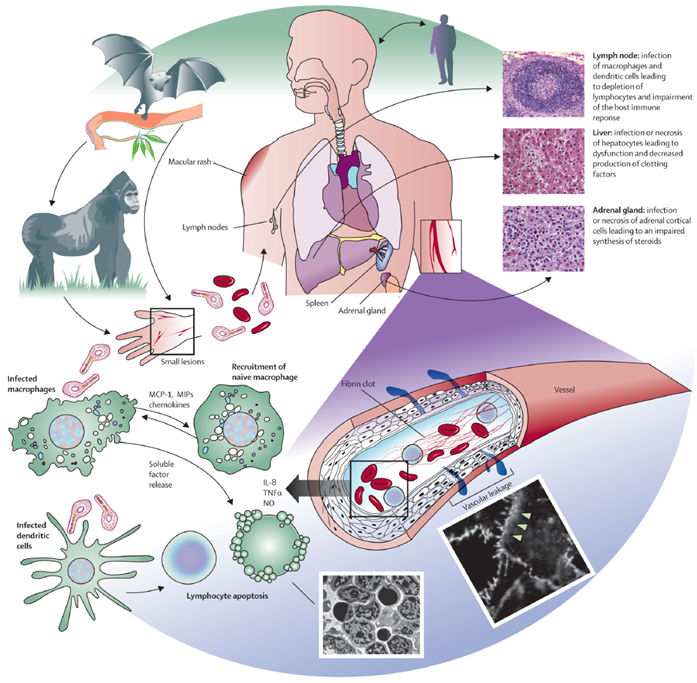
Figure 3. Model of Ebola virus disease pathogenesis.
(Source: doi:10.1016/S0140-6736(10)60667-8)
To date, no approved vaccines or specific therapeutics exist for Bundibugyo virus (BDBV). Existing countermeasures developed against Zaire ebolavirus (EBOV) — including Ervebo (rVSV-ZEBOV), Inmazeb (REGN-EB3), and Ebanga (mAb114) — offer limited or no cross-protection against BDBV. Clinical management therefore relies heavily on supportive care, including fluid resuscitation, electrolyte management, organ support, and symptom control, all of which significantly reduce mortality when implemented promptly. Rapid RT-PCR diagnostics and whole-genome sequencing remain the cornerstone of outbreak surveillance. Research efforts are concentrated on the GP glycoprotein, particularly its conserved regions, and on the development of pan-ebolavirus vaccine platforms and antibody combinations.
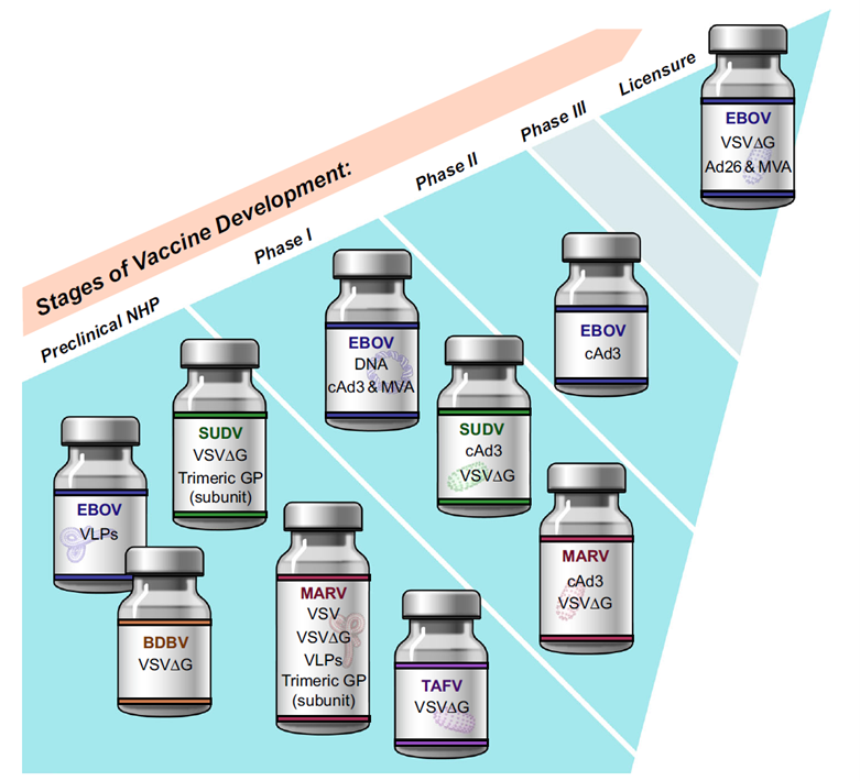
Figure 4. Development stages of Ebola virus vaccines in the United States.
(Source: https://doi.org/10.1038/s41541-024-00985-y)
The rVSVΔG/BDBV-GP vaccine — a recombinant vesicular stomatitis virus-based construct — has demonstrated post-exposure protection against BDBV challenge. In a cynomolgus macaque model, animals vaccinated as late as 20–23 minutes after BDBV infection showed a survival rate of 5 out of 6 (83%), compared to an expected natural survival rate of only 21% in this model — a striking therapeutic effect. Separately, a single dose of rVSV-EBOV confers 100% protection in non-human primates and approximately 50% protection even when administered within 24 hours of EBOV challenge. In surviving animals, the liver and spleen showed no significant pathological lesions, whereas fatal cases exhibited necrotizing hepatitis and widespread viral antigen-positive infiltration. Although VSV-BDBV remains in preclinical testing with no human trials initiated, the animal data provide a compelling rationale for accelerated clinical development.
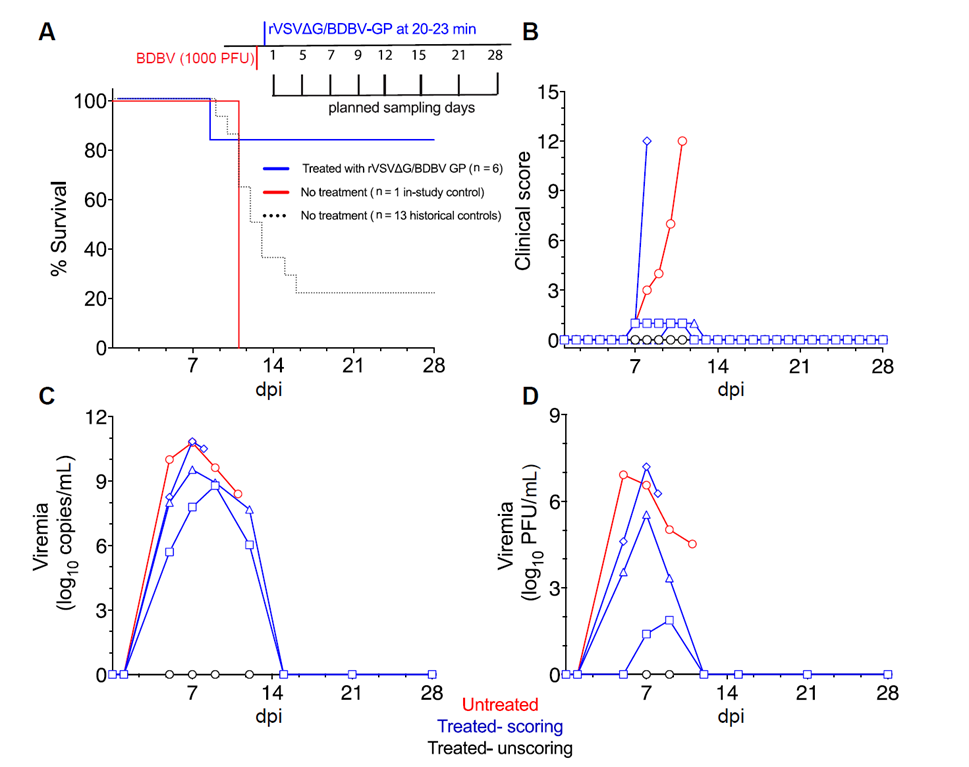
Figure 5. Survival outcomes and viral load comparisons in cynomolgus macaques following BDBV infection and rVSVΔG/BDBV-GP post-exposure treatment.
(Source: https://doi.org/10.1093/infdis/jiad207)
Beyond the VSV backbone, multivalent vaccine platforms based on adenovirus serotype 26 and modified vaccinia Ankara (Ad26-filo/MVA-BN-filo) have demonstrated protection against Sudan virus and Marburg virus in non-human primate studies. The multivalent design principles underpinning these platforms are directly extendable to provide coverage against BDBV. The key advantage of this approach is the ability to simultaneously confer broad protection across multiple filovirus species, reducing the complexity and cost of developing pathogen-specific countermeasures.
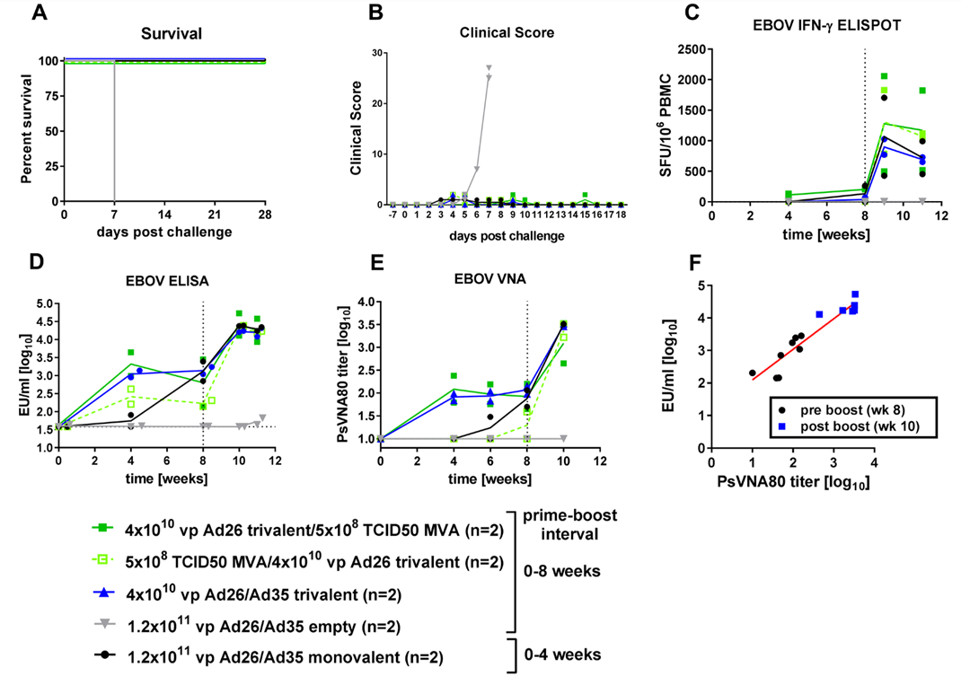
Figure 6. Immunogenicity of heterologous trivalent vaccine regimens (Ad26-Ad35, Ad26-MVA-BN-Filo, or MVA-BN-Filo-Ad26) versus Ad26-Ad35 monovalent vaccine and protection against EBOV Kikwit challenge.
(Source: https://doi.org/10.1371/journal.pone.0192312)
Crystal structure analysis of the Sudan ebolavirus (SUDV) NP–VP35 complex has revealed a previously uncharacterized interaction interface defined by a precisely formed β-sheet. Affinity binding studies confirm that this β-sheet is essential for maintaining high-affinity interaction between VP35 and the hydrophobic pocket of NP. Electron microscopy further demonstrates that this binding interface is critical for NP oligomerization and assembly within human cells. Structure-guided mutagenesis has identified key residues conserved across the filovirus family — residues that represent ideal targets for pan-filovirus antiviral drug development.
Disrupting the NP–VP35 interaction destabilizes the viral replication complex, blocking transcription and replication of the viral genome — making this interface an attractive target for broad-spectrum antifiloviral drug discovery.
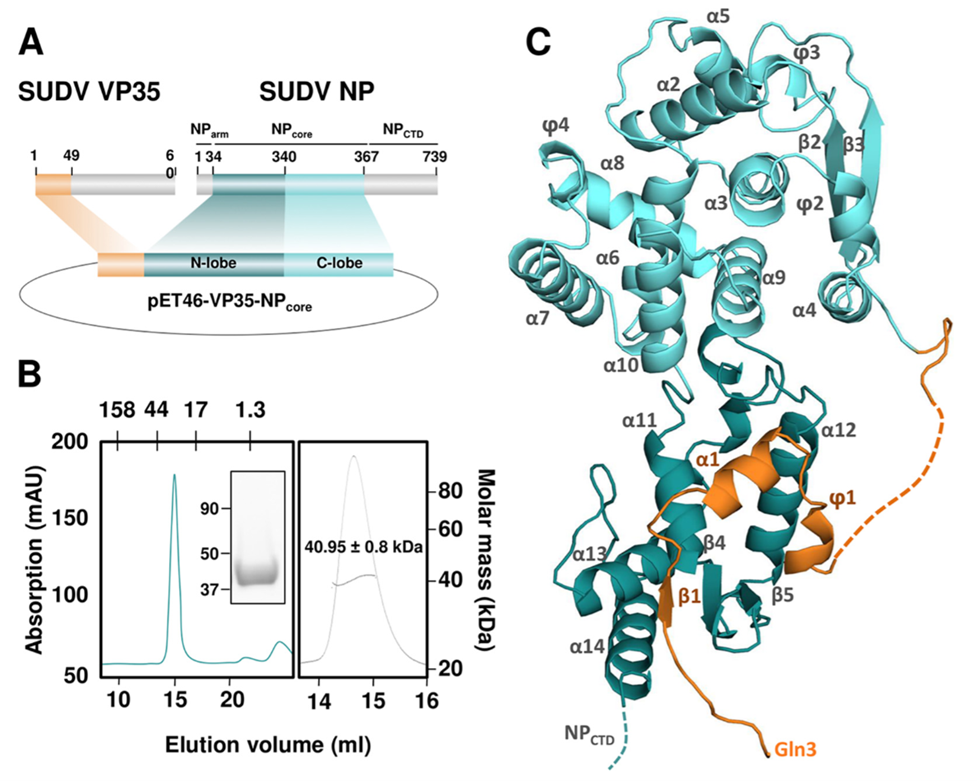
Figure 7. Crystal structure of the SUDV VP35–NP core complex.
(Source: https://doi.org/10.1128/mBio.00734-19)
Natural products isolated from myxobacteria — including Cystobactamid 919-1, Cystobactamid 934-2, and 2-Hydroxysorangiadenosine — have shown stable binding interactions with both VP35 and VP40 in molecular docking studies. Molecular dynamics simulations further confirm the stability of the Cystobactamid 919-1–VP35 and 2-Hydroxysorangiadenosine–VP40 complexes, supporting the therapeutic potential of these natural scaffolds.
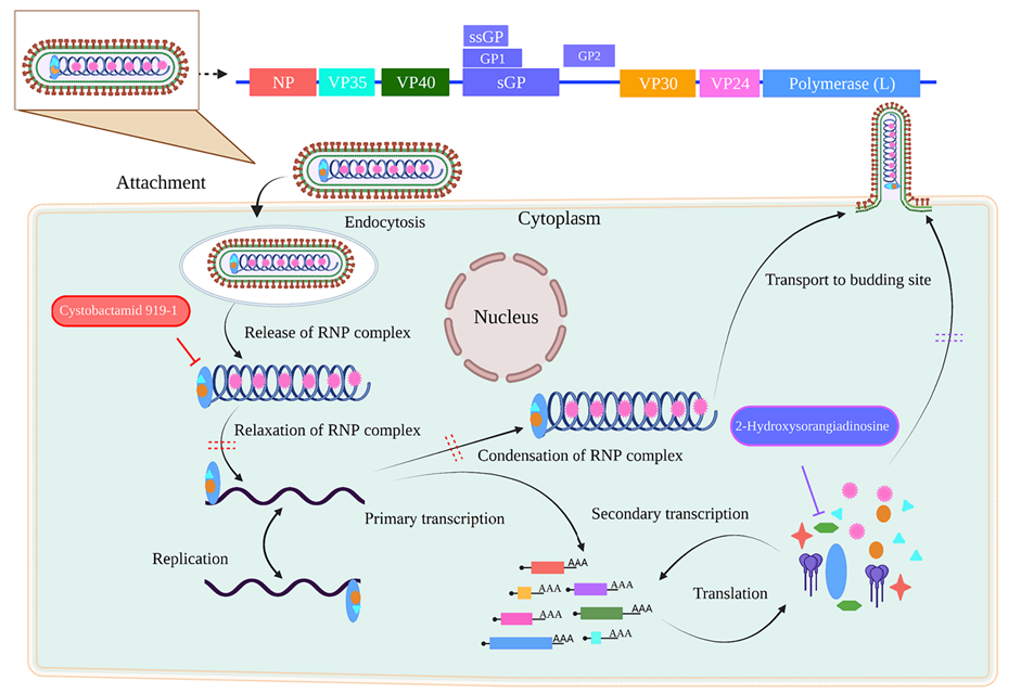
Figure 8. Proposed mechanisms by which myxobacterial natural products inhibit EBOV.
(Source: https://doi.org/10.3390/biom14060660)
As the primary driver of viral assembly, VP40 is an attractive target: blocking its interaction with nuclear envelope proteins disrupts budding and the release of new virions. Inhibiting the interferon-antagonistic activity of VP35, in turn, restores innate immune signaling, enhancing viral clearance. The layered, multi-target approach targeting both proteins substantially increases the probability of therapeutic success.
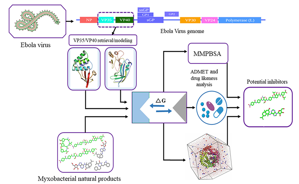
Figure 9. Identification of potential Ebola virus VP35 and VP40 inhibitors from myxobacterial natural products.
(Source: https://doi.org/10.3390/biom14060660)
BDBV GP is the primary antigen for all current vaccine strategies. Compared to EBOV, BDBV GP may adopt a distinct upstream receptor-binding domain conformation, offering opportunities for strain-specific vaccine design. The highly conserved NPC1-binding site provides a structural basis for developing broadly neutralizing monoclonal antibodies capable of cross-neutralizing multiple ebolavirus species — assets of particular value for emergency prophylactic and therapeutic interventions.
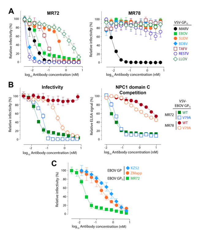
Figure 10. Monoclonal antibodies targeting the conserved GP1 receptor-binding site exhibit pan-filovirus neutralizing activity.
(Source: doi:10.1128/mBio.02154-15)
The ongoing transmission of BDBV in the DRC and Uganda underscores the urgent need for a comprehensive pan-filovirus defense framework. Priority actions include: stockpiling multivalent vaccines and treatment regimens active against BDBV, Sudan virus, and Marburg virus; building decentralized regional diagnostic capacity to eliminate dependence on single reference laboratories; advancing multinational research collaborations to accelerate vaccine and drug development; deepening environmental surveillance and studies of animal reservoir hosts; and establishing pre-negotiated emergency vaccine deployment protocols between WHO and national governments. Central to all of these efforts is a deepened understanding of filovirus molecular biology — including the structure, interactions, and immunological targets of key viral proteins.
abinScience offers a comprehensive portfolio of high-quality recombinant proteins and antibodies to support filovirus research at every stage. Our catalog includes recombinant nucleoproteins (NP), polymerase cofactors (VP35), matrix proteins (VP40), and glycoproteins (GP) from BDBV, EBOV, Sudan virus, and related species, as well as species- and pan-ebolavirus-specific monoclonal and polyclonal antibodies targeting each of these key antigens. Whether your work focuses on vaccine immunogenicity assessment, diagnostic reagent development, or the screening and characterization of therapeutic antibodies, our products are designed to support your research and translational needs. Click any catalog number below to access the full product details.
| Catalog No. | Product Name |
|---|---|
| VK057011 | BDBV Envelope glycoprotein Recombinant Protein (C-Fc) |
| VK057021 | BDBV Envelope glycoprotein Recombinant Protein (C-His) |
| VK057012 | BDBV Envelope glycoprotein Recombinant Protein (N-His) |
| VK058012 | BDBV Matrix Recombinant Protein VP40 (N-His) |
| VK059012 | BDBV Nucleoprotein Recombinant Protein (N-His) |
| VK061012 | BDBV VP24 Recombinant Protein (N-His) |
| VK060012 | BDBV VP30 Recombinant Protein (N-His) |
| VK578021 | Bombali virus/BOMV GP1 Recombinant Protein (C-Fc) |
| VK578011 | Bombali virus/BOMV GP1 Recombinant Protein (C-His) |
| VK578012 | Bombali virus/BOMV GP1 Recombinant Protein (N-His) |
| VK623051 | REBOV GP1 Recombinant Protein (C-Fc) |
| VK623041 | REBOV GP1 Recombinant Protein (C-His) |
| VK623022 | REBOV GP1 Recombinant Protein (N-His) |
| VK518012 | REBOV Nucleoprotein Recombinant Protein (N-His) |
| VK783042 | REBOV VP40/Matrix Recombinant Protein (N-His) |
| VK623071 | Recombinant SEBOV GP1 Protein, C-Fc |
| VK623061 | Recombinant SEBOV GP1 Protein, C-His |
| VK623032 | Recombinant SEBOV GP1 Protein, N-His |
| VK512022 | Recombinant SEBOV Nucleoprotein, N-His |
| VK783032 | Recombinant SEBOV VP40 Protein, N-His |
| VK623091 | Recombinant TAFV GP1 Protein, C-Fc |
| VK623081 | Recombinant TAFV GP1 Protein, C-His |
| VK623042 | Recombinant TAFV GP1 Protein, N-His |
| VK512012 | Zaire ebolavirus NP/Nucleoprotein Recombinant Protein (C-His) |
| VK623011 | ZEBOV GP/GP1 2 Recombinant Protein (C-His) |
| VK623031 | ZEBOV GP1 Recombinant Protein (C-Fc) |
| VK623021 | ZEBOV GP1 Recombinant Protein (C-His) |
| VK623012 | ZEBOV GP1 Recombinant Protein (N-His) |
| VK095022 | ZEBOV VP24 Recombinant Protein (N-His) |
| VK661012 | ZEBOV VP24 Recombinant Protein (N-His-SUMO) |
| VK661022 | ZEBOV VP35 Recombinant Protein (N-His) |
| VK783022 | ZEBOV VP40 Recombinant Protein (N-His) |
| VK095012 | ZEBOV VP40 Recombinant Protein (N-His-SUMO) |
| VK783012 | ZEBOV VP40 Recombinant Protein (N-His-SUMO) |
| Catalog No. | Product Name |
|---|---|
| VK518010 | InVivoMAb Anti-REBOV/SEBOV/TAFV/ZEBOV NP/Nucleoprotein Antibody (MJ20) |
| VK623050 | InVivoMAb Anti-SEBOV/ZEBOV GP/Envelope glycoprotein Antibody (Iv0198) |
| VK623040 | InVivoMAb Anti-ZEBOV GP/Envelope glycoprotein Antibody (6D8) |
| VK623010 | InVivoMAb Anti-ZEBOV GP/Envelope glycoprotein Antibody (Iv0195) |
| VK623020 | InVivoMAb Anti-ZEBOV GP/Envelope glycoprotein Antibody (Iv0196) |
| VK623030 | InVivoMAb Anti-ZEBOV GP/Envelope glycoprotein Antibody (Iv0197) |
| VK057014 | Anti-BDBV Envelope glycoprotein Polyclonal Antibody |
| VK058014 | Anti-BDBV Matrix Protein VP40 Polyclonal Antibody |
| VK059013 | Anti-BDBV Nucleoprotein Antibody (SAA2994) |
| VK059014 | Anti-BDBV Nucleoprotein Polyclonal Antibody |
| VK061014 | Anti-BDBV VP24 Polyclonal Antibody |
| VK060014 | Anti-BDBV VP30 Polyclonal Antibody |
| VK578014 | Anti-BOMV Envelope glycoprotein GP1 Polyclonal Antibody |
| VK623076 | Anti-Ebolavirus GP/Glycoprotein Reference Antibody (Ansuvimab, RUO) |
| VK623066 | Anti-Ebolavirus GP/Glycoprotein Reference Antibody (Atoltivimab, RUO) |
| VK623036 | Anti-Ebolavirus GP/Glycoprotein Reference Antibody (Cosfroviximab, RUO) |
| VK623016 | Anti-Ebolavirus GP/Glycoprotein Reference Antibody (Larcaviximab, RUO) |
| VK623056 | Anti-Ebolavirus GP/Glycoprotein Reference Antibody (Maftivimab, RUO) |
| VK623026 | Anti-Ebolavirus GP/Glycoprotein Reference Antibody (Odesivimab, RUO) |
| VK623046 | Anti-Ebolavirus GP/Glycoprotein Reference Antibody (Porgaviximab, RUO) |
| VK059023 | Anti-pan-Ebola virus Nucleoprotein Antibody (SAA2995) |
| VK059033 | Anti-pan-Ebola virus Nucleoprotein Nanobody (SAA3007) |
| VK623116 | Anti-pan-Ebolavirus GP/Envelope glycoprotein Reference Antibody (ADI-15742, RUO) |
| VK125013 | Anti-pan-Filovirus GP/Envelope glycoprotein Antibody (m21D10) |
| VK623044 | Anti-REBOV GP1 Polyclonal Antibody |
| VK518014 | Anti-REBOV Nucleoprotein Polyclonal Antibody |
| VK783034 | Anti-REBOV VP40/Matrix Protein Polyclonal Antibody |
| VK623106 | Anti-REBOV/SEBOV/TAFV/ZEBOV GP/Envelope glycoprotein Reference Antibody (ADI-15878, RUO) |
| VK623033 | Anti-SEBOV GP/Envelope glycoprotein Antibody (16F6) |
| VK623034 | Anti-SEBOV GP1 Polyclonal Antibody |
| VK512024 | Anti-SEBOV Nucleoprotein Polyclonal Antibody |
| VK591013 | Anti-SEBOV RNA-directed RNA polymerase L/Protein L Nanobody (SAA1489) |
| VK783024 | Anti-SEBOV VP40 Polyclonal Antibody |
| VK518013 | Anti-Sudan ebolavirus/SEBOV NP/Nucleoprotein Recombinant Antibody (SAA1402) |
| VK623024 | Anti-TAFV GP1 Polyclonal Antibody |
| VK512014 | Anti-Zaire ebolavirus NP/Nucleoprotein Polyclonal Antibody |
| VK623013 | Anti-Zaire ebolavirus/ZEBOV GP/GP1, 2 Recombinant Nanobody (SAA1248) |
| VK623053 | Anti-ZEBOV GP/Envelope glycoprotein Neutralization Recombinant Antibody (KZ52) |
| VK623043 | Anti-ZEBOV GP/Envelope glycoprotein Recombinant Antibody (GPE118) |
| VK623023 | Anti-ZEBOV GP/Envelope glycoprotein Recombinant Antibody (mAb100) |
| VK623086 | Anti-ZEBOV GP/Glycoprotein Reference Antibody (ANP-015, RUO) |
| VK623096 | Anti-ZEBOV GP/Glycoprotein Reference Antibody (Zmapp, RUO) |
| VK623014 | Anti-ZEBOV GP1 Polyclonal Antibody |
| VK518023 | Anti-ZEBOV NP/Nucleoprotein Recombinant Antibody (KZ51) |
| VK095014 | Anti-ZEBOV VP24 Polyclonal Antibody |
| VK661014 | Anti-ZEBOV VP35 Polyclonal Antibody |
| VK661013 | Anti-ZEBOV VP35/Polymerase cofactor VP35 Recombinant Antibody (F9) |
| VK783014 | Anti-ZEBOV VP40 Polyclonal Antibody |
| VK783044 | Anti-ZEBOV VP40 Polyclonal Antibody |
| VK783013 | Anti-ZEBOV VP40/Matrix protein VP40 Recombinant Antibody (DSTL094) |
+86-027-65523339
中国武漢市深敦寺路666号C棟、武漢、430206


 中文
中文 English
 한국어
한국어 日本語
日本語 Español
 Français
 Русский