As flow cytometry workflows become increasingly standardized, more antibody products are offered in convenient, pre-diluted "Test" formats. Compared to traditional bulk packaging (which is typically microgram-based), the "Test" format eliminates tedious pre-experimental calculations and optimization efforts, making it highly attractive for routine marker detection and rapid assay setup.
However, in real-world research, many scientists find that even when following the manufacturer’s recommended "Test" volume, staining performance can still vary significantly across sample types and experimental objectives.
This does not imply a flaw in the "Test" format design. Rather, flow cytometry is highly sensitive to a variety of biological and technical variables, including sample microenvironments, target antigen density, and panel design. While a "Test" volume provides a pre-validated, standardized starting point, optimizing your antibody titer for your specific experimental system remains a critical step in achieving top-tier data quality.
1. The Design Logic and Practical Value of Test-Formatted Antibodies
1.1 Reduce the cost of establishing the experimental
With traditional bulk-packaged antibodies, researchers must go through a tedious optimization process before launching a new experiment—including serial dilutions, calculating optimal staining volumes, and evaluating signal-to-noise ratios. Test-formatted packaging provides a pre-validated, standardized starting point, allowing researchers to skip initial optimization groundwork and initiate experiments promptly.
1.2 Enhancing Operational Convenience and Workflow Efficiency
The Test format offers distinct efficiency advantages in several key scenarios:
- • Rapid validation of novel markers and clones: Ideal for exploratory targets or preliminary screens of new antibody clones.
- • Precious, volume-limited samples: Perfect for clinical biopsies, rare immune cell subsets, or early exploratory pilots where sample conservation is paramount and extensive titration is not feasible.
- • Routine detection of highly expressed markers: For well-characterized, stably and highly abundant markers (such as CD4 and CD8 in peripheral blood), the recommended Test volume is typically sufficient for daily quality control or routine immunophenotyping.
1.3 Improving Batch-to-Batch Consistency
Fixed Test volumes eliminate systematic pipetting and dilution errors across different laboratory personnel. This minimizes variability between technical replicates, laying a solid foundation for experimental reproducibility.
2. Why the Recommended "Test" Volume Isn't Always the Optimal Condition
Manufacturers validate Test volumes using highly standardized systems—typically healthy human peripheral blood mononuclear cells (PBMCs), highly expressed constitutive antigens, and single-color staining. Real-world research, however, introduces multi-dimensional heterogeneity that a single fixed volume cannot fully accommodate. Key confounding factors include:
2.1 Dynamic Fluctuations in Antigen Density
The optimal working concentration of an antibody is directly tied to the number of target antigen copies on the cell surface.
- • High-abundance, constitutively expressed antigens (e.g., CD3, CD4, CD8) provide abundant binding sites and a wide tolerance range for antibody dosage. Standard Test volumes easily yield clear population resolution.
- • Low-abundance targets (e.g., activation markers, immune checkpoints, intracellular transcription factors) and weakly positive cell populations have very low copy numbers, narrowing the signal window. A fixed Test volume can easily lead to insufficient signal (false negatives) or elevated non-specific binding (high background), directly compromising population resolution (stain index).
2.2 Sample Complexity and Matrix Effects
The cellular state and microenvironment of different sample types can significantly alter staining efficiency and background noise:
- • Tissue digests, bone marrow, and tumor samples often contain a high percentage of dead cells and complex Fc receptor expression, posing a much higher risk of non-specific binding (NSB) than fresh PBMCs. Consequently, the same antibody volume can lead to significantly higher background.
- • Primary cells versus cell lines, or resting versus activated cells, exhibit fundamental differences in antigen expression. As a result, the saturation concentration of the antibody will vary.
2.3 Spillover Spreading in Multicolor Panels
Test volume validation is typically performed using single-color staining. However, multicolor flow panels introduce complex system interactions:
- • Fluorophore spillover and the resulting compensation can degrade the effective resolution of a detector. Weakly expressed targets often require a higher signal intensity to overcome spillover spreading.
- • Co-incubating multiple antibodies can introduce steric hindrance or binding competition. The optimal concentration determined in a single-color system does not automatically translate to a multicolor panel.
- • High-dimensional immunophenotyping demands far superior population resolution than qualitative detection. A single recommended Test volume is rarely optimized to balance the performance of an entire panel.
3. Why Flow Cytometry Assays Require Titration Optimization
The core objective of antibody titration is not simply to "find the lowest dosage" to reduce reagent costs, but to identify the optimal balance point between specific signal intensity and non-specific background noise — to maximize data quality.
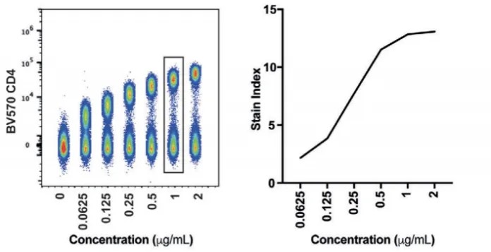
Figure 1. Flow cytometry antibody titration
3.1 Achieving the Optimal Signal-to-Noise Ratio (Stain Index)
- • Under-titration (too low): The fluorescence signal of positive cells is insufficient, making it difficult to distinguish weakly expressing populations from negative ones, which can result in false negatives.
- • Over-titration (too high): Non-specific binding increases, raising the background fluorescence and decreasing population resolution. It can also exacerbate spectral spillover, complicating compensation.
- • ptimal titer: The optimal titer is achieved when specific positive signals are fully saturated, yielding the lowest non-specific background and the highest population resolution (Stain Index).
3.2 Enhancing Detection Sensitivity for Low-Expression Markers
Titration optimization is particularly critical for low-abundance targets such as transcription factors, activation markers, immune checkpoints, and rare cell subsets. Because these targets yield limited signal and are highly sensitive to experimental variables, precise titration significantly enhances detection sensitivity, preventing signal loss or background interference due to improper antibody dosing.
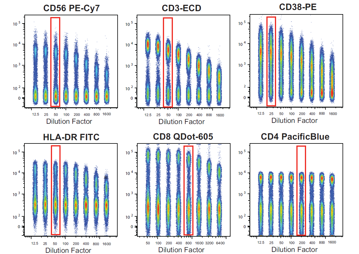
Figure 2. Representative titration profiles of various flow cytometry antibodies
3.3 Safeguarding Overall Multicolor Panel Performance
In complex multicolor panels, potential interactions between different antibodies—including spectral overlap, epitope competition, and buffer compatibility—can impact performance. Systematically optimizing the dosage of each antibody helps lower the overall background, improve population resolution, and ultimately fine-tune the performance of the entire panel.
4. In What Scenarios May the Recommended "Test" Volume Not Be the Optimal Choice?
While Test volumes suffice for most routine assays, further titration optimization is highly recommended in the following scenarios to guarantee data quality:
- • Detecting low-expression or weakly positive targets: When identifying weak positive populations, rare subsets, or activation markers, a fixed Test volume may not yield the optimal signal-to-noise ratio. Titration is necessary to maximize detection sensitivity.
- • Designing complex multicolor panels: For panels featuring multiple low-abundance markers, diverse fluorophore combinations, or high-dimensional immunophenotyping, systematic titration is essential to minimize spectral interference and maximize population resolution.
- • Switching sample matrix systems: When transitioning from PBMCs to tumor tissue, bone marrow, digested tissues, or in vitro cultured cells, the sample matrix, cellular state, and antigen expression profiles change. The applicability of the standard Test volume must be re-evaluated through titration on the new sample type.
- • High-precision quantitative analysis: If your research goal goes beyond simple positive/negative gating—such as resolving subtle differences in antigen expression, conducting relative quantification, or discovering novel cell subsets—titration is vital to ensure robust, reliable data.
5. How to Understand the Relationship Between Test Volume and Titration Optimization
Test-formatted packaging and titration optimization are not mutually exclusive; rather, they are consecutive, complementary phases in the flow cytometry workflow:
- • Test Volume: Serves as a validated, standardized starting point, to help researchers quickly establish assays, obtain preliminary data, and significantly reduce pre-experimental workload.
- • Titration Optimization: Builds upon this standardized starting point, fine-tuning the antibody concentration for a specific experimental system to further improve detection performance and data quality.
For routine marker screening, daily runs of established panels, or rapid validation assays, the recommended Test volume is usually more than enough. However, for complex multicolor panel design, low-abundance target detection, and high-precision quantification, performing titration optimization on top of the recommended Test volume is key to securing highly reliable data.
Summary
Test-formatted antibodies provide a standardized and convenient solution for flow cytometry, reducing setup costs while enhancing workflow efficiency and consistency. However, because flow cytometry is deeply influenced by antigen density, sample type, panel design, and experimental goals, a single, universal recommended Test volume cannot fit every scenario.
Recognizing the role and limitations of Test volumes, making informed decisions about when to perform titration based on specific research needs, and conducting systematic optimization when necessary are fundamental to obtaining robust, high-quality flow cytometry data.
Looking for Reliable Flow Cytometry Antibodies to Support your Experimental Workflows?
abinScience Flow Cytometry Antibodies are subjected to rigorous quality control to ensure consistent fluorescent performance and clear discrimination of cell populations, supporting robust and reproducible flow cytometry analyses.
Learn More about abinScience Flow Cytometry Antibodies
About Us
abinScience was founded in 2023 as a strategic venture of AtaGenix (established 2011), dedicated to delivering premium life science reagents that accelerate discovery.
abinScience flow cytometry antibody products cover commonly used detection markers, with a wide variety to meet the research needs of multiple species(
Human,
Mouse,
Rat,
Dog,
Hamster,
Monkey, etc.)We provide stable and reliable support for scientific research.



 中文
中文 English
 한국어
한국어 日本語
日本語 Español
 Français
 Русский


