1. From Observation to Intervention: A Paradigm Shift in Immunology Research
In modern life sciences, the widespread adoption of single‑cell RNA sequencing (scRNA‑seq), high‑parameter flow cytometry, and spatial transcriptomics has granted researchers an unprecedented microscopic perspective. We can now meticulously map the abundance, differentiation states, and spatial distributions of dozens of immune cell subsets within the tumor microenvironment (TME), infectious foci, or autoimmune lesions.
However, while generating massive datasets of "cellular expression atlases," researchers face a classic and challenging methodological hurdle: accumulating descriptive observational data does not automatically establish functional causality.
In physiological or pathological models, the marked local enrichment or depletion of a specific immune cell subset usually reflects one of three distinct biological logics:
- Primary Driver: The cell directly drives disease progression or mediates protective immunity.
- Bystander: The cell passively responds to local inflammatory signals without exerting a substantive effect on key phenotypes.
- Negative Compensatory Regulator: The cell functions as a negative feedback mechanism established by the host to maintain tissue homeostasis and limit hyper‑inflammation.
Descriptive parameters—such as cell counts, frequency shifts, or spatial co‑localization of phenotypic markers—are inherently insufficient to distinguish among these three roles. To transition from "phenotypic description" to "mechanistic dissection," researchers must rely on functional intervention experiments. By selectively depleting or functionally blocking targeted immune cell populations and observing downstream effects on whole‑organism phenotypes and broader immune networks, researchers can establish robust causal inference.
Among these approaches, antibody‑mediated in vivo immune cell depletion stands out as a highly selective, dynamically controllable functional tool that serves as a vital bridge between clinical observation and mechanistic discovery.
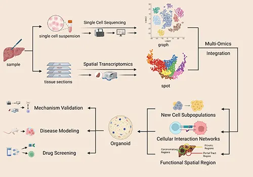
Figure 1. Workflow for investigating the immune microenvironment in chronic liver disease (DOI: 10.3389/fimmu.2026.1743439)
2. Antibody‑Mediated In Vivo Cell Depletion: A Key Tool Bridging Phenotypic Observation and Functional Validation
2.1 Fc Effector Mechanisms Driving Target Cell Clearance
In antibody‑mediated in vivo depletion, highly specific monoclonal antibodies (mAbs) targeting lineage‑specific surface markers are administered to experimental animals. Upon binding to the target cell antigen, the mAb engages host effector mechanisms to clear target cells via three main pathways:
- Antibody‑Dependent Cellular Cytotoxicity (ADCC): The Fc region of the bound mAb engages Fcγ receptors (FcγRs) on NK cells or macrophages, triggering the release of perforin, granzymes, or reactive oxygen species (ROS) to induce target cell apoptosis.
- Antibody‑Dependent Cellular Phagocytosis (ADCP): Cells of the reticuloendothelial system (e.g., splenic and hepatic macrophages) recognize antibody‑opsonized cells via FcγRs, engulfing and degrading them directly.
- Complement‑Dependent Cytotoxicity (CDC): Antibody binding initiates the classical complement cascade, forming membrane attack complexes (MACs) that lyse target cell membranes.
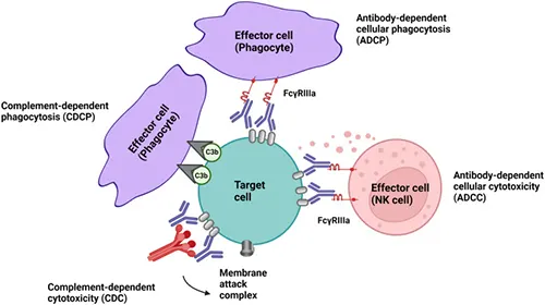
Figure 2. Key effector mechanisms of antibody‑mediated in vivo immune cell depletion (DOI: 10.3389/fimmu.2022.953649)
2.2 Experimental Advantages Complementing Genetic Models
Compared with genetic models (e.g., constitutive Knockout or Cre‑LoxP conditional systems), antibody‑mediated in vivo depletion offers distinct advantages:
| Feature / Dimension |
Genetic Knockout Models |
In Vivo mAb Depletion |
| Onset & Dynamics |
Continuous/embryonic or inducible; relatively slow onset |
Acute, rapid clearance; takes effect shortly post‑administration |
| Developmental Compensation |
Prone to developmental immune compensation, which can mask true phenotypes |
Bypasses developmental compensation, reflecting true adult homeostatic function |
| Temporal Control |
Requires complex inducing agents (e.g., Tamoxifen) |
Flexible administration schedule (pre‑, during, or post‑disease induction) |
| Strain Compatibility |
Frequently restricted to specific genetically engineered mouse strains |
Broadly applicable across wild‑type (WT), transgenic, and disease mouse models |
3. Targeting Distinct Immune Cell Subsets: From Functional Validation to Network Dissection
Immune cell depletion is not merely about cell removal; it is a hypothesis‑driven directional perturbation. Selecting which population to deplete tests a fundamental scientific question: does the target biological process persist in the absence of this cell population?
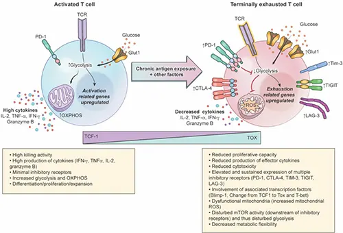
Figure 3. Characteristics of functional and exhausted T cells (DOI: 10.1016/j.trecan.2022.12.008)
3.1 Validating Effector Dependency: Cytotoxic Roles in Oncology and Infection
In cancer and infection research, CD8⁺ T cells are frequently observed enriching at pathological sites, with their abundance correlating with therapeutic efficacy or clinical prognosis. However, correlation alone does not confirm whether cytotoxic T cells are indispensable for the observed outcome. In vivo CD8⁺ T cell depletion offers direct functional validation: by removing CD8⁺ T cells, researchers can observe whether anti‑tumor responses diminish or viral control fails. A significant loss of efficacy supports a CD8⁺‑dependent mechanism, whereas an unchanged response points toward alternative or compensatory effector mechanisms (e.g., NK cells or humoral immunity).
3.2 Dissecting Regulatory Dynamics: How Helper and Regulatory Subsets Shape Responses
Not all immune cells act as direct effectors; many regulate the orientation and intensity of the broader response. When CD4⁺ T cell ratios shift in diseased tissue or when helper signals are suspected of driving an immune response, CD4⁺ T cell depletion helps dissect these regulatory hierarchies. Removing CD4⁺ T cells tests whether response initiation requires helper signals, whether CD8⁺ T cell activation and memory formation are compromised, and how the entire immune network rebalances in their absence.
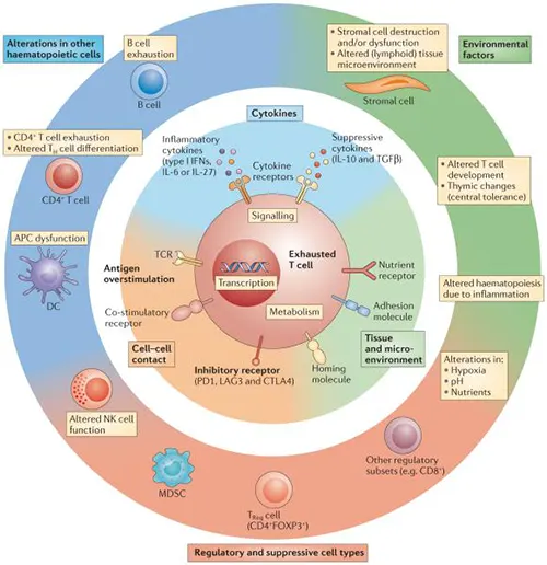
Figure 4. Mechanisms of T cell exhaustion (DOI: 10.1038/nri3862)
3.3 Distinguishing Bystanders from Drivers: Functional Roles of Infiltrating Myeloid Cells
Massive infiltration of myeloid cells—particularly neutrophils—is a hallmark of infection, inflammation, and cancer. Yet increased cell numbers do not inherently signify disease promotion; these cells may be passive recruits or active disease drivers. Ly‑6G‑mediated neutrophil depletion provides a tool to differentiate these roles: selectively removing neutrophils reveals whether the pathological phenotype changes. Crucially, target selection dictates which cells are eliminated: the 1A8 clone specifically recognizes Ly‑6G (targeting neutrophils), whereas the RB6‑8C5 clone binds both Ly‑6G and Ly‑6C, additionally affecting monocytes and subsets of CD8⁺ T cells. Researchers must align target selection with their specific research question and consider marker specificity during data interpretation.
3.4 Deconstructing Network Contributions: Decoupling Innate and Adaptive Immunity
The immune system operates as a coordinated network where phenotypes arise from multi‑cellular cooperation. When isolating the unique contribution of a single subset or evaluating innate‑adaptive crosstalk, NK cell depletion is a key tool. As core innate effectors, NK cells contribute to tumor surveillance and metastasis control, though their activity depends heavily on the tissue microenvironment. Depleting NK cells reveals whether innate immunity provides an independent functional contribution at specific disease stages and whether NK cells can compensate when adaptive immunity is impaired.
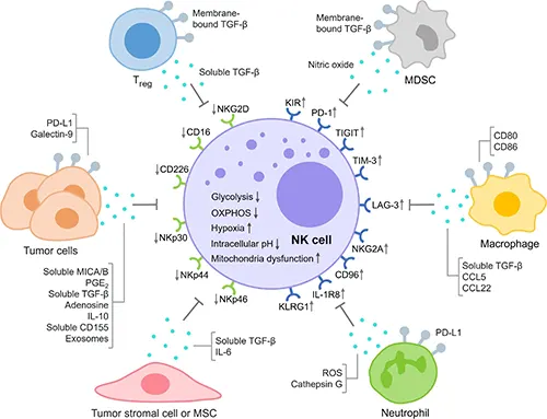
Figure 5. Key mechanisms driving NK cell exhaustion in the TME (DOI: 10.3389/fimmu.2023.1303605)
4. Key Pitfalls in Data Interpretation: Avoiding Methodological Traps
Interpreting immune depletion experiments demands scientific rigor. Assuming that "cell depletion altering a phenotype" implies "that cell is the sole direct cause" can lead to erroneous conclusions. The following factors warrant careful assessment:
-
Tissue Heterogeneity in Depletion Efficiency
Assessing depletion efficiency solely via peripheral blood sampling and assuming complete systemic knockout across all organs is a common oversight. Vascular permeability, tissue microenvironment barriers, and local macrophage density lead to significant tissue‑specific depletion heterogeneity. For instance, while CD8⁺ T cell depletion may exceed 95% in blood and spleen, clearance in deep lymph nodes, the central nervous system, or solid tumor cores may fall below 70%. Failing to evaluate target tissue clearance locally risks mistaking residual functional cells for a "non‑functional" cell population.
-
Epitope Competition and Artifacts in Flow Cytometry
When evaluating depletion efficiency by flow cytometry, using a fluorescently labeled detection antibody that targets the exact same epitope as the injected depleting mAb can yield misleading results. Residual circulating mAb in the animal competitively blocks the detection antibody, resulting in a loss of fluorescent signal that can be misread as complete cell depletion. To ensure reliable verification, researchers should use non‑competing antibody clones or indirect gating strategies (e.g., gating CD3⁺CD4⁻ populations to quantify CD8⁺ T cell loss).
-
Immune Network Remodeling and Compensatory Dynamics
The immune system is a dynamic, interconnected network; eliminating a core cell node often triggers secondary effects, such as rebound granulopoiesis or cytokine redistribution. For example, depleting T cells frees up homeostatic cytokines like IL‑7 and IL‑15, which can drive non‑specific compensatory expansion of residual NK or γδ T cells. Consequently, phenotypes observed post‑depletion should be validated alongside cytokine profiling and high‑dimensional flow cytometry to rule out secondary network remodeling artifacts.
5. From Cell Removal to Systemic Perturbation: Driving Next‑Generation Systems Immunology
In modern immunology, the goal of cell depletion extends beyond binary questions of cell necessity. Depletion functions as a system‑level perturbation tool to interrogate intercellular communication and cross‑talk networks. By pairing cell‑specific depletion with single‑cell multi‑omics, researchers can track the molecular and cellular evolution of the microenvironment, building high‑dimensional causal maps of immune regulation.
abinScience InVivo Cell Depletion Research Tools
To support functional validation and mechanistic studies, abinScience offers high‑quality InVivo antibodies targeting key cell depletion markers:
| Catalog No. |
Product Name |
| MF998109 |
InVivo Plus Anti‑Mouse CD4 Antibody (GK1.5) |
| MF815010 |
InVivoMAb Anti‑Mouse CD8 alpha Antibody (Iv0165) |
| MP370200 |
InVivoMAb Anti‑Mouse TCR β Antibody (H57‑597) |
| MB996010 |
InVivoMAb Anti‑Mouse CD19 & CD3 epsilon Antibody (Iv0224) |
| MY035010 |
InVivoMAb Anti‑Mouse M‑CSF R/CD115 Recombinant Antibody (AFS98) |
| MF996010 |
InVivoMAb Anti‑Mouse CD25/IL‑2R alpha Antibody (PC61/PC‑61.5.3) |
| MS870039 |
InVivo Plus Anti‑Mouse PD‑1 Antibody (RMP1‑14) |
| MV974029 |
InVivo Plus Anti‑Mouse PD‑L1 Antibody (10F.9G2) |
| MB651020 |
InVivoMAb Anti‑Mouse CTLA‑4 Antibody (9H10) |
| MS739040 |
InVivoMAb Anti‑Mouse TIGIT Antibody (10A7) |
| MB613010 |
InVivoMAb Anti‑Mouse LAG‑3 & PD‑1 Antibody (Iv0273) |
Click here to view InVivo antibody products and InVivo Plus antibody products
References
- [1] Liu, Q., Wang, C., Ye, C., Zhang, H., Li, T., Wei, W., Wang, S., & Zhang, H. (2026). Integrated multi‑omics, spatial profiling and organoid modeling drive transformative advances in chronic liver disease and hepatocellular carcinoma immunomicroenvironment research. Frontiers in immunology, 17, 1743439. https://doi.org/10.3389/fimmu.2026.1743439.
- [2] Mariottini, A., Muraro, P. A., & Lünemann, J. D. (2022). Antibody‑mediated cell depletion therapies in multiple sclerosis. Frontiers in immunology, 13, 953649. https://doi.org/10.3389/fimmu.2022.953649.
- [3] Watowich, M. B., Gilbert, M. R., & Larion, M. (2023). T cell exhaustion in malignant gliomas. Trends in cancer, 9(4), 270–292. https://doi.org/10.1016/j.trecan.2022.12.008.
- [4] Jia, H., Yang, H., Xiong, H., & Luo, K. Q. (2023). NK cell exhaustion in the tumor microenvironment. Frontiers in immunology, 14, 1303605. https://doi.org/10.3389/fimmu.2023.1303605.
- [5] Wherry, E. J., & Kurachi, M. (2015). Molecular and cellular insights into T cell exhaustion. Nature reviews. Immunology, 15(8), 486–499. https://doi.org/10.1038/nri3862.


 中文
中文 English
 한국어
한국어 日本語
日本語 Español
 Français
 Русский


