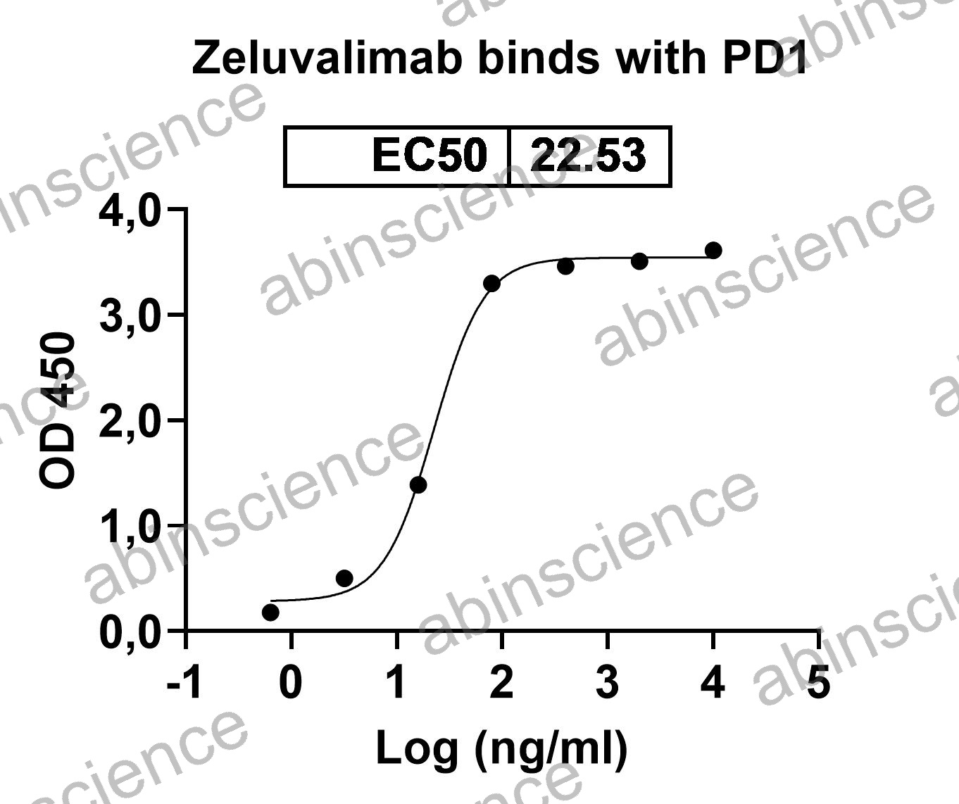
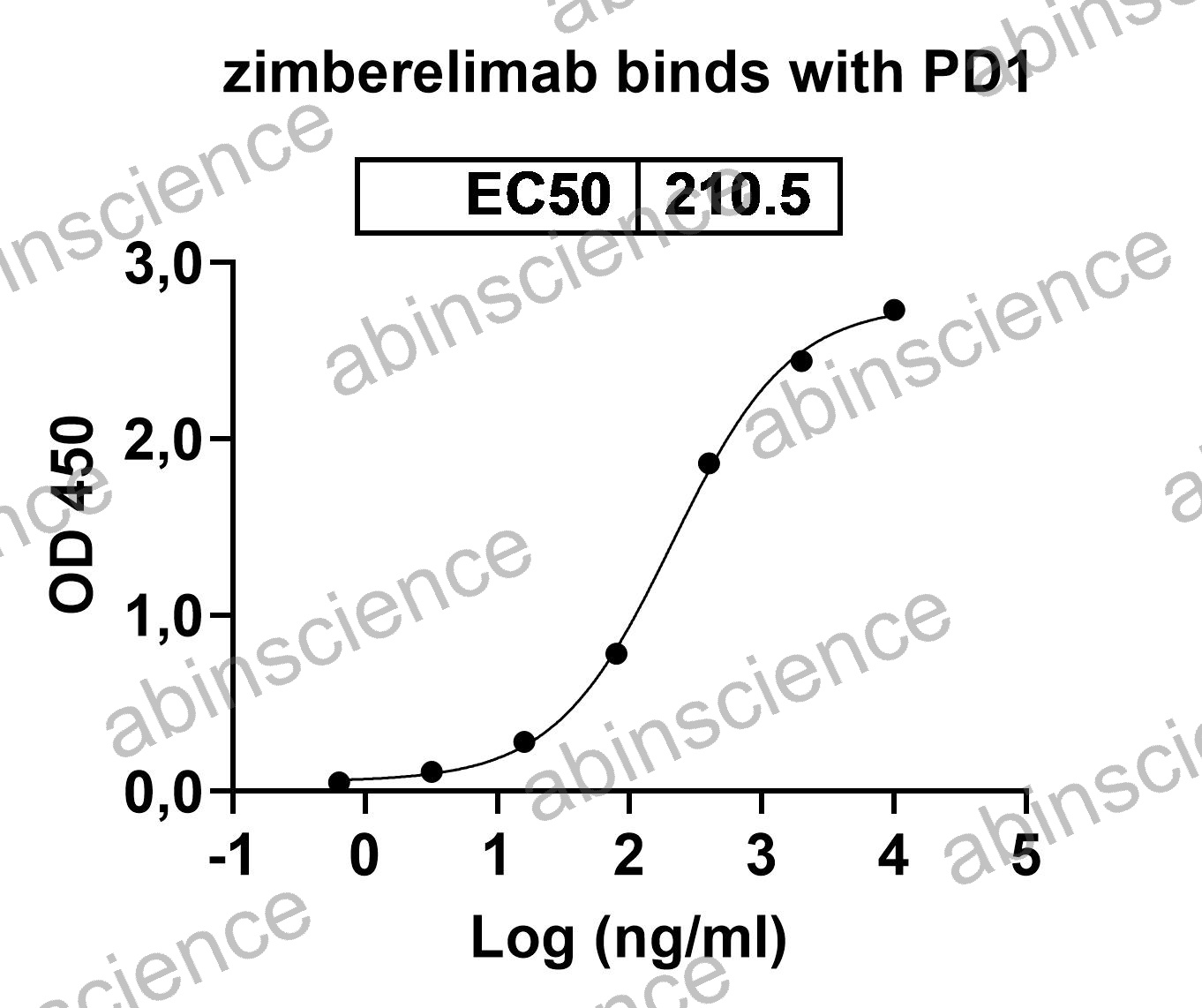
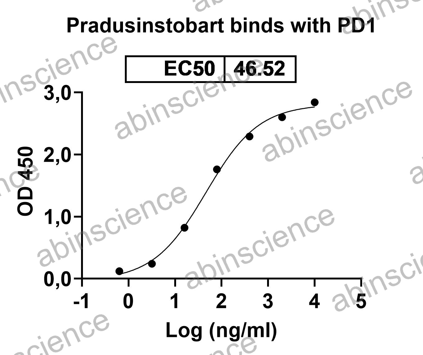
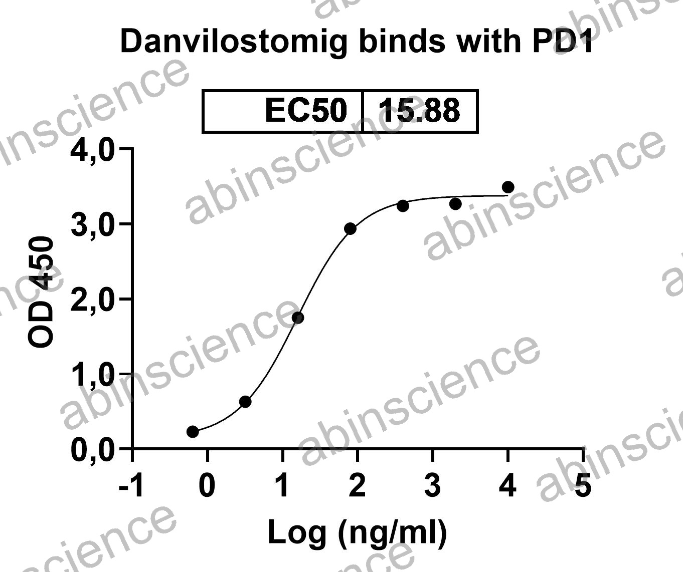
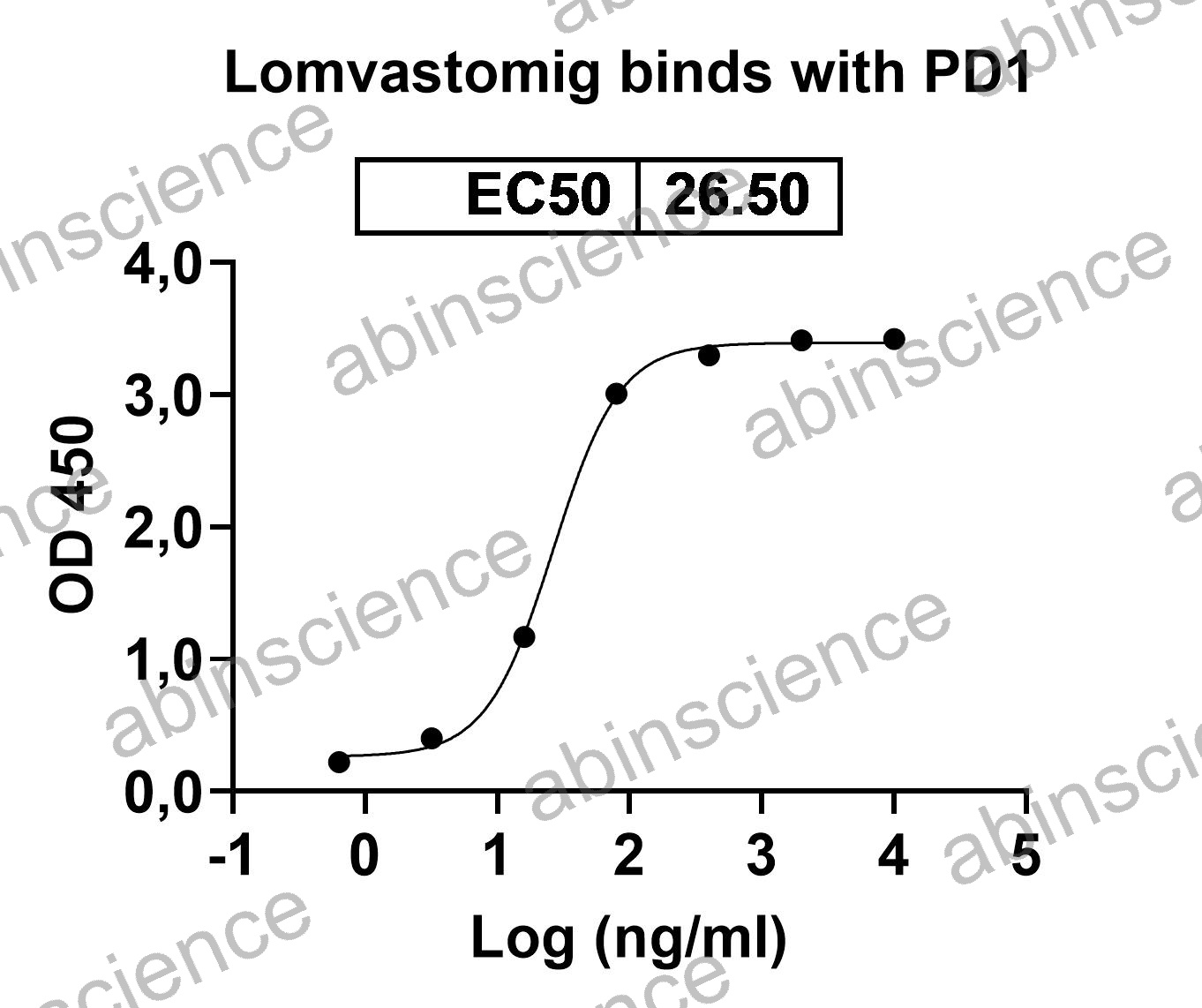
PD-1 (Programmed Cell Death Protein 1 / CD279) is an inhibitory immune checkpoint receptor on T cells that, upon engagement by its ligands PD-L1 (CD274) and PD-L2 (CD273), suppresses T-cell effector functions in the tumor microenvironment. Therapeutic antibodies blocking the PD-1/PD-L1 axis — including Pembrolizumab (Keytruda) and Nivolumab (Opdivo) — have transformed treatment of melanoma, NSCLC, and many other cancers. abinScience offers recombinant PD-1 proteins, anti-PD-1 antibodies, biosimilar reference standards, ELISA kits, and PD-1/PD-L1 blocking assay reagents.
PD-1 Biology and Mechanism
PD-1 is a ~55 kDa type I transmembrane receptor belonging to the CD28/CTLA-4 immunoglobulin superfamily. It is expressed on activated T cells, B cells, NK cells, and myeloid cells. PD-1 contains an immunoreceptor tyrosine-based inhibitory motif (ITIM) and an immunoreceptor tyrosine-based switch motif (ITSM) in its cytoplasmic tail, which recruit the phosphatase SHP-2 upon ligand binding.
When PD-L1 on tumor cells binds PD-1 on T cells, SHP-2 dephosphorylates key TCR signaling intermediates, attenuating T-cell proliferation, cytokine production (IFN-γ, TNF-α, IL-2), and cytotoxic killing. Unlike CTLA-4, which acts primarily during T-cell priming in the lymph node, PD-1 functions as an "exhaustion checkpoint" in peripheral tissues and the tumor microenvironment, making PD-1 blockade particularly effective at reactivating tumor-infiltrating lymphocytes (TILs).
Key Research Applications
Anti-PD-1 Antibody Screening Recombinant PD-1 proteins (His, Fc, Avi-tagged, biotinylated) for therapeutic antibody discovery, affinity maturation, and epitope binning. Human, mouse, and cynomolgus PD-1 for cross-species reactivity assessment.
PD-1/PD-L1 Blocking Assays PD-1-Fc and PD-L1 proteins for competitive ELISA-based and cell-based reporter blockade assays. Measure the ability of candidate antibodies to disrupt PD-1/PD-L1 interaction and restore T-cell activation.
Biosimilar Comparability Research-grade Pembrolizumab and Nivolumab biosimilars for analytical comparability studies, forced degradation, charge variant analysis, and as positive controls in bioassay development.
PK/ADA & Immunogenicity Assays Anti-PD-1 antibody pairs and PD-1 calibrator proteins for sandwich ELISA-based pharmacokinetic, anti-drug antibody, and neutralizing antibody assay development in Pembrolizumab/Nivolumab clinical programs.
PD-1 Product Selection Guide
| Application |
Recommended Product |
Format |
| SPR/BLI binding kinetics |
Recombinant PD-1 protein |
Avi-tagged or biotinylated |
| PD-1/PD-L1 blocking assay |
PD-1-Fc + PD-L1 protein |
Fc-chimera |
| ELISA / serum PD-1 quantification |
PD-1 ELISA kit or antibody pair |
Matched capture/detection |
| Flow cytometry / TIL profiling |
Anti-PD-1 antibody |
PE, APC, BV421 conjugated |
| WB / IHC detection |
Anti-PD-1 monoclonal antibody |
Unconjugated |
| PK/ADA ELISA |
Anti-idiotype antibody pair + PD-1 calibrator |
Capture/detection pair |
| Analytical comparability |
Pembrolizumab or Nivolumab biosimilar |
Research-grade RUO |
| Cross-species preclinical work |
Cynomolgus / mouse PD-1 protein |
His-tagged or Fc-chimera |
PD-1 FAQs
What is the difference between PD-1 and PD-L1 blockade?
Anti-PD-1 antibodies (Pembrolizumab, Nivolumab) block PD-1 from interacting with both PD-L1 and PD-L2, providing broader inhibition of the PD-1 pathway. Anti-PD-L1 antibodies (Atezolizumab, Durvalumab) selectively block the PD-1/PD-L1 interaction while leaving PD-1/PD-L2 signaling intact, and may also block PD-L1/CD80 (B7-1) interaction. The choice depends on the tumor's ligand expression profile and the desired breadth of checkpoint blockade.
How do I set up a PD-1/PD-L1 blocking bioassay?
The standard approach uses PD-1-expressing Jurkat-NFAT-Luc reporter cells co-cultured with PD-L1-expressing CHO or aAPC cells. PD-1/PD-L1 engagement suppresses NFAT-driven luciferase expression; anti-PD-1 or anti-PD-L1 blocking antibodies release this inhibition in a dose-dependent manner. Use our recombinant PD-1 and PD-L1 proteins as calibrators and Pembrolizumab/Nivolumab biosimilars as positive controls.
What is the difference between Pembrolizumab and Nivolumab biosimilars?
Both are anti-human PD-1 monoclonal antibodies but differ in their epitope, isotype, and binding characteristics. Pembrolizumab is a humanized IgG4 (S228P stabilized) that binds the C'D loop of PD-1. Nivolumab is a fully human IgG4 that binds the N-terminal loop of PD-1. They have distinct epitopes and do not compete for binding. Our biosimilars replicate the published sequences and are produced in CHO cells for analytical comparability use.
Do you offer cynomolgus monkey PD-1 for preclinical studies?
Yes. Cynomolgus monkey (Macaca fascicularis) PD-1 protein is available in His-tagged and Fc-chimera formats. Human PD-1 and cynomolgus PD-1 share approximately 94% sequence identity in the extracellular domain, making cynomolgus monkeys the primary non-human primate model for anti-PD-1 toxicology studies. Cross-reactivity confirmation by SPR is recommended before entering GLP studies.
How should I detect PD-1 expression on tumor-infiltrating T cells?
For flow cytometry, use a fluorochrome-conjugated anti-PD-1 antibody (PE or APC recommended for bright signal) in combination with CD3, CD4, and CD8 markers for T-cell subset gating. For IHC on FFPE sections, use an unconjugated anti-PD-1 monoclonal antibody with antigen retrieval (Tris-EDTA pH 9.0 recommended). Include a PD-1-positive tonsil section as a positive control.
Key References
1. Sharpe AH, Pauken KE. (2018) The diverse functions of the PD1 inhibitory pathway. Nat Rev Immunol. 18(3):153-167. PMID: 28990585
2. Robert C, et al. (2015) Pembrolizumab versus Ipilimumab in advanced melanoma. N Engl J Med. 372(26):2521-2532. PMID: 25891173
3. Tan S, et al. (2017) Crystal structure of PD-1/Pembrolizumab complex reveals the molecular basis for therapeutic antibody recognition. Nat Commun. 8:14369. PMID: 28098143


 中文
中文 English
 한국어
한국어 日本語
日本語 Español
 Français
 Русский