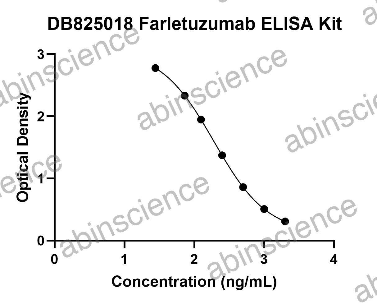
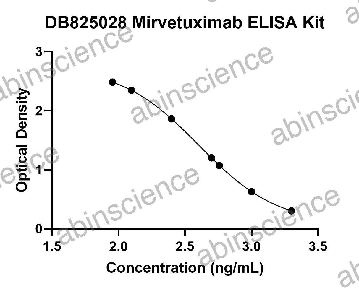
FOLR1 (Folate Receptor Alpha / FRα / FBP) is a GPI-anchored glycoprotein that mediates cellular uptake of folate and is overexpressed on the surface of epithelial ovarian cancer, endometrial cancer, NSCLC, and triple-negative breast cancer. The FDA-approved ADC Mirvetuximab soravtansine (Elahere) targets FRα-positive ovarian cancer, and multiple next-generation anti-FRα ADCs and CAR-T therapies are in development. FOLR1 expression is also a validated companion diagnostic biomarker (IHC) for patient selection. abinScience offers recombinant FOLR1 proteins, anti-FOLR1 antibodies, biosimilar reference standards, and ELISA kits.
FOLR1 Biology and Therapeutic Significance
FOLR1 is a ~38 kDa glycosylphosphatidylinositol (GPI)-anchored protein that binds folic acid and reduced folates with high affinity (Kd ~0.1-1 nM) and mediates their internalization through receptor-mediated endocytosis. In normal tissues, FOLR1 expression is restricted to the apical (luminal) surface of polarized epithelial cells in the kidney proximal tubule, choroid plexus, and lung — where it is inaccessible to circulating antibodies due to tight junctions.
In tumors, epithelial polarity is disrupted, and FOLR1 becomes accessible across the entire cell surface. This creates a favorable therapeutic window: anti-FRα antibodies can reach tumor-expressed FOLR1 but not normal tissue FOLR1 (which is sequestered behind intact tight junctions). Combined with efficient receptor-mediated internalization, this makes FOLR1 an ideal ADC target. The folate receptor family includes FRα (FOLR1), FRβ (FOLR2, expressed on activated macrophages), FRγ (FOLR3, secreted), and FRδ (FOLR4) — therapeutic antibodies must be selective for FRα to avoid off-target effects.
Key Research Applications
ADC Development & Internalization Recombinant FOLR1 proteins for anti-FRα antibody screening and characterization. FOLR1-expressing stable cell lines for ADC internalization assays, cytotoxicity studies, and payload release evaluation.
IHC & Companion Diagnostics Anti-FOLR1 antibodies validated for IHC on FFPE tissue. FRα IHC scoring is used clinically as a companion diagnostic to select patients for Mirvetuximab soravtansine treatment in platinum-resistant ovarian cancer.
Biosimilar & PK/ADA Assays Mirvetuximab biosimilar reference standards for analytical comparability. Anti-FOLR1 antibody pairs and calibrator proteins for pharmacokinetic and anti-drug antibody ELISA development.
Folate-Drug Conjugate Research FOLR1 proteins for studying folate-conjugated small molecules, imaging agents, and nanoparticles that exploit folate receptor-mediated endocytosis for tumor-targeted drug delivery.
FOLR1 Product Selection Guide
| Application |
Recommended Product |
Format |
| SPR/BLI binding kinetics |
Recombinant FOLR1 protein |
Avi-tagged or biotinylated |
| ADC internalization / cytotoxicity |
FOLR1-expressing stable cell line |
Engineered overexpression |
| IHC (FFPE tissue) |
Anti-FRα monoclonal antibody |
Unconjugated, IHC-validated |
| Flow cytometry |
Anti-FOLR1 antibody |
PE or APC conjugated |
| Soluble FRα quantification |
FOLR1 ELISA kit |
Sandwich ELISA |
| Folate receptor selectivity panel |
FOLR1 + FOLR2 proteins |
His-tagged (matched format) |
| PK/ADA ELISA |
Anti-FOLR1 antibody pair + calibrator |
Capture/detection pair |
| Analytical comparability |
Mirvetuximab biosimilar |
Research-grade RUO |
FOLR1 FAQs
What is the difference between FRα (FOLR1) and FRβ (FOLR2)?
FRα (FOLR1) is expressed on epithelial tumor cells — ovarian, endometrial, NSCLC — and is the primary target for anti-FRα ADCs. FRβ (FOLR2) is expressed on activated macrophages, including tumor-associated macrophages (TAMs), and is a separate therapeutic target for macrophage-directed therapies. Anti-FRα therapeutic antibodies must demonstrate selectivity over FRβ to ensure tumor-cell-directed killing. When testing antibody candidates, include both FOLR1 and FOLR2 recombinant proteins in your binding panel.
Why is FOLR1 considered a safe ADC target despite normal tissue expression?
In normal tissues, FOLR1 is expressed on the apical (luminal) surface of polarized epithelial cells — specifically in kidney proximal tubules, choroid plexus, and lung. Tight junctions prevent circulating antibodies from reaching the apical surface. In tumors, epithelial polarity is lost, exposing FOLR1 on the basolateral surface accessible to blood-borne antibodies. This anatomical inaccessibility in normal tissues — combined with high tumor expression — creates a wide therapeutic window for anti-FRα ADCs.
How is FRα expression scored for companion diagnostics?
For Mirvetuximab soravtansine, FRα expression is scored by IHC using the PS2+ scoring method: staining intensity (0, 1+, 2+, 3+) × percentage of positive cells, with a threshold of ≥75% of cells at ≥2+ intensity defining "FRα-high" status. Only FRα-high patients are eligible for treatment. Different anti-FRα clinical programs may use different scoring algorithms, so confirm the specific companion diagnostic criteria for your study.
What is soluble FRα (sFRα) and can it interfere with anti-FRα therapy?
Soluble FRα is shed from the tumor cell surface by GPI-anchor cleavage. Elevated serum sFRα levels are associated with advanced ovarian cancer and may act as an antigen sink — binding circulating anti-FRα therapeutic antibodies before they reach the tumor, potentially reducing efficacy. Our FOLR1 ELISA kit can measure sFRα in serum to assess this risk and to monitor sFRα as a pharmacodynamic biomarker during treatment.
Which tumors express FOLR1 most strongly?
The highest FOLR1 expression is seen in high-grade serous ovarian cancer (70-80% of cases are FRα-positive), followed by endometrial cancer, NSCLC (especially adenocarcinoma), triple-negative breast cancer, and mesothelioma. FRα expression is generally low or absent in colorectal, gastric, and hematologic malignancies. IHC screening is recommended to confirm FRα positivity before including tumor models in anti-FRα drug development programs.
Key References
1. Vergote I, et al. (2023) Mirvetuximab soravtansine in FRα-positive, platinum-resistant ovarian cancer. N Engl J Med. 389(23):2162-2174. PMID: 37870962
2. Cheung A, et al. (2016) Targeting folate receptor alpha for cancer treatment. Oncotarget. 7(32):52553-52574. PMID: 27248175
3. Scaranti M, et al. (2020) Exploiting the folate receptor alpha in oncology. Nat Rev Clin Oncol. 17(6):349-359. PMID: 32152484


 中文
中文 English
 한국어
한국어 日本語
日本語 Español
 Français
 Русский