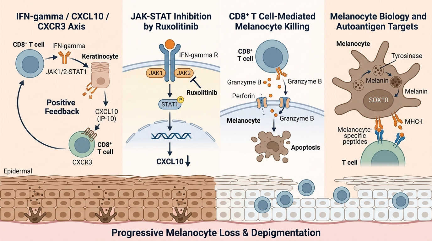
CXCL10 (IP-10) / CXCR3 — The CXCL10/CXCR3 chemokine axis is the central T cell recruitment pathway in vitiligo. IFN-γ-induced CXCL10 from keratinocytes recruits CXCR3+ CD8+ T cells to the epidermis, creating a positive-feedback loop that sustains melanocyte destruction. Serum CXCL10 is the leading disease activity biomarker. abinScience offers anti-CXCL10 reference antibodies (Eldelumab, NI-0801), anti-CXCL10 recombinant antibodies (human SAA0413, mouse SAA0412), and anti-CXCR3/CD183 recombinant antibodies (4 clones: V3G6, 5H7, 1C6, 7H5) with fluorescent conjugates for flow cytometric T cell phenotyping and chemotaxis assays.
→ Browse CXCL10 & CXCR3 antibodies
IFN-γ / JAK-STAT — IFN-γ is the master cytokine driving vitiligo pathogenesis, inducing CXCL10 production, upregulating melanocyte MHC-I expression, and promoting cytotoxic T cell activation. Ruxolitinib (JAK1/2 inhibitor) blocks downstream IFN-γ signaling via the JAK-STAT1 pathway. Anti-IFN-γ reference antibodies, recombinant antibodies with fluorescent conjugates, recombinant IFN-γ proteins, and anti-JAK1/JAK2 polyclonal antibodies with recombinant proteins support cytokine profiling, JAK-STAT signaling pathway dissection, and ruxolitinib mechanism-of-action studies.
→ Browse IFN-γ & JAK antibodies
Granzyme B / Perforin — CD8+ cytotoxic T cells kill melanocytes via the granzyme B/perforin pathway. Granzyme B cleaves intracellular substrates to trigger apoptosis, while perforin forms pores in the target cell membrane for granzyme B delivery. Anti-granzyme B recombinant antibodies (clones Fv02, scFv17) and anti-perforin recombinant antibody (clone SAA0104) are available with fluorescent conjugates (FITC, PE, APC, PerCP) for perilesional T cell cytotoxicity profiling by flow cytometry and IHC on skin biopsies.
→ Browse granzyme B & perforin antibodies
Melanocyte Biology (Tyrosinase / SOX10) — Tyrosinase (TYR) is the rate-limiting enzyme in melanin biosynthesis and a melanocyte-specific autoantigen. SOX10 is a transcription factor essential for melanocyte lineage specification and survival. Anti-tyrosinase recombinant antibody (clone SAA1969), anti-TYRP1 reference antibody (Flanvotumab) and recombinant antibody (clone TA99), and anti-SOX10 polyclonal antibody support melanocyte quantification in skin biopsies (IHC), melanocyte differentiation research, and autoantigen characterization in vitiligo.
→ Browse melanocyte biology antibodies


 中文
中文 English
 한국어
한국어 日本語
日本語 Español
 Français
 Русский