Everything you need to plan, run, and troubleshoot antibody- and protein-based experiments — from Western blot and ELISA to IHC, IP, and flow cytometry. All guides are written by scientists and paired with abinScience's catalog of 24,000+ antibodies, 13,000+ recombinant proteins, and 1,000+ ELISA kits.
Western Blot
Step-by-Step WB Protocol — Sample prep through detection, with recommended antibody dilutions
WB Troubleshooting Guide — No band, multiple bands, high background, and uneven transfer fixes
Loading Control Selection — Beta-actin vs GAPDH vs histone H3: how to choose by target MW and compartment
Phospho-Western Blot Protocol | Nuclear/Cytoplasmic Fractionation | Blocking Reagent Selection
Immunohistochemistry (IHC)
IHC Staining Protocol: Paraffin & Frozen Sections — Complete walkthrough from deparaffinization to mounting
Antigen Retrieval: Citrate vs EDTA vs Enzymatic — Target-based recommendation table for 15+ common markers
IHC Troubleshooting | IHC Panel Design by Tumor Type | Multiplex IHC/IF for TME Profiling
ELISA
ELISA Protocol: Sandwich & Indirect — Coating, blocking, detection, and 4PL curve fitting
ELISA Troubleshooting — High background, low signal, poor standard curve, and edge effects
Indirect vs Sandwich vs Competitive ELISA | Sandwich ELISA Antibody Pairing | Multiplex Immunoassay Troubleshooting
Immunofluorescence (IF)
IF Staining Protocol & Antibody Selection | Blocking & Permeabilization Guide
Immunoprecipitation (IP) & ChIP
IP Protocol: Bead Selection, Antibody Amount & Elution — Protein A/G vs Nano-Trap, lysis buffer, heavy chain avoidance
Co-IP Troubleshooting | ChIP Protocol | Cell Lysis & Protein Extraction
Flow Cytometry
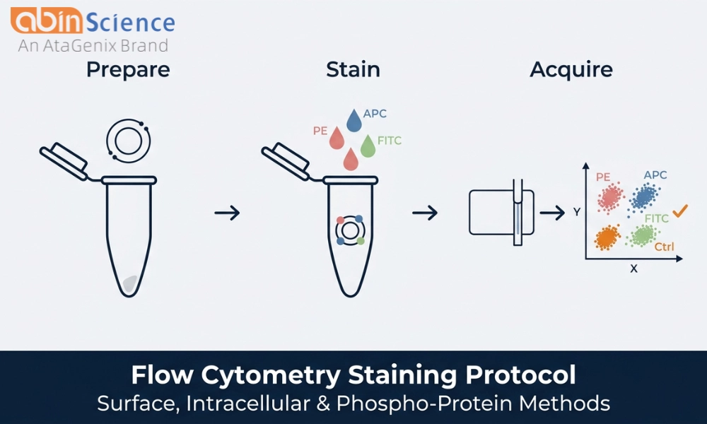
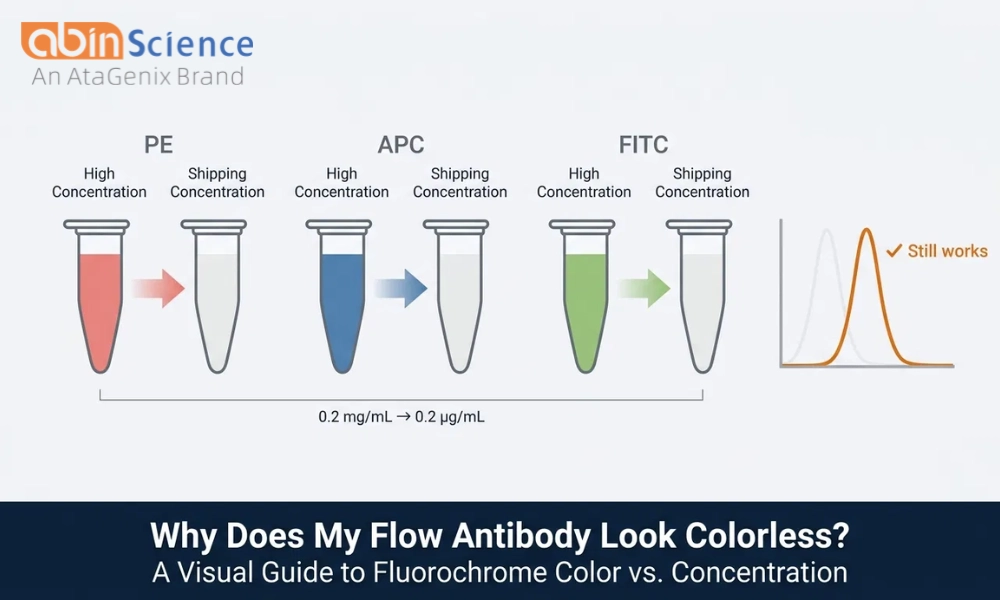
FC Staining: Surface vs Intracellular | Fluorochrome Conjugate Matching | Isotype Controls vs FMO
PBMC Isolation Protocol | TIL Analysis Panel Design | Full Flow Cytometry Knowledge Center (70+ guides) →
Antibody Selection & Fundamentals
Monoclonal vs Polyclonal Antibodies — How to choose by application (WB, IHC, IF, FC, ELISA)
Antibody Validation: 5 Orthogonal Strategies — KO/KD, orthogonal, independent Ab, peptide competition, expression pattern
Antibody Formats: IgG vs F(ab')₂ vs Fab vs scFv vs Nanobody | Nanobodies (VHH)
Secondary Antibody Selection | Affinity vs Avidity | Lot-to-Lot Consistency | KO-Validated Meaning
Antibody Storage & Handling | Storage Temperature & Aliquoting | Dilution Optimization
Antibody Structure: Regions & Domains | What Is a Clone Number? | Fc Tag Guide
Recombinant Proteins
Protein Handling: Storage, Reconstitution & Stability | Protein Concentration Methods | Inclusion Body Refolding
Biosimilar & ADA Testing
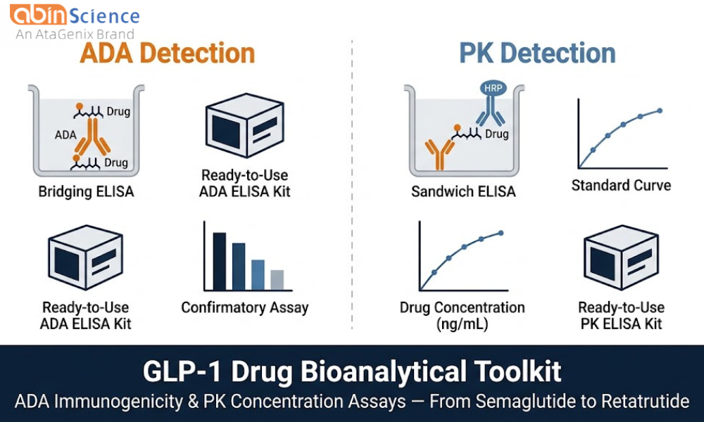
ADA ELISA Kit Selection: Bridging, Direct & Competitive — PK + ADA + NAb trios for 21 drug targets
GLP-1 ADA Assay FAQ | Anti-Idiotype Antibody Selection | PK/ADA Assay Development
What Are Research Biosimilars? | Biosimilar vs Originator | Endotoxin Level for In Vivo Use
Neutralization Assays & Binding Kinetics
Neutralization Assay Protocol: Pseudovirus & sVNT | Neutralization Assay Guide | SPR vs BLI Binding Kinetics
Oncology Research & Therapeutic Targets
Oncology Research Reagents | Immune Checkpoint Inhibitors | ADC Drug Targets | Tumor Microenvironment
Targets: PD-1/PD-L1 | CTLA-4 | LAG-3, TIGIT & TIM-3 | OX40 & 4-1BB | CD47/SIRPα
Targets: HER2 | EGFR | CD20 | VEGF/VEGFR | TNF-α
Cancer types: Lung | Breast | Colorectal | Melanoma | Lymphoma & Leukemia | Pancreatic | HCC | GBM
Other Resources
Cytokines & Chemokines Classification | Stable Cell Line Development | Cancer Cell Line Authentication
PEG FAQ | Protein Database Resources (UniProt) | CD4+CD8+ Double-Positive T Cells


 中文
 English
 한국어
 日本語
 Español
 Français
 Русский