Recent outbreaks of Acute Encephalitis Syndrome (AES) caused by Chandipura virus (CHPV) in India have once again pushed this long-neglected arbovirus to the forefront of global biosecurity and public health research. According to surveillance data from Indian health authorities and the World Health Organization (WHO), as of August 2026, multiple cases of unexplained pediatric encephalitis and laboratory-confirmed infections have been reported across several Indian states, accompanied by persistently high case fatality rates.
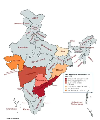
Figure 1. Outbreak Trends and Case Fatality Rates of CHPV Across Indian States
First isolated in 1965, CHPV is an Old World rhabdovirus and not a novel viral genotype. However, unlike most common arboviruses that induce only mild fever or rash, CHPV rapidly breaches host defenses within 24 to 48 hours post-infection, invading the central nervous system to trigger fulminant encephalitis and exceptionally high mortality. Why does a virus discovered decades ago still lack targeted therapeutics and vaccines? What molecular mechanisms enable such efficient barrier penetration and neurovirulence?
I. Rapid Outbreaks and High Mortality: The Unique Biological Puzzle of CHPV
Taxonomically, Chandipura virus belongs to the genus Vesiculovirus within the family Rhabdoviridae, sharing the family with classic experimental model viruses such as Vesicular Stomatitis Virus (VSV) and Rabies Virus (RABV). CHPV is primarily transmitted through the bites of sandflies (Phlebotomus spp.).
Compared with other common viruses, CHPV displays distinct pathogenic features:
- • Extremely Rapid Clinical Progression: The timeline from non-specific prodromal symptoms (such as sudden high fever and vomiting) to severe neurological manifestations—including convulsions, altered consciousness, coma, and death—spans a mere 24 to 72 hours. Such explosive neurological damage is rare among negative-sense RNA viruses.
- • Marked Host Age Preference: Epidemiological and clinical observations reveal that severe illness and fatal cases are almost exclusively concentrated in children and adolescents under 15 years of age. This strict age preference suggests a critical window of vulnerability in the immature host blood-brain barrier (BBB) structure or innate immune response network.
From a molecular and cellular biology perspective, CHPV pathogenesis raises core scientific questions: How does the virus breach the blood-brain barrier? How does it replicate rapidly within neurons while evading host antiviral responses? Investigating these mechanisms is essential not only for understanding CHPV itself, but also for providing an ideal model to study the pathogenic mechanisms of broad-spectrum neurotropic viruses.
II. Penetration and Neuroinvasion: Neurotropism and BBB Crossing
A central feature of CHPV infection is its strong neurotropism. After sandfly bites inject the virus into subcutaneous tissues, CHPV rapidly replicates in local endothelial cells and lymphatic tissues, establishing high-titer viremia. Subsequently, the virus initiates invasion into the central nervous system (CNS).
Two primary hypotheses and supportive findings explain how CHPV enters the CNS:
- • Blood-Brain Barrier (BBB) Disruption Route: High-titer peripheral viral replication triggers a massive release of pro-inflammatory cytokines (such as TNF-α and IL-6), causing a "cytokine storm". This severe inflammatory response widens endothelial intercellular junctions and increases barrier permeability, allowing direct viral invasion into the brain parenchyma. Recent studies also suggest that viral proteins may induce selective endocytosis in brain microvascular endothelial cells to mediate direct transcytosis.
- • Retrograde Axonal Transport Route: Similar to Rabies Virus, some evidence indicates that CHPV can infect peripheral nerve terminals and travel via retrograde axonal transport along nerve fibers directly to central neuronal cell bodies.
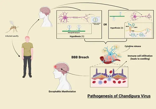
Figure 2. Pathogenesis of CHPV
Once inside brain tissue, the virus rapidly targets neurons. Within the neuronal cytoplasm, CHPV activates endogenous stress pathways, causing excessive accumulation of reactive oxygen species (ROS). This induces widespread neuronal apoptosis and necrosis via extrinsic and intrinsic apoptotic pathways (such as the caspase cascade), ultimately leading to severe cerebral edema and neurological failure.
III. Genomic Organization and Key Structural Protein Analysis
CHPV is a single-stranded negative-sense RNA virus (-ssRNA) with a genome size of approximately 11 kb, encoding five structural proteins in sequential order: Nucleoprotein (N), Phosphoprotein (P), Matrix protein (M), Glycoprotein (G), and Large protein/RNA polymerase (L).
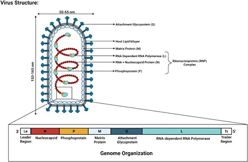
Figure 3. Genomic Organization of CHPV (DOI: 10.1016/j.idnow.2025.105179)
These five proteins work orchestratively to carry out the complete viral life cycle, including attachment, entry, transcription, replication, assembly, and budding:
1. Glycoprotein (G Protein): Receptor Binding and Viral Entry Key
Structure & Expression: The G protein is the sole transmembrane glycoprotein expressed on the viral envelope, adopting a trimeric conformation.
Function & Mechanism: It recognizes specific host cell surface receptors and mediates virus entry via endocytosis. Inside the acidic endosomal environment, the G protein undergoes conformational rearrangement, triggering fusion between the viral envelope and the endosomal membrane to release the nucleocapsid into the host cytoplasm.
Research Value: As the primary target for neutralizing antibodies, the G protein is crucial for vaccine development and entry inhibitor screening.
2. Nucleoprotein (N Protein): Genome Protection and Replication Template
Structure & Expression: The N protein is the major structural component of the viral ribonucleoprotein (RNP) complex.
Function & Mechanism: Newly synthesized N proteins encapsidate the single-stranded RNA genome with high affinity, forming a helical, nuclease-resistant nucleocapsid (RNP). Beyond physical RNA protection, the dynamic association between the N protein and viral RNA serves as a key switch regulating the viral polymerase's shift between transcription and replication modes.
Research Value: Due to its high abundance in virions and conserved sequence, the N protein serves as an essential target for diagnostic and analytical assays.
3. Phosphoprotein (P Protein): Molecular Hub of the Polymerase Complex
Structure & Expression: The P protein is an intrinsically disordered, highly phosphorylated cofactor protein.
Function & Mechanism: Acting as a bridge during replication, the P protein binds RNA-free N protein to prevent non-specific aggregation while simultaneously binding the L protein to accurately position it on the N-RNA template for transcription and replication. Additionally, the P protein suppresses host innate immune pathways (e.g., inhibiting interferon activation) to facilitate immune evasion.
4. Matrix Protein (M Protein): Virion Assembly, Budding, and Host Shut-off
Structure & Expression: The M protein is a small structural protein situated on the inner surface of the viral envelope.
Function & Mechanism: Operating during mid-to-late infection, the M protein interacts with both the N-RNA complex and the cytoplasmic tail of the G protein to drive virion assembly and budding at the plasma membrane. Crucially, the M protein exhibits potent cytotoxicity, blocking host mRNA nuclear export and transcription machinery to shut down host antiviral protein synthesis (host shut-off) and trigger apoptosis.
IV. Frontiers and Challenges in CHPV Basic Research
Despite clear knowledge of CHPV's genomic architecture, fundamental questions in molecular virology and immunology remain:
- • Identification of Specific Entry Receptors: While G protein-mediated endocytosis is known, specific surface receptors facilitating CHPV binding and uptake (such as low-density lipoprotein receptor family members investigated in recent studies) require broader experimental validation and structural confirmation.
- • Countermeasures Against Stress Granules (SGs) and Host Stress: Host cells assemble stress granules during infection to halt translation, but CHPV induces inclusion bodies (IBs) that sequester or dismantle SGs. The structural protein interaction network driving this subversion process warrants further mapping.
- • Molecular Targets for BBB Permeability Interventions: Identifying small molecules or biologics that block CHPV from crossing the blood-brain barrier is essential for developing early intervention strategies against AES.
Addressing these biological questions requires high-quality, specific molecular tools such as recombinant proteins and monoclonal/polyclonal antibodies. In vitro recombinant expression and functional Western blot analysis targeting distinct epitopes remain vital steps toward unraveling CHPV pathogenesis.
abinScience Chandipura Virus Research Products
To support life science researchers in advancing virology, immunology, and pathogenesis studies on Chandipura virus (CHPV), abinScience has developed a high-quality portfolio of recombinant proteins and antibodies targeting key structural proteins (G, N, P, and M proteins). These tools facilitate structural biology, host-pathogen interactions, and diagnostic assay development.
Recombinant Proteins
| Catalog No. |
Product Name |
| VK396011 |
CHPV G/Glycoprotein Recombinant Protein (C-His) |
| VK396021 |
CHPV G/Glycoprotein Recombinant Protein (C-Fc) |
| VK396012 |
CHPV G/Glycoprotein Recombinant Protein (N-His) |
| VK546012 |
CHPV P/Phosphoprotein Recombinant Protein (N-His) |
| VK535012 |
CHPV M/Matrix protein Recombinant Protein (N-His) |
| VK773012 |
CHPV N/Nucleoprotein Recombinant Protein (N-His) |
Antibodies
| Catalog No. |
Product Name |
| VK396014 |
Anti-CHPV G/Glycoprotein Polyclonal Antibody |
| VK546014 |
Anti-CHPV P/Phosphoprotein Polyclonal Antibody |
| VK535014 |
Anti-CHPV M/Matrix protein Polyclonal Antibody |
| VK773014 |
Anti-CHPV N/Nucleoprotein Polyclonal Antibody |
References
- [1] Akingbola, A., Adegbesan, A., Adegoke, K., Chuku, J., Ojo, O., Mariaria, P., Alao, U., Salami, R. A., & Oladunjoye, M. (2025). Chandipura Virus Resurgence in India: Insights Into Diagnostic Tools, Antiviral Development, and Public Health Implications. Global health, epidemiology and genomics, 2025, 1015031. https://doi.org/10.1155/ghe3/1015031
- [2] Dangi, R. R., Vageriya, V., Sharma, A., & Dabhi, P. (2026). Chandipura Virus: A Neglected Sandfly-Borne Neurotropic Virus in Children - Insights From Seven Decades of Evidence. Reviews in medical virology, 36(2), e70119. https://doi.org/10.1002/rmv.70119
- [3] Aderao, G. N., Nikhil, K. C., Sarkar, S., Patel, S. K., Kanaka, K. K., Mhaske, V. S., Kumar, A., & Emran, T. B. (2025). Emergence of Chandipura viral encephalitis in India: a strategic approach to combat a fatal viral epidemic. Annals of medicine and surgery (2012), 88(1), 542–547. https://doi.org/10.1097/MS9.0000000000004421
- [4] Lodha, L., Swaminathan, A., John, A., Kunhikannan, S., Sundar, B., Sathishkumar, A., Nair, K. J., & Pattanaik, A. (2025). Chandipura virus: A comprehensive review. Infectious diseases now, 55(8), 105179. https://doi.org/10.1016/j.idnow.2025.105179
- [5] Kanabar, B., Malek, S., & Piparva, K. (2024). Temporal Trends in Outbreaks of Chandipura Viral Infection in India: A Systematic Review. Cureus, 16(8), e68097. https://doi.org/10.7759/cureus.68097


 中文
中文 English
 한국어
한국어 日本語
日本語 Español
 Français
 Русский


