The core operating logic of the immune system lies in maintaining a delicate dynamic balance between efficient clearance of foreign entities and the avoidance of autoimmune tissue damage. On one hand, upon activation, effector immune cells such as T cells execute immune clearance functions through proliferation, differentiation, and the release of cytokines and cytotoxic molecules. On the other hand, multiple negative regulatory pathways are concurrently initiated to strictly limit the magnitude, duration, and tissue-damage risk of immune activation. Immune checkpoints serve as the core nodes within this balanced regulatory network.
Different checkpoint pathways act on distinct receptors, ligands, cell subsets, and stages of the immune response, collectively shaping the magnitude and duration of immune responses by modulating costimulatory signaling, receptor signal transduction, intercellular ligand accessibility, and immune cell fate. Therefore, when upregulation of a checkpoint molecule is observed in research, the critical scientific question to address is not simply "whether it is present," but rather "how much of a functional regulatory role it actually plays in the given immune phenotype".
This question is particularly crucial in functionally restricted states such as immune exhaustion, discussed in our previous issue: when sustained high expression of inhibitory receptors such as PD-1 and TIGIT is identified, expression levels merely provide correlative clues. Only targeted functional intervention experiments can further validate the underlying regulatory mechanisms. Consequently, immune checkpoint blockade (ICB) is not only a classic therapeutic/intervention strategy in immunology, but also a core experimental tool for dissecting in vivo immunomodulatory mechanisms.
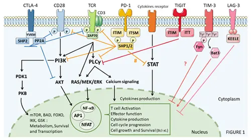
Figure 1. Immune Checkpoints in Cancer Cells (DOI: 10.3390/cancers13184573)
1. Why Do Immune Checkpoints Require Functional Blockade? Moving from Expression Profiling to Mechanism Validation
1.1 High Checkpoint Expression: A Phenotypic Marker or Functional Driver?
Immune checkpoints are not simple "on/off switches" for immune suppression; rather, they are key regulatory nodes for maintaining immune response homeostasis. Under physiological conditions, coinhibitory receptors limit excessive immune activation, maintaining a dynamic equilibrium between effective target clearance and prevention of immunopathology. However, under conditions of persistent antigen stimulation, chronic inflammation, or complex tissue microenvironments, multiple checkpoint pathways frequently undergo dynamic changes simultaneously.
Consequently, observing elevated checkpoint expression via transcriptomics, single-cell RNA sequencing, flow cytometry, or multiplex imaging merely demonstrates an association with the current cellular state. It cannot independently prove that a specific pathway is the core driver of immune dysfunction. In other words, while high checkpoint expression offers valuable research clues, it does not directly constitute causal evidence.
Based on this, mechanistic studies typically need to address three fundamental questions:
- (1) Is high expression of this checkpoint molecule an over-stimulation marker following repeated T cell activation, or a functional driver that directly forces cells into a suppressed state?
- (2) Within complex tissue microenvironments, what is the relative contribution of this specific pathway to the overall immune phenotype? Are there redundant compensatory effects from other signaling pathways?
- (3) If ligand binding to this checkpoint is specifically blocked, can the functional status of immune cells—such as cytokine secretion, proliferative capacity, and cytotoxicity—undergo reversible recovery?
1.2 From Correlation to Causality: The Value of Functional Interventions
Functional blockade provides a critical experimental path to transition from correlative observations to functional validation. By utilizing blocking antibodies against specific receptors or ligands, ligand binding or inhibitory signal transduction can be disrupted, followed by evaluation of changes in cell proliferation, cytokine secretion, cytotoxicity, differentiation status, and metabolic reprogramming. Furthermore, combining blockade with flow cytometry, in situ tissue analysis, transcriptomic, or epigenetic assays helps determine whether checkpoint blockade induces functional remodeling of specific cell populations or the immune microenvironment.
Thus, the experimental logic of checkpoint blockade research can be summarized as: Expression Observation → Targeted Blockade Intervention → Phenotypic & Functional Remodeling Analysis. For in vivo studies, the core value of functional blockade lies in its ability to evaluate pathway effects within a relatively complete cellular composition and tissue microenvironment, moving beyond simple descriptions at the molecular expression level.
2. Classic Immune Checkpoint Pathways: Core Mechanisms and Research Paradigms
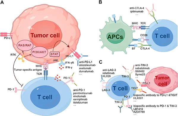
Figure 2. Immune Checkpoint Pathways and Mechanisms of Action of Blocking Antibodies (DOI: 10.3389/fimmu.2024.1491330)
2.1 PD-1/PD-L1: A Classic Model for Dissecting T Cell Dysfunction and Remodeling
PD-1 is one of the most extensively studied immune checkpoint receptors, primarily expressed on activated and chronically antigen-stimulated T cells, as well as on B cells and NK cells. Its ligand, PD-L1, is broadly expressed on antigen-presenting cells (APCs) and various parenchymal tissue cells. Upon ligand engagement, PD-1 recruits intracellular phosphatases via its immunoreceptor tyrosine-based inhibitory motifs to downregulate downstream signaling, attenuating positive inputs such as CD28-mediated costimulation and ultimately suppressing T cell activation and effector function.
In immune exhaustion studies, elevated PD-1 expression serves as a hallmark phenotypic marker. However, the key mechanistic questions remain: after relieving PD-1-mediated inhibition, which cell subsets respond? Is this response primarily characterized by proliferation, cytokine secretion, cytotoxicity, or shifts in cell differentiation states? Consequently, PD-1 blockade is often integrated with multicolor flow cytometry, functional assays, and transcriptomic/epigenetic profiling to dissect the heterogeneity of responses across exhausted T cell subsets.
Notably, exhausted T cells do not constitute a homogeneous population. Among them, TCF-1⁺ progenitor-like (precursor) exhausted T cells exhibit robust self-renewal capacity and the potential to replenish effector T cell pools. Functional interventions targeting PD-1 thus answer not only whether changes occur post-blockade, but also which specific cell subsets drive functional remodeling and what time-course kinetics govern this process.
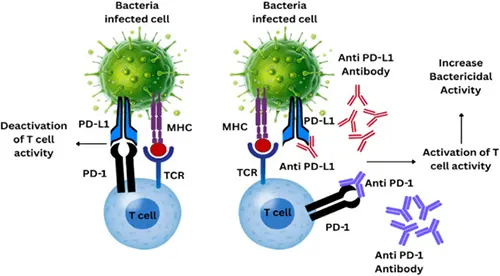
Figure 3. Immune Checkpoint PD1, PDL1 inhibition (DOI: 10.1016/j.clicom.2025.11.003)
2.2 CTLA-4: Competition for CD28 Costimulation and Treg-Mediated Immunomodulation
CTLA-4 shares ligands (CD80/CD86) with CD28 but binds them with significantly higher affinity, thereby competitively limiting the strength of CD28-mediated costimulatory signals. Unlike PD-1, which operates predominantly during the peripheral effector phase of activated T cells, CTLA-4 plays a more prominent regulatory role during initial T cell priming within lymphoid tissues.
The regulatory mechanism of CTLA-4 is also tightly linked to regulatory T cells (Tregs). Tregs constitutively express high levels of CTLA-4, which interacts with CD80/CD86 on APCs to induce ligand endocytosis and degradation, thereby remodeling the local costimulatory microenvironment. Therefore, CTLA-4 blockade serves as an essential tool for investigating the tripartite relationship between early T cell activation, CD28 costimulatory signaling, and Treg-mediated immune regulation.
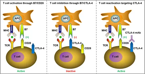
Figure 4. Mechanisms of CTLA-4-Mediated T Cell Suppression (DOI: 10.1038/s41419-023-06389-5)
3. Exhaustion-Associated Checkpoints: TIGIT, TIM-3, and LAG-3
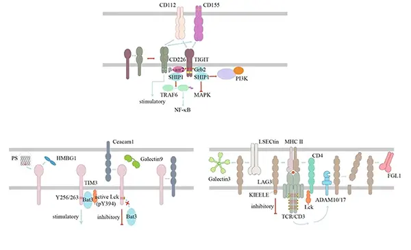
Figure 5. Mechanisms of Action of Exhaustion-Associated Checkpoints (DOI: 10.1038/s41422-020-0343-4)
3.1 TIGIT: Coinhibitory-Costimulatory Balance along the CD155/CD226 Axis
TIGIT is expressed on T cells, NK cells, and certain regulatory lymphocyte subsets, with CD155 (PVR) and CD112 serving as its primary ligands. A key feature of TIGIT research is its co-regulation of the same ligand axis alongside the costimulatory receptor CD226. Elevated TIGIT signaling competitively impairs CD226-mediated activating signals, thereby dampening T cell and NK cell effector functions.
Consequently, TIGIT blockade is particularly suited for studying the dynamic balance between coinhibition and costimulation. In T cell or NK cell study models, combining blockade intervention with measurements of cytotoxicity, cytokine production, and CD226 expression enables the independent role of the TIGIT pathway under specific immune conditions to be defined, as well as its crosstalk with other checkpoint pathways.
3.2 TIM-3: Bridging Exhaustion Phenotypes to Functional Validation
TIM-3 (HAVCR2/CD366) is a checkpoint receptor strongly associated with dysfunctional T cells and is also expressed on NK cells, Tregs, and myeloid cell subsets. Compared to PD-1, TIM-3 possesses a more complex ligand network, including Galectin-9, CEACAM1, HMGB1, and phosphatidylserine (PtdSer). Because these interactions occur across varied cell types and microenvironmental contexts, TIM-3 function is highly context-dependent.
Under chronic antigen stimulation, TIM-3 is frequently co-expressed with PD-1. PD-1⁺TIM-3⁺ double-positive T cell populations are commonly investigated via TIM-3 blockade assays to verify the pathway's actual contribution to proliferation, cytokine release, and cytotoxicity. As such, TIM-3 serves as a pivotal research tool bridging phenotypic observation of exhaustion with functional mechanistic validation.
3.3 LAG-3: From MHC II Interactions to Checkpoint Network Synergies
LAG-3 (CD223) is a key coinhibitory receptor expressed on activated and functionally impaired T cells, as well as subsets of Tregs and NK cells. MHC class II (MHC II) is its canonical ligand, while novel ligands such as FGL1 have also gained significant research interest. LAG-3 function cannot be simplified to a linear "ligand binding leads to inhibition" model; its intracellular signaling and spatial organization within the immunological synapse remain active areas of investigation.
In in vivo mechanistic studies, LAG-3 blockade enables assessment of the pathway's independent contribution to specific T cell states and functions. When LAG-3 is co-expressed with checkpoints like PD-1, comparing single-agent vs. dual-blockade outcomes helps elucidate synergistic and compensatory dynamics within the checkpoint network.
4. Broader Immune Checkpoints: From Core Pathways to Regulatory Networks
Beyond PD-1/PD-L1, CTLA-4, TIGIT, TIM-3, and LAG-3, current research is expanding into a broader array of immunomodulatory molecules, including VISTA, BTLA, CD96, NKG2A, B7-H3, B7-H4, and CD200R. These pathways span T cells, NK cells, B cells, and myeloid cells, regulating immune tolerance, effector functions, and tissue microenvironments through distinct receptor-ligand interactions.
Table 1. Comparative Research Focus Across Key Immune Checkpoints
| Checkpoint |
Molecule Type |
Primary Expressing Cells |
Representative Ligands / Interactions |
Core Research Focus |
| PD-1 / PD-L1 |
Coinhibitory receptor / ligand axis |
T, B, NK cells, and various tissue cells |
PD-L1, PD-L2 |
T cell dysfunction, functional remodeling, and microenvironment regulation |
| CTLA-4 |
Coinhibitory receptor |
Activated T cells, Tregs |
CD80, CD86 |
CD28 costimulation competition, Treg-mediated local immunomodulation |
| TIGIT |
Coinhibitory receptor |
T cells, NK cells, etc. |
CD155, CD112; forms regulatory network with CD226 |
Coinhibitory/costimulatory balance and T/NK effector functions |
| TIM-3 |
Coinhibitory-associated receptor |
Impaired T cells, NK cells, myeloid cells |
Galectin-9, CEACAM1, HMGB1, PtdSer |
Exhaustion-associated functional states and synergistic effects with other checkpoints |
| LAG-3 |
Coinhibitory receptor |
Activated/impaired T cells, Tregs, NK cells |
MHC II, FGL1, etc. |
T cell functional regulation and synergy with PD-1 |
| VISTA |
Coinhibitory-associated pathway |
Myeloid cells, T cells, etc. |
PSGL-1, VSIG-3; ligand dynamics under ongoing investigation |
Myeloid suppression, immune tolerance, and microenvironment modulation |
| BTLA |
Coinhibitory receptor |
T cells, B cells, and other lymphocytes |
HVEM |
Immune homeostasis, lymphocyte activation, and tissue immune regulation |
| CD96 |
Coinhibitory-associated receptor |
NK cells, T cells |
CD155, CD112 |
NK/T cell effector functions and crosstalk with TIGIT/CD226 network |
| NKG2A |
Coinhibitory receptor |
NK cells, subset of CD8⁺ T cells |
HLA-E |
Cytotoxic lymphocyte inhibition and tissue immune surveillance |
| CD200R |
Coinhibitory receptor |
Myeloid cells, etc. |
CD200 |
Myeloid activation threshold and tissue immune homeostasis |
5. In Vivo Immune Checkpoint Blockade: From Pathway Intervention to Functional Validation
The scientific value of immune checkpoint blockade extends far beyond merely increasing a particular immune metric. From a mechanistic standpoint, its true significance lies in selectively disrupting a known regulatory node to determine whether cellular phenotypes, functional outputs, and microenvironmental dynamics respond predictably. Only through such functional perturbations can it be definitively verified whether altered checkpoint expression is merely an incidental phenotypic marker or an active driver of the observed immune phenotype.
Therefore, a comprehensive translational research pipeline from expression profiling to functional validation typically follows this sequence: initial screening of candidate checkpoints via flow cytometry, single-cell sequencing, or in situ imaging; targeted intervention using functional blocking antibodies; and systematic evaluation of outcomes via cell subset profiling, effector molecule quantification, proliferation/cytotoxicity assays, and metabolic analysis.
abinScience In Vivo Functional Antibodies for Immune Checkpoint Research
abinScience offers a comprehensive portfolio of InVivo antibodies targeting key immune checkpoints to empower research into immunomodulatory mechanisms and checkpoint blockade experiments, providing robust tools for in vivo functional validation.
| Catalog No. |
Product Name |
| MS870039 |
InVivo Plus Anti-Mouse PD-1 Antibody (RMP1-14) |
| MV974029 |
InVivo Plus Anti-Mouse PD-L1 Antibody (10F.9G2) |
| MB651020 |
InVivoMAb Anti-Mouse CTLA-4 Antibody (9H10) |
| MS739040 |
InVivoMAb Anti-Mouse TIGIT Antibody (10A7) |
| MV029010 |
InVivoMAb Anti-Mouse TIM-3 Antibody (B8.2C12) |
| MV612030 |
InVivoMAb Anti-Mouse CD276/B7-H3 Antibody (MJ18) |
| MB613010 |
InVivoMAb Anti-Mouse LAG-3 & PD-1 Antibody (Iv0273) |
| MS739010 |
InVivoMAb Anti-Mouse PD-1 & TIGIT Antibody (Iv0247) |
| MP543010 |
InVivoMAb Anti-Mouse VISTA/B7-H5/PD-1H & PD-L1 Antibody (Iv0246) |
| MW342010 |
InVivoMAb Anti-Mouse OX40/TNFRSF4 & PD-L1 Antibody (Iv0244) |
| MB651040 |
InVivoMAb Anti-Mouse CTLA-4 & OX40/TNFRSF4 Antibody (Iv0227) |
| MY422010 |
InVivoMAb Anti-Mouse PD-L1 & CD28 Antibody (Iv0245) |
Click here to explore our complete portfolio of InVivo antibody products and InVivo Plus antibody products
References
- [1] Meng, L., Wu, H., Wu, J., Ding, P., He, J., Sang, M., & Liu, L. (2024). Mechanisms of immune checkpoint inhibitors: insights into the regulation of circular RNAS involved in cancer hallmarks. Cell Death & Disease, 15(1), 3. https://doi.org/10.1038/s41419-023-06389-5.
- [2] Hu, Y., Wang, Y., Min, K., Zhou, H., & Gao, X. (2024). The influence of immune checkpoint blockade on the outcomes of allogeneic hematopoietic stem cell transplantation. Frontiers in Immunology, 15, 1491330. https://doi.org/10.3389/fimmu.2024.1491330.
- [3] He, X., & Xu, C. (2020). Immune checkpoint signaling and cancer immunotherapy. Cell Research, 30(8), 660–669. https://doi.org/10.1038/s41422-020-0343-4.
- [4] Pisibon, C., Ouertani, A., Bertolotto, C., Ballotti, R., & Cheli, Y. (2021). Immune Checkpoints in Cancers: From Signaling to the Clinic. Cancers, 13(18), 4573. https://doi.org/10.3390/cancers13184573.
- [5] Wykes, M. N., & Lewin, S. R. (2018). Immune checkpoint blockade in infectious diseases. Nature Reviews Immunology, 18(2), 91–104. https://doi.org/10.1038/nri.2017.112.
- [6] Sarkar, S., Chowdhury, S., Kar, K., Chakraborty, P., & Das, A. (2025). Immunological interventions in bacterial infections: From mechanisms to clinical applications. Clinical Immunology Communications, 8, 124-137. https://doi.org/10.1016/j.clicom.2025.11.003.


 中文
中文 English
 한국어
한국어 日本語
日本語 Español
 Français
 Русский


