West Nile virus (WNV) activity across Europe has continued to draw significant attention in 2026. On October 15, the European Food Safety Authority (EFSA) and the European Centre for Disease Prevention and Control (ECDC) jointly released their fourth monthly WNV monitoring report. Data show that as of October 7, 18 European countries had cumulatively reported 1,855 locally acquired infections and 141 deaths, setting a historical record for the geographical spread of viral activity. A subsequent weekly report published on October 23 further indicated that as of October 21, 1,965 locally acquired infections had been reported across 20 European countries.
The expanding activity range of WNV has renewed scientific focus on this pathogen. As a representative member of the genus Orthoflavivirus within the family Flaviviridae, WNV is not a newly discovered pathogen; its genome structure, protein composition, and fundamental replication mechanisms have been investigated extensively. However, recent molecular virology studies demonstrate that the biological characteristics of WNV cannot be fully explained solely by the virus's own genetic information and protein functions. Complex molecular interactions exist between the virus and host cells, encompassing multiple dimensions such as the cellular membrane environment, lipid metabolism, protein homeostasis, and structural shifts in viral proteins. These findings not only broaden our understanding of WNV but also offer novel perspectives for deeply comprehending the molecular mechanisms associated with viral infection.
WNV is a single-stranded positive-sense RNA virus enveloped in a lipid membrane. Like all viruses, WNV lacks the cellular systems independently required for protein synthesis and energy metabolism; its biological activities rely entirely on the host cell to provide the necessary molecular environment. The relationship between WNV and host cells exhibits clear multi-layered characteristics. At the cellular structure level, the virus induces the reorganization of endoplasmic reticulum (ER)-derived membrane structures; at the metabolic level, virus-associated activities are closely linked with lipid synthesis, membrane lipid distribution, and plasma membrane properties; at the protein homeostasis level, the synthesis and processing of viral proteins also impact protein folding and stress regulation systems within the endoplasmic reticulum.
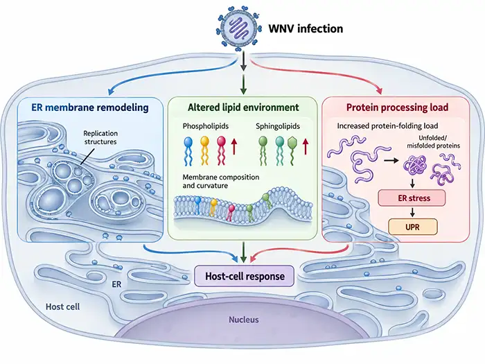
Figure 1. Multi-level changes in host cells induced by WNV infection
Meanwhile, host cells do not passively accept these changes. The cell's inherent protein quality control, metabolic regulation, and stress response systems influence the stability and function of virus-associated molecules; certain host molecules may even exhibit virus-restricting or virus-supporting roles under varying conditions. Consequently, the relationship between WNV and the host is not a simple one-way control mechanism, but rather the result of ongoing interactions between viral molecules and the cellular environment.
Upon infecting host cells, WNV not only generates viral proteins but also reshapes the cell's original structures and functions. Among these, the remodeling of endoplasmic reticulum membrane structures and changes in the protein-processing environment are two representative phenomena. They are both closely associated with WNV biological activities and reflect the reciprocal influence between the virus and host cells.
The endoplasmic reticulum (ER) is one of the cellular structures undergoing pronounced alterations during WNV infection. Research has revealed that WNV can induce the rearrangement of ER membranes to form specialized membrane structures, concentrating virus-associated proteins and RNA into localized compartments. Non-structural proteins such as NS4A and NS4B participate in this process, indicating that the virus actively utilizes its own proteins to modify the host cell membrane environment rather than relying solely on pre-existing cellular architectures.
Studies have found that WNV infection alters the composition of certain intracellular phospholipids and sphingolipids, and the lipid proportions in the viral particle envelope also differ from the overall membrane lipid profile of the host cell. These phenomena indicate that WNV's utilization of the host cell membrane system extends beyond a simple increase in membrane mass, involving membrane reorganization and compositional shifts. For fundamental WNV research, the linkage between viral proteins, host membrane structures, and the lipid environment represents a critical dimension for understanding virus-host interactions.
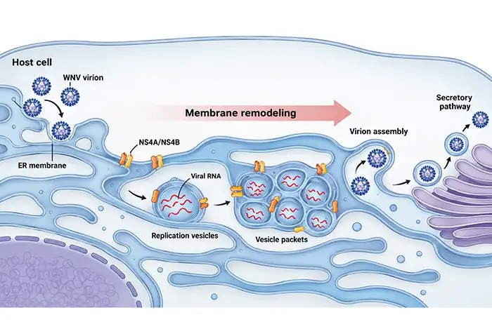
Figure 2. Induction of endoplasmic reticulum membrane remodeling by WNV infection
Beyond cellular membrane structures, WNV infection also impacts the protein-processing environment of host cells. The ER is responsible for protein folding, modification, and quality control, and the massive amounts of viral proteins produced during WNV infection must utilize these cellular machinery for processing. When the protein-processing load within the ER increases, cells may activate the Unfolded Protein Response (UPR) to regulate protein synthesis and folding, thereby maintaining normal cellular functions.
WNV infection can activate partial ER stress-related signaling pathways and alter the expression of molecules associated with protein quality control. These responses may help cells adapt to the pressure of viral protein processing while potentially influencing the formation and stability of virus-related molecules. If stress intensifies persistently, it may further compromise cell survival. Therefore, ER stress is not a straightforward process of solely promoting or inhibiting WNV activity; its effects are modulated by factors such as the viral strain, cell type, and infection state.
Interactions between WNV and host cells involve not only changes in the cellular environment but also the conformational and structural states of viral molecules.
WNV particles possess a lipid envelope, and the spatial arrangement of their surface proteins is not completely uniform across all particles. During viral particle maturation, the processing of the precursor membrane protein (prM) is coupled with the conformational rearrangement of the envelope protein (E); however, prM cleavage may be incomplete, leaving a proportion of uncleaved prM retained on certain viral particles. Consequently, a single viral population may comprise particles with varying degrees of maturity and surface structural differences.
WNV studies have confirmed that the maturation state of viral particles influences the accessibility of certain E protein epitopes, thereby altering their sensitivity to specific antibodies. For antibodies recognizing poorly exposed epitopes, the arrangement of surface proteins on the particle can directly dictate whether antibodies can adequately access their respective binding sites. Notably, the relationship between a viral particle's maturation state and its biological properties is not a simple binary correlation, as partially immature particles may retain infectivity under specific conditions.
These findings demonstrate that WNV viral particles do not form a homogeneous population composed of structurally identical molecular entities. The maturation state, protein arrangement, and local conformation of viral particles collectively shape their functional properties, making structural biology a vital component in deciphering WNV molecular functions.
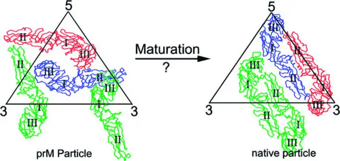
Figure 3. Maturation rearrangement of West Nile virus particles (DOI: 10.1128/JVI.00037-07)
In addition to structural differences on viral particle surfaces, certain WNV proteins can adopt multiple molecular assembly states. The non-structural protein NS1 is a prime example. NS1 can exist as a membrane-associated dimer or form higher-order structures such as secretory hexamers. Across different states, the hydrophobic regions, protein surfaces, and intermolecular interaction modes of NS1 vary, enabling adaptation to diverse molecular environments.
Structural analyses have revealed that NS1 contains hydrophobic surfaces associated with membrane interaction and structural domains capable of forming stable protein complexes. Secretory NS1 can also form complexes with lipids, exhibiting structural traits distinct from its membrane-associated counterpart. These characteristics provide a rationale for why a single viral protein can exhibit distinct molecular properties across different spatial environments.
NS1 is not the only WNV protein exhibiting distinct molecular states, but it exemplifies a universally applicable virological principle: a protein's biological function is dictated not solely by its coding sequence, but is closely tied to its folding pathway, post-translational modifications, molecular assembly, and local environment.
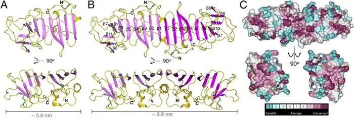
Figure 4. Monomeric/dimeric structure of WNV NS1 (DOI: 10.1073/pnas.1322036111)
Another key characteristic of WNV research is that molecular changes across different tiers do not always exhibit simple one-to-one correlations. Linkages exist among viral protein expression and processing, cellular membrane environmental shifts, protein homeostasis regulation, and the structural states of viral particles; however, alterations at a single level cannot fully represent the entire virological process. For example, protein expression levels do not strictly correlate with proper folding, assembly, and functional status, while shifts in cellular stress signaling alone cannot fully account for the specific role of an individual viral protein.
Similarly, alterations in a metabolic or stress pathway within host cells do not necessarily imply that the change is directly triggered by a specific viral protein. Cellular responses induced by viral infection typically involve multiple interconnected processes, necessitating multi-tiered experimental evidence to establish functional relationships. Therefore, the significance of molecular research on WNV lies not only in discovering specific protein functions, but also in elucidating the molecular conditions governing these functions and their correlation with overall viral biological phenotypes.
From the remodeling of host membrane environments and the regulation of ER protein homeostasis to the structural heterogeneity of viral particles and proteins, WNV research unveils intricate molecular connections between viruses and host cells. The biological properties of the virus are constrained by its genetic information and protein structure while remaining subject to the metabolic state, molecular processing pathways, and local environment of the host cell.
These findings establish WNV as a prominent model for investigating interactions between positive-sense single-stranded RNA viruses and host cells. Studies on viral proteins advance our understanding of the nexus between structure and molecular function, whereas analyses of host membrane systems, lipid metabolism, and protein homeostasis shed light on how viruses exploit and modulate intracellular molecular networks. Evidence from diverse research directions is mutually complementary, fostering a more comprehensive perspective on WNV molecular biology.
Concurrently, viral proteins and their molecular states remain essential foundational materials for related research. Recombinant viral proteins, specific antibodies, and immunoassay tools support viral protein characterization, antigen recognition, and associated molecular studies, providing experimental support for further exploring WNV biological properties.
abinScience offers a diverse portfolio of research tools for foundational life science studies on WNV and related pathogens, including recombinant proteins, antibodies, ELISA detection kits, and antibody pair sets.
| Catalog No. | Product Name |
|---|---|
| VK554012 | West Nile Virus/WNV NS1 Recombinant Protein (N-His) |
| VK554011 | West Nile Virus/WNV NS1 Recombinant Protein (C-His) |
| VK554021 | West Nile Virus/WNV NS1 Recombinant Protein (C-Fc) |
| Catalog No. | Product Name |
|---|---|
| VK554014 | Anti-WNV NS1 Polyclonal Antibody |
| VK554033 | Anti-WNV Envelope protein E Recombinant Antibody (CR4274) |
| VK554013 | Anti-WNV Envelope protein E Recombinant Antibody (CR4353) |
| VK554023 | Anti-WNV Envelope protein E/DIII domain Recombinant Antibody (CR4374) |
| VK589263 | Anti-DENV-2 Envelope protein E/DII domains Recombinant Antibody (E53#) |
| VK554043 | Anti-WNV Envelope protein E Recombinant Antibody (E53) |
| VK558033 | Anti-Flavivirus Group Antigen Recombinant Antibody (4G2) |
| VK554053 | Anti-WNV Envelope protein E Recombinant Antibody (E16) |
| VK740063 | Anti-Flavivirus Envelope protein E/DI-II domain Recombinant Antibody (ZV-13) |
| VK563083 | Anti-Flavivirus NS1 Recombinant Antibody (2B7) |
| VK813013 | Anti-DENV-2 Envelope Protein E/EDII Recombinant Antibody (2A10) |
| VK813023 | Anti-DENV-2 Envelope Protein E/EDII Recombinant Antibody (2A10) |
| VK589273 | Anti-DENV-2 Envelope protein E/EDIII domain Recombinant Antibody (E16#) |
| VK731033 | Anti-SLEV Envelope protein E Recombinant Antibody (6B6C-1) |
| Catalog No. | Product Name |
|---|---|
| VK554018 | West Nile Virus/WNV NS1 Antigen ELISA Kit |
| VK554028 | Anti-West Nile Virus/WNV NS1 IgG ELISA Kit |
| VK554038 | Anti-West Nile Virus/WNV NS1 IgM ELISA Kit |
| VK554048 | West Nile virus/WNV GP1/Genome Antigen ELISA Kit |
| Catalog No. | Product Name |
|---|---|
| VK554908 | West Nile Virus/WNV NS1 Antibody Pair Set |
| VK554918 | West Nile virus/WNV GP1/Genome Antibody Pair Set |
お問い合わせ
+86-027-65523339
中国武漢市深敦寺路666号C棟、武漢、430206


 中文
 English
 한국어
 日本語
 Español
 Français
 Русский