The immune microenvironment is a dynamic system governed by continuous intercellular communication and functional remodeling. In animal models of cancer, infection, and inflammation, standard descriptive metrics—such as increased immune cell infiltration, elevated inflammatory cytokine expression, altered local immune states, and tissue pathology shifts—capture what changes within the microenvironment, but rarely explain what drives those changes.
Cytokines and chemokines serve as primary signaling mediators bridging individual cells and local tissue microenvironments. Cytokines regulate immune cell activation, differentiation, survival, and functional state, whereas chemokines and their receptors primarily govern cell recruitment and spatial positioning. When a signaling molecule correlates with a target phenotype, it becomes a candidate node for downstream invivo functional validation—shifting the research paradigm from passive signal observation to active signaling perturbation. This active perturbation logic forms the cornerstone of mechanistic studies in immune microenvironment regulation.
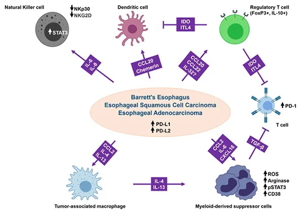
Figure 1. Mechanisms of cytokine‑ and chemokine‑mediated immune evasion in the tumor microenvironment (TME) (DOI: 10.1186/s12943‑020‑01294‑3).
1. Tracing Intercellular Communication from Cellular Phenotypes
When experimental data reveal altered immune cell counts within a target tissue, static cell frequency profiling is insufficient to explain the underlying biology. For instance, an influx of myeloid cells may stem from continuous peripheral recruitment, local expansion, or prolonged cell survival. Furthermore, once recruited into tissue, cells undergo further functional remodeling driven by the local cytokine milieu.
Deconstructing a distinct immune phenotype requires addressing two fundamental questions:
● Which cell subsets undergo phenotypic shifts?
● Which specific signaling pathways drive these shifts?
Localized chemokine signals direct specific immune populations into tissues, where regional inflammatory cytokines subsequently reshape their functional programs. Macroscopic tissue inflammation, immunosuppression, or effector responses represent the cumulative outcome of these multi‑layered signals. Establishing whether a candidate signal actively drives a phenotype requires moving beyond transcriptomic or proteomic correlation analyses to establish causal validation through invivo functional intervention.
2. Distinct Functional Roles: Cell State Modulation vs. Spatial Localization
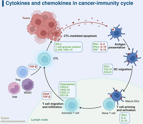
Figure 2. Cytokines and chemokines in the cancer‑immunity cycle (DOI: 10.1038/s41392‑024‑01868‑3).
Functional classification dictates appropriate intervention strategies and guides tool selection:
- • Cytokines Regulate Cell State: Cytokines bind specific receptors to trigger intracellular signaling cascades, altering transcriptional programs and cell function. For example, type I interferons (IFNs) act via the IFN receptor axis to induce interferon‑stimulated genes (ISGs), reshaping both innate and adaptive immune responses. Similarly, interleukins and tumor necrosis factors modulate immune cell activity and tissue homeostasis.
- • Chemokines Direct Spatial Positioning: Chemokine‑receptor signaling pathways orchestrate leukocyte egress from circulation into tissue compartments and govern intratissue positioning. The TME heavily relies on this spatial regulation, where distinct chemokine profiles determine the recruitment efficiency, spatial distribution, and cellular interactions of lymphocytes and monocytes.
Consequently, altered cell abundance and altered cellular function represent distinct biological processes. If cellular infiltration increases without obvious functional shifts, chemokine/receptor pathways represent primary targets for invivo blocking studies. Conversely, if cell counts remain stable while local inflammation or functional reprogramming increases, cytokine neutralizing antibodies should be deployed to block functional activity.
3. Moving Beyond Correlation: Establishing Functional Causality
Concomitant increases in cytokine levels and inflammation, or elevated chemokine expression paired with immune cell infiltration, highlight candidate pathways but do not prove causality.
Immune microenvironmental networks exhibit extensive functional redundancy, shared upstream/downstream regulation, and compensatory mechanisms. A single phenotype may be driven by redundant pathways, and molecular shifts may represent secondary consequences rather than primary drivers. Expression correlations alone cannot distinguish whether a candidate signal acts as an active driver or a passive bystander.
Transforming correlative observations into functional proof requires precise, targeted invivo perturbation. Functional invivo antibodies convert an observed correlation into a controllable experimental variable. For example, in a model showing sustained IL‑17A elevation, deploying an InVivo grade anti‑IL‑17A antibody specifically attenuates downstream signaling, enabling investigators to evaluate subsequent impacts on inflammatory cell populations, secondary mediators, and tissue pathology.
4. Multi‑Layered Mechanisms: Evaluating Multi‑Level Biological Responses
When performing antibody interventions, evaluating biological efficacy requires assessing coherent responses across multiple physiological tiers rather than relying on isolated endpoints. This approach necessitates high‑specificity, high‑purity invivo intervention reagents to avoid off‑target confounding effects.
Invivo mechanistic validation should evaluate three interconnected tiers:
● Signal Level: Verification of targeted pathway inhibition and attenuation of downstream molecular mediators.
● Cellular Level: Quantitative and qualitative shifts in immune cell abundance, infiltration, activation, and functional phenotype.
● Tissue Level: Macro‑level changes in tissue inflammation, damage, fibrosis, or tumor progression.
For instance, in IL‑17A blockade studies, concurrent reductions in inflammatory cellular infiltrates, secondary inflammatory mediators, and tissue pathology collectively validate IL‑17A's functional contribution. Similarly, target validation for CCL2 using InVivoMAb Anti‑Human CCL2 demonstrates reduced tissue infiltration of CCR2‑positive inflammatory monocytes, accompanied by corresponding shifts in myeloid composition and tissue pathology.
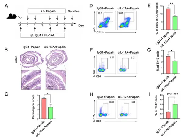
Figure 3. IL‑17A blockade attenuates asthma‑induced intestinal injury in mice (DOI: 10.1002/JLB.3MA0622‑426RR).
The primary value of invivo functional intervention lies in demonstrating whether signaling perturbation drives phenotypic changes along a biologically logical hierarchy. Coordinated changes across cell ratios, functional states, cytokine expression, and histopathology provide robust mechanistic proof.
5. Navigating Signaling Networks: Compensatory and Systemic Responses
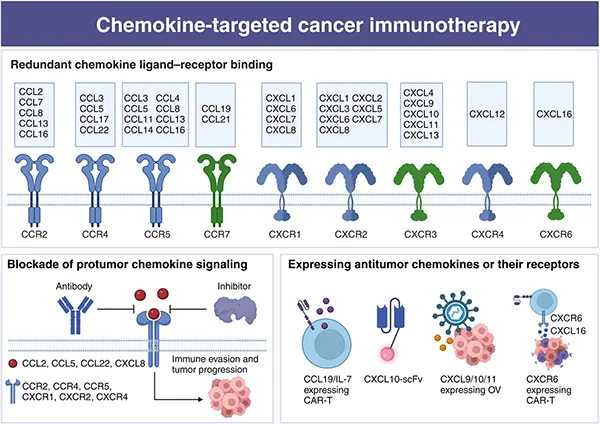
Figure 4. Chemokine ligand‑receptor interactions and their implications for cancer immunotherapy (DOI: 10.1038/s41392‑024‑01868‑3).
Cytokines and chemokines rarely function in isolation; individual cells release multiple factors simultaneously and respond to complex combinations of external stimuli. In the TME, chemokine networks regulate cell entry, spatial distribution, and localized cellular crosstalk.
Targeting a single signaling axis invivo frequently triggers compensatory responses:
- ● Inhibiting one chemokine axis may lead alternative chemokines to maintain cell recruitment.
- ● Neutralizing a specific inflammatory cytokine may result in parallel inflammatory pathways sustaining partial tissue reactivity.
Deconstructing these complex signaling networks requires a multi‑target invivo antibody portfolio. Ligand‑level tracking reveals local signaling changes; receptor profiling identifies responsive target cells; downstream effector analysis confirms functional pathway engagement; and tissue histopathology links signaling events to organ‑level outcomes.
Invivo antibodies serve as precision tools to perturb specific network nodes. By systematically altering individual targets within complex biological systems, researchers can map node‑specific functional contributions without oversimplifying microenvironmental complexity.
6. Experimental Workflow: From Signal Discovery to Functional Validation
Cytokine‑driven functional modulation and chemokine‑driven cell trafficking interact continuously within the tissue microenvironment. A systematic invivo validation pipeline proceeds as follows:
Observe Microenvironmental Shift ➔ Identify Candidate Signals ➔ Formulate Mechanistic Hypotheses ➔ Apply Targeted InVivo Antibody Intervention ➔ Evaluate Multilevel Responses (Molecular, Cellular, Tissue) ➔ Establish Functional Contribution ➔ Map Network Dynamics (Synergy, Feedback, Compensation)
By identifying candidate factors and actively perturbing them using targeted InVivo antibodies, researchers convert descriptive cytokine and chemokine data into functionally validated signaling nodes. Note that single‑antibody interventions should be interpreted alongside target expression profiles, model biology, intervention efficiency, and broader molecular metrics.
abinScience InVivo Cytokine and Chemokine Research Tools
To support rigorous invivo functional validation, abinScience offers a comprehensive portfolio of high‑purity, low‑endotoxin InVivo and InVivo Plus functional antibodies designed for cytokine pathway modulation and chemokine‑mediated microenvironment research.
Cytokine Pathway Modulation
| Catalog No. |
Product Name |
| MS856019 |
InVivo Plus Anti‑Mouse IL‑17A Antibody (BZN035) |
| MS856020 |
InVivoMAb Anti‑Mouse IL‑17A Antibody (17F3) |
| MV466019 |
InVivo Plus Anti‑Mouse IL‑23 p19 Antibody (G23‑8) |
| MB823109 |
InVivo Plus Anti‑Mouse IFN‑alpha/beta R1 Antibody (MAR1‑5A3) |
| MC614019 |
InVivo Plus Anti‑Mouse IL‑10R alpha Antibody (1B1.3A) |
| MY328020 |
InVivoMAb Anti‑Mouse IL‑6 Antibody (MP5‑20F3) |
| MF813010 |
InVivoMAb Anti‑Mouse IFN‑gamma Antibody (XMG1.2) |
| HF879010 |
InVivoMAb Anti‑Human TNF‑alpha Antibody (Iv0050) |
| HB769010 |
InVivoMAb Anti‑Human IL‑12/IL‑23 p40 Antibody (Iv0026) |
| HF977010 |
InVivoMAb Anti‑Human TGF‑beta 1 Antibody (Iv0069) |
Chemokine‑Mediated Recruitment & Microenvironment Modulation
| Catalog No. |
Product Name |
| HY280019 |
InVivo Plus Anti‑Human/Mouse/Rat CCL2/MCP1 Antibody (2H5) |
| HY280010 |
InVivoMAb Anti‑Human CCL2/MCP‑1 Antibody (Iv0077) |
| MW620019 |
InVivo Plus Anti‑Mouse CCR3 Antibody (6S2‑19‑4) |
| HY312010 |
InVivoMAb Anti‑Human CCL3/MIP‑1 alpha Antibody (Iv0078) |
| HY346010 |
InVivoMAb Anti‑Human CCL5/RANTES Antibody (Iv0079) |
| HY554010 |
InVivoMAb Anti‑Human IL‑8/CXCL8 Antibody (Iv0023) |
| HC487010 |
InVivoMAb Anti‑Human CXCL9/MIG Antibody (Iv0087) |
| HY524010 |
InVivoMAb Anti‑Human CXCL10/IP‑10/CRG‑2 Antibody (Iv0088) |
| HW376010 |
InVivoMAb Anti‑Human CXCL12/SDF‑1 beta Antibody (Iv0090) |
| HX143010 |
InVivoMAb Anti‑Human CX3CL1/Fractalkine Antibody (Iv0091) |
Browse InVivo Antibody Products
Browse InVivo Plus Antibody Products


 中文
中文 English
 한국어
한국어 日本語
日本語 Español
 Français
 Русский


