OMIP-005 is specifically developed for preclinical vaccine research in rhesus macaques, aiming to reliably detect antigen-responsive T cells. It offers two optimized variants: an 8-colour core panel targeting key cytokines like IFNγ, IL-2, and TNF, and an 11-colour extended one. Adapting to fresh/cryopreserved PBMCs and tissues, it enables cytokine measurement and memory phenotyping, with proven reproducibility in formal qualification, consistently delivering stable results across different sample preservation methods.
Detecting antigen-specific T cells in macaques is remarkably challenging: target cells often exist at extremely low frequencies, leading to weak signals easily masked by background noise. Issues like activation-induced CD3 internalization and nonspecific antibody binding also arise, easily leading to false negatives. Traditional panels lack sample adaptability and fail to integrate T cell function and phenotype analysis, further complicating data interpretation in multi-center studies.
OMIP-005 breaks through these bottlenecks with a "sensitivity-comprehensiveness-standardization" integrated design. It optimizes signal amplification via antibody clone screening (prioritizing reagents with minimal cross-reactivity), uses intracellular CD3 staining to avoid false negatives from CD3 internalization, and links cytokine production with memory phenotypes. Its unified protocol ensures data comparability across samples, making it an ideal and reliable tool for preclinical vaccine studies.
OMIP-005 panel enables concurrent assessment of T cell activation, cytokine production, and memory differentiation within a single assay. This integrated readout provides a clearer view of how functional responses align with T cell subset composition, offering a more coherent framework for interpreting complex vaccine-induced immune responses.
| Target | Fluorochrome | Function | abinScience Recommendation |
|---|---|---|---|
| Live/Dead | Aqua Blue | Exclude dead cells | — |
| CD3 | APC-Cy7 | T-cell lineage marker | View CD3 antibodies |
| CD4 | QD 605 | View CD4 antibodies | |
| CD8 | Pacific Blue | View CD8 antibodies | |
| CD69 | ECD | Exclude unactive cells and reduce background interference | View CD69antibodies |
| IFN-γ | APC | Detection of cytokines | View IFN-γ antibodies |
| IL-2 | PE | View IL-2 antibodies | |
| TNF | FITC | View TNF antibodies | |
| CD45RA | PE-Cy7 | Detection of memory cell subtypes | — |
| CD28 | PE-Cy5 | View CD28 antibodies | |
| CCR7 | Alexa 680 | View CCR7 antibody |
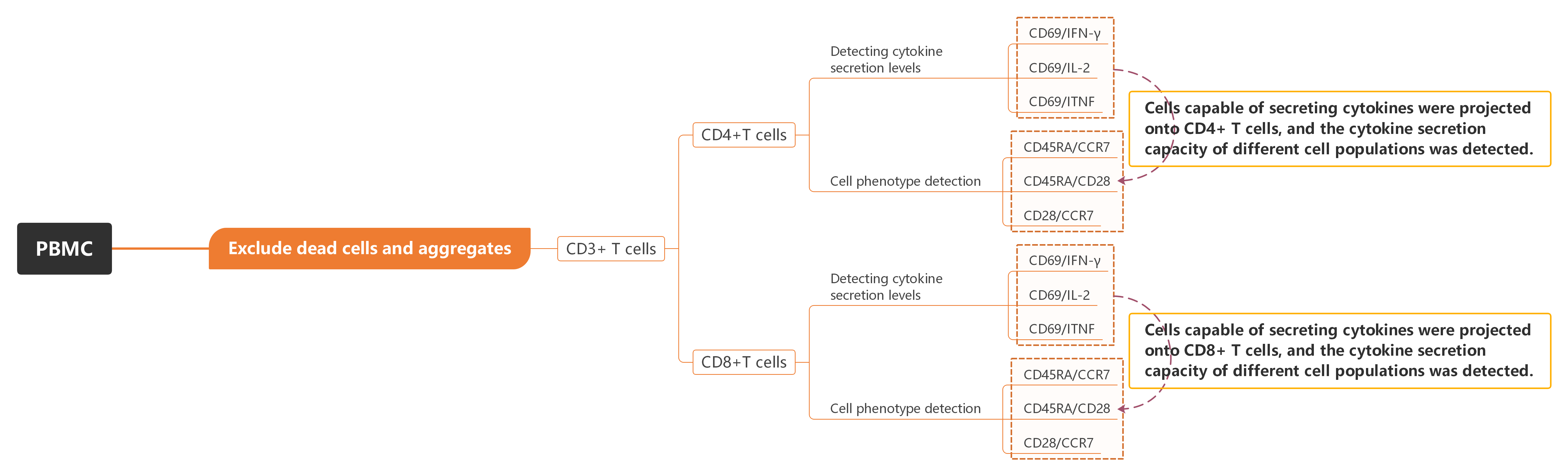
Figure 1. Overview of OMIP-005 Gating Strategy
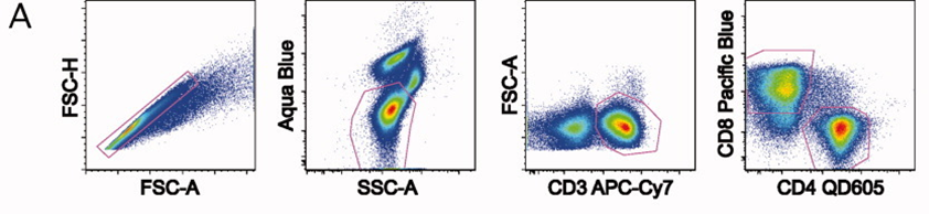
Initial gating was performed using FSC/SSC parameters to exclude aggregates. Dead cells were excluded, CD4+ and CD8+ T cell populations were identified by CD3/CD4/CD8.
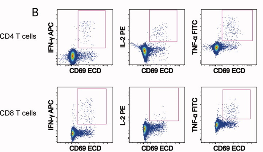
Within the CD4 and CD8 T cell subsets, antigen-responsive cells were identified by intracellular expression of IFNγ, IL-2 and TNF following stimulation. CD69 expression was used in combination with cytokine staining to support the identification of activated, cytokine-producing T cells and to reduce background.
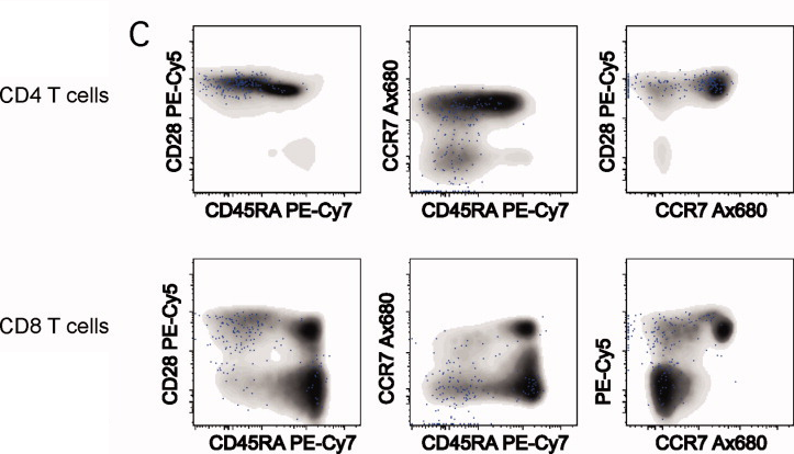
Memory differentiation of cytokine-positive CD4 and CD8 T cells was analysed based on CD28, CD45RA and CCR7 expression. Cytokine-producing cells were overlaid onto the total CD4 or CD8 T cell populations to visualise their distribution across distinct memory subsets.
1). Exclude cell aggregates, dead cells, and identify CD3+ T cells, followed by the distinction of CD4+ and CD8+ T cells.
2). In both CD4+ and CD8+ T cells, identify cytokine-secreting cells (CD69 is used to exclude unactivated cells as background interference).
3). Differentiate CD4+ and CD8+ T cell phenotypes using CD45RA, CD28, and CCR7 markers (grey markers), and project cytokine-secreting cells (blue) from panel B onto panel C to observe the cytokine secretion capabilities of different cell types.
4.1 Innovative Stratified Design with Efficient Expandability
By using 8-colour flow cytometry to detect cytokine levels in T cells, the panel is expanded with 3 additional markers (CD45RA, CD28, CCR7), enabling further differentiation of memory T cell subsets (such as TCM and TEM). This allows for precise measurement of cytokine secretion characteristics in each subset.
4.2 Background Control Strategy: "Precision Exclusion" Using CD69 Activation Marker
OMIP-005 introduces CD69 as a specific activation marker for T cells. By applying a "precision exclusion" strategy to eliminate unactivated cells, this approach effectively reduces background noise, ensuring more accurate analysis of activated T cells.
4.3 Iconic Experimental Design Laying the Foundation for the Subsequent OMIP Panels
As the first “cytokine + subset” dual-dimensional detection panel in the OMIP series, its design logic (such as multi-parameter combination optimization and background control strategies) provides a methodological template for subsequent OMIP panels, such as OMIP-052 and OMIP-075.
OMIP-005 is a worthwhile experimental protocol for primate immunology and preclinical vaccine research, providing a reference-worthy scheme for rhesus macaque antigen-responsive T cell detection, key cytokine measurement, T cell memory phenotype analysis, HIV-related investigations, and standardized data interpretation in multi-center studies.
OMIP-005 presents an innovative multi-parameter flow cytometry design that successfully integrates cytokine detection with the analysis of memory T cell subsets, providing powerful technical support for immunology research. Additionally, the successful implementation of this panel has laid the groundwork for the design and optimization of future OMIP panels, offering a valuable experimental template for researchers in related fields.
abinScience offers validated flow cytometry antibodies (multi-target, multi-species), including monkey-reactive ones for rhesus macaque research.
[1] Foulds KE, Donaldson M, Roederer M. OMIP-005: Quality and phenotype of antigen-responsive rhesus macaque T cells. Cytometry A. 2012;81(5):360-361.

As a strategic venture of AtaGenix (established 2011), abinScience was founded in 2023 to deliver premium life science reagents that accelerate discovery. Our flow cytometry antibody products cover commonly used detection markers, with a wide variety to meet the research needs of multiple species (Human/Mouse/Rat/Dog/Hamster/Monkey, etc.). We provide stable and reliable support for scientific research.
+86-027-65523339
중국 우한시 심둔사로 666번지 C동, 우한, 430206


 中文
中文 English
 한국어
한국어 日本語
日本語 Español
 Français
 Русский