Celiac disease (CD) is a genetic, lifelong autoimmune disorder in which the immune system mistakenly attacks the small intestinal lining when a person consumes gluten proteins found in wheat, barley, and rye. This leads to villous atrophy and malabsorption. It affects about 1% of the global population, with a slightly higher prevalence in females. Symptoms vary widely: classic presentations include diarrhea, weight loss, bloating, and anemia, while non-classic forms may involve skin issues, osteoporosis, neurological symptoms, or even asymptomatic (silent) disease. The gold-standard diagnosis remains small intestinal biopsy, showing villous atrophy, crypt hyperplasia, and lymphocytic infiltration. Although a strict, lifelong gluten-free diet (GFD) is currently the only standard treatment, recent scientific advances are opening the door to a "post-GFD era."
The majority of celiac disease cases are driven by genetics: approximately 95% of patients carry the HLA-DQ2 or HLA-DQ8 alleles, which encode molecules capable of efficiently presenting deamidated gluten peptides. Gluten peptides cross the intestinal epithelium into the lamina propria, where they are deamidated by tissue transglutaminase 2 (TG2), increasing their binding affinity to HLA-DQ2/8. Antigen-presenting cells then activate CD4+ T cells, which release IFN-γ, TNF-α, and IL-15, causing epithelial damage and villous atrophy. Autoantibodies targeting TG2 and deamidated gluten peptides further amplify the inflammatory response. Innate immunity plays a role as well, with epithelial cells releasing IL-15, which upregulates NKG2D on CD8+ T cells and triggers cytotoxic damage. Genome-wide association studies have also identified non-HLA susceptibility loci, including IL2/IL21, IL18RAP, SH2B3, PTPN2, UBE2L3, and TNFAIP3, which collectively implicate T-cell signaling, cytokine pathways, and NF-κB regulation in disease susceptibility.
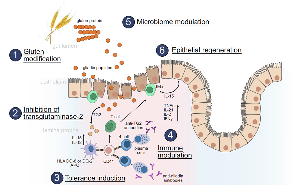
Figure 1. Pathophysiology of celiac disease and current investigational therapeutic strategies
Genetics alone do not determine disease — environmental and microbial factors also play critical roles.
Gut dysbiosis: Studies show increased pathogenic bacteria (e.g., Escherichia coli, Staphylococcus) and reduced beneficial species (e.g., Bifidobacterium) in celiac patients. Certain protease-secreting bacteria degrade gluten into more immunogenic peptides, while bacterial accessory proteins may act as adjuvants, heightening immune sensitivity to gluten.
Viral infection and loss of immune tolerance: Infections such as reovirus or human herpesviruses may disrupt intestinal immune homeostasis. Viral-triggered proinflammatory signals (e.g., IFN-α) can mislead the immune system into recognizing otherwise tolerated gluten antigens as threats, breaking tolerance and triggering disease.
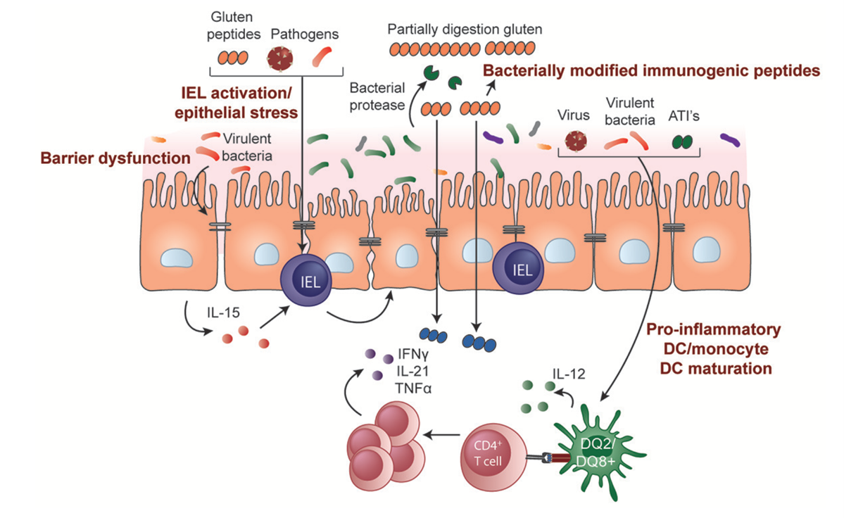
Figure 2. Potential roles of microbiota and environmental triggers in celiac disease pathogenesis
The following table highlights major research targets in celiac disease, focusing on different steps in the pathogenic pathway and offering potential therapeutic strategies.
| Target | Representative Drug | Mechanism of Action |
|---|---|---|
| TG2 inhibition | ZED1227 (TAK-227) | TG2 is the key enzyme that converts gluten peptides into highly immunogenic forms. ZED1227 directly inhibits its activity, blocking immune activation at the molecular level. Phase 2b data (CEC-004/CEL) presented at UEG Week 2025 demonstrated histological improvement in symptomatic celiac patients. |
| IL-15 blockade | PRV-015 (ordesekimab) | Blocks epithelial damage and CD8+ T-cell activation, reducing the inflammatory cascade. The Phase 2b PROACTIVE trial (NCT04424927) has been completed, evaluating efficacy in non-responsive celiac disease patients on a gluten-free diet. |
| Gluten-degrading enzymes | Latiglutenase (ALV003) | Oral enzyme preparations break down immunogenic gluten fragments into harmless small peptides in the stomach. Phase 2b results were mixed, with histological improvement observed primarily in seropositive patients. |
| Intestinal barrier modulation | Larazotide acetate (AT-1001) | Restores tight junctions between epithelial cells, preventing harmful gluten peptides from crossing into deeper tissues. Larazotide showed symptom improvement in earlier trials but did not meet the primary endpoint in its Phase 3 study; further clinical evaluation is ongoing. |
| Immune tolerance induction | TAK-101 (TIMP-GLIA) | Uses biodegradable nanoparticle technology to encapsulate gluten protein (gliadin) and deliver it to liver and spleen immune cells via intravenous injection, reprogramming the immune system to recognize gluten as a non-threatening antigen and potentially restoring immune tolerance. Phase 2 dose-ranging studies are ongoing. |
| JAK/STAT inhibition | Ritlecitinib (preclinical rationale) | JAK3/TEC family kinase inhibitor that blocks cytokine signaling, reducing Th17 differentiation and inflammation. Currently approved for alopecia areata; application to celiac disease remains at the preclinical/mechanistic rationale stage. |
The following products from abinScience target core celiac disease research pathways, including the TG2 autoantigen, HLA-DQ antigen presentation, IL-15-mediated innate immunity, gliadin detection, and GWAS-implicated susceptibility loci. Click the catalog numbers to go directly to the product pages.
| Catalog No. | Product Name |
|---|---|
| HB247016 | Anti-Human TGM2 Reference Antibody (Zampilimab, RUO) |
| HW541016 | Anti-Human IL-15 Reference Antibody (Humax-IL15, RUO) |
| HW541036 | Anti-Human IL-15 Reference Antibody (DISC0280, RUO) |
| HM817016 | Anti-Human HLA-DQB1 Reference Antibody (DQB1-FL8, RUO) |
| PR115025 | Anti-Wheat Alpha/beta-gliadin MM1 Monoclonal Antibody (1A447) |
| PR098013 | Anti-Wheat Alpha/beta-gliadin MM1 Recombinant Antibody (SAA0518) |
| Catalog No. | Product Name |
|---|---|
| HB247012 | Recombinant Human TGM2 Protein, N-His |
| HB247011 | Recombinant Human TGM2 Protein, C-His |
| HM817012 | Recombinant Human HLA-DQB1 Protein, N-His |
| HF996011 | Recombinant Human CD25/IL2RA Protein, C-His |
| HF996012 | Recombinant Human CD25/IL2RA Protein, N-His-KSI |
| HB936011 | Recombinant Human IL12A/IL-12 p35/NKSF1 Protein, C-Strep |
| HV658012 | Recombinant Human IL21 Protein, C-His |
| HF719012 | Recombinant Human CD218b/IL18RAP Protein, N-His |
| HF719011 | Recombinant Human CD218b/IL18RAP Protein, C-Strep |
| HF550012 | Recombinant Human TGM6 Protein, N-His |
| HB969012 | Recombinant Human PTPN2 Protein, N-His |
| HV649012 | Recombinant Human SH2B3 Protein, N-His |
| HX198012 | Recombinant Human UBE2L3 Protein, N-His |
| HB289012 | Recombinant Human TNFAIP3 Protein, N-GST |
| HW412012 | Recombinant Human CD195/CCR5 Protein, N-GST |
References:
1. Lebwohl B, Sanders DS, Green PHR. Coeliac disease. Lancet. 2018;391(10115):70-81. doi: 10.1016/S0140-6736(17)31796-8
2. Schuppan D, Mäki M, Gliadin Z, et al. A Randomized Trial of a Transglutaminase 2 Inhibitor for Celiac Disease. N Engl J Med. 2021;385(1):35-45. doi: 10.1056/NEJMoa2032441
3. Kelly CP, Murray JA, Leffler DA, et al. TAK-101 Nanoparticles Induce Gluten-Specific Tolerance in Celiac Disease: A Randomized, Double-Blind, Placebo-Controlled Study. Gastroenterology. 2021;161(1):66-80. doi: 10.1053/j.gastro.2021.03.014
4. Leffler DA, Kelly CP, Green PH, et al. Larazotide acetate for persistent symptoms of celiac disease despite a gluten-free diet: a randomized controlled trial. Gastroenterology. 2015;148(7):1311-1319. doi: 10.1053/j.gastro.2015.02.008
5. Trynka G, Hunt KA, Bockett NA, et al. Dense genotyping identifies and localizes multiple common and rare variant association signals in celiac disease. Nat Genet. 2011;43(12):1193-1201. doi: 10.1038/ng.998
6. Ferranti P, Ferrara C, Mancuso M, et al. New therapies in celiac disease. Curr Opin Gastroenterol. 2025;41(3):124-131. doi: 10.1097/MOG.0000000000001080
+86-027-65523339
中国武漢市深敦寺路666号C棟、武漢、430206


 中文
中文 English
 한국어
한국어 日本語
日本語 Español
 Français
 Русский