Rhesus macaques serve as a critical nonhuman primate model for immunology due to the high homology of their immune system with humans. They play an indispensable role in SIV infection studies, vaccine evaluation, and mechanistic immune research. Natural killer (NK) cells, as core effectors of the innate immune system, are pivotal regulators of viral infections and immune responses. However, the heterogeneity of NK cell subsets and their functional states in Rhesus macaques presents significant complexity, making it difficult to capture dynamic changes using conventional single-marker approaches.
During infection or immune interventions, combinations of KIR receptors, CD16/CD56 differentiation status, and activation/proliferation markers define the functional diversity and responsiveness of NK cell subsets. To accurately characterize these subsets and their functional states, a high-resolution, multi-marker analysis tool is required. Such a tool can capture comprehensive information on NK cell lineage, differentiation, activation, and proliferation even from limited samples.
OMIP-028 integrates lineage markers including CD8α, NKG2A/C, KIR3DL01, and KIR3DL05 with differentiation markers CD16 and CD56, enabling systematic delineation of major NK cell subsets. By combining these with four activation/proliferation markers—CD69, HLA-DR, PD-1, and Ki-67—the panel provides refined functional characterization for each subset. It offers a robust platform for NK cell analysis in SIV infection and vaccine immunology studies, and can monitor NK cell functional diversity induced by infection or immune interventions.
OMIP-028 provides a multicolor flow cytometry solution to quantify Rhesus macaque NK cell subsets and their activation/proliferation phenotypes. Optimized antibody-fluorochrome combinations allow simultaneous monitoring of up to 12 NK cell subsets, while Boolean analysis of the four activation/proliferation markers defines 16 phenotypic subsets. This enables tracking of activation and proliferation dynamics across different physiological and pathological conditions, and dissects viral impacts on NK cell subset distribution and functional maturation.
| Target | Fluorochrome | Function | abinScience Recommendation |
|---|---|---|---|
| Live Dead | Near infrared | Viability | — |
| CD45 | BV786 | Lymphocyte marker | View CD45 antibodies |
| CD3 | Alexa Fluor 700 | T cell marker | View CD3 antibodies |
| CD20 | Alexa Fluor 700 | B cell marker | View CD20 antibodies |
| CD14 | Alexa Fluor 700 | Monocyte marker | View CD14 antibodies |
| CD8 | BV510 | NK cell marker | View CD8 antibodies |
| CD16 | BV711 | View CD16 antibodies | |
| CD56 | FITC | View CD56 antibodies | |
| NKG2A/C | PE-Cy7 | View NKG2A/C antibodies | |
| KIR3DL01 | PE | View KIR3DL01 antibodies | |
| KIR3DL05 | BV421 | — | |
| NKp46 | PE-Cy5 | NK cell differentiation | View NKp46 antibodies |
| CD279 (PD-1) | BV605 | View CD279 antibodies | |
| CD69 | PE-Texas Red | Activation | View CD69 antibodies |
| HLA-DR | BV650 | View HLA-DR antibodies | |
| Ki-67 | Alexa Fluor 647 | Proliferation | — |

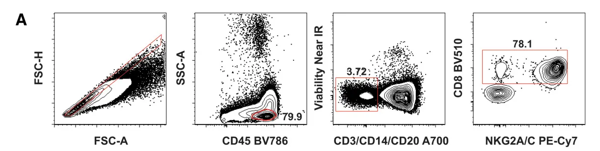
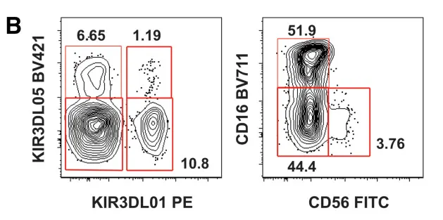
Figure 1. Overview of OMIP-028 Gating Strategy
Exclude doublets via FSC-A/H, CD45+ lymphocytes gated, dead cells, T cells, monocytes, and B cells excluded, followed by CD8 and NKG2A/C gating to identify NK cells.
KIR3DL01 and KIR3DL05 divided NK cells into four different populations, further subdivided by CD16 and CD56 expression; NKp46 expression differences are analyzed among these subsets.
Activation and proliferation analyzed using CD69, HLA-DR, PD-1, and Ki-67.
1). PBMCs were first gated to exclude doublets (FSC-A/H), then CD45+ lymphocytes were selected. Dead cells and T, B, and monocyte populations were excluded, followed by CD8 and NKG2A/C gating to identify NK cells.
2). Total NK cells were separated into four populations based on KIR3DL01 and KIR3DL05 expression, and further into three subsets (CD16+, CD56+, CD16-CD56-) to analyze NKp46 levels.
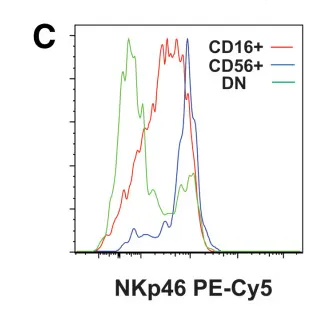
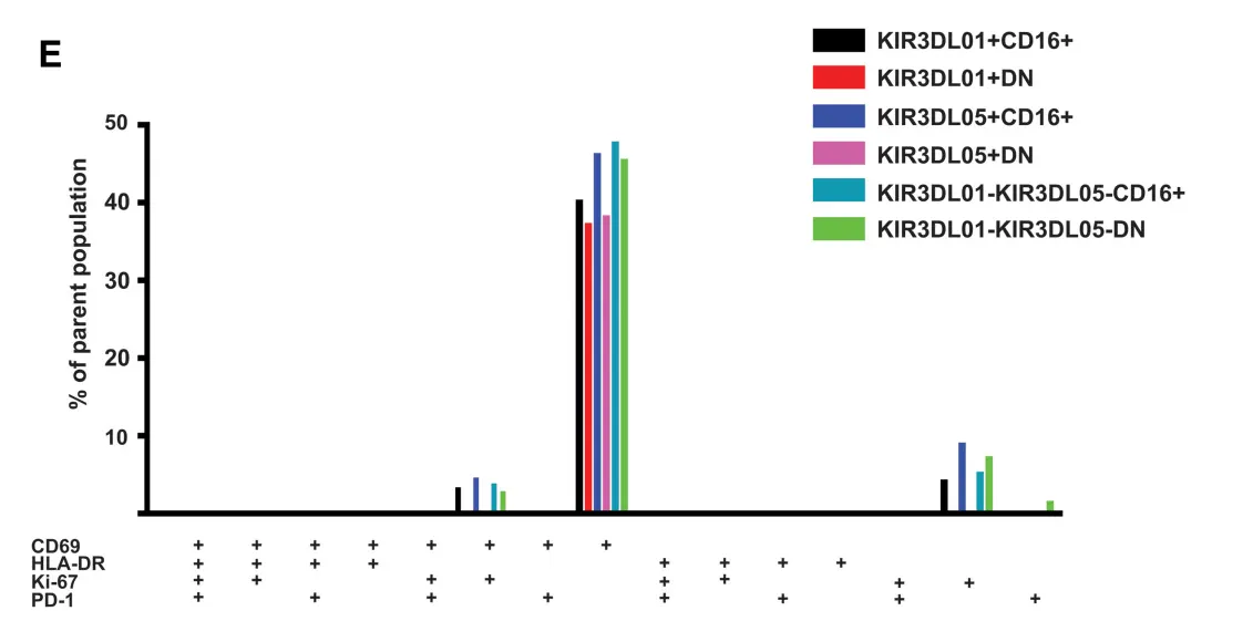
3). Activation and proliferation within the three NK cell subsets were analyzed via CD69, HLA-DR, PD-1, and Ki-67.
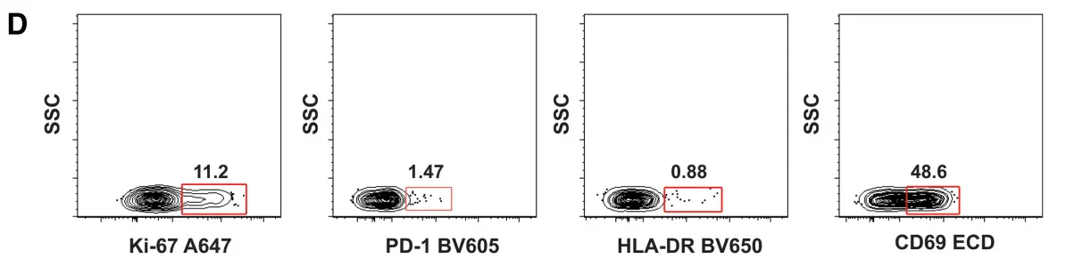
4). Boolean gating of the four activation/proliferation markers generated 16 functional states (single-positive, double-positive, triple-positive, quadruple-positive), depicting NK cell activation, proliferation, and regulatory status.
4.1 Species-Specific NK Cell Definitions
Rhesus macaque NK cells differ significantly from humans in CD56 and CD16 expression; for example, CD16 is also expressed on monocytes, making human-based gating strategies inappropriate. OMIP-028 employs a combination of CD3, CD8α, and NKG2A/C, together with KIR receptors and CD16/CD56, to construct a Rhesus-specific NK cell analysis framework. This approach ensures accurate interpretation of NK cell responses in infection or vaccine studies and provides a methodological basis for cross-species comparisons.
4.2 High-Resolution NK Cell Analysis
OMIP-028 enables simultaneous resolution of 12 structural NK cell subsets and 16 functional states in a single tube. Structural subsets are first defined by KIR3DL01 and KIR3DL05 expression into four categories, then further divided by CD16/CD56 differentiation to yield 12 detailed subsets. Functional states are enumerated by Boolean combinations of CD69, HLA-DR, PD-1, and Ki-67, capturing diversity in activation, proliferation, and regulation. This hierarchical design overcomes limitations of single-marker analysis and allows single-cell level mapping of NK cell structure and function.
4.3 Support for Infection Models and Vaccine Studies
OMIP-028 tracks dynamic changes in NK cell subsets during SIV/HIV infection, such as expansion of the CD16-CD56- population and diversification of functional states, reflecting systemic effects of infection on NK cell distribution and function. This high-resolution tracking allows quantitative assessment of NK subset activation, proliferation, and regulatory states during vaccine evaluation and immune interventions, supporting mechanistic insights and optimization of immunization strategies.
By integrating lineage markers, differentiation markers, and activation/proliferation markers, OMIP-028 provides high-resolution phenotyping of Rhesus macaque NK cells. In a single-tube assay, 12 structural and 16 functional NK cell subsets can be resolved, enabling tracking of NK cell dynamics under infection or immune interventions. Its high adaptability and reproducibility make it a reliable tool for dissecting NK cell functional heterogeneity in infectious disease, vaccine research, and fundamental immunology, supporting cross-species comparisons and mechanistic exploration of immune regulation.
abinScience offers high-quality Flow Cytometry Antibodies for NK cell-related differentiation and function, supporting research on NK cell functional heterogeneity.
[1] Pomplun N, Weisgrau KL, Evans DT, Rakasz EG. OMIP-028: activation panel for Rhesus macaque NK cell subsets. Cytometry A. 2015 Oct;87(10):890-3. doi: 10.1002/cyto.a.22727. Epub 2015 Jul 28. PMID: 26218174; PMCID: PMC4758211.

As a strategic venture of AtaGenix (established 2011), abinScience was founded in 2023 to deliver premium life science reagents that accelerate discovery. Our flow cytometry antibody products cover commonly used detection markers, with a wide variety to meet the research needs of multiple species (Human/Mouse/Rat/Dog/Hamster/Monkey, etc.). We provide stable and reliable support for scientific research.
+86-027-65523339
中国武漢市深敦寺路666号C棟、武漢、430206


 中文
中文 English
 한국어
한국어 日本語
日本語 Español
 Français
 Русский