
 中文
 English
 한국어
 日本語
 Español
 Français
 Русский


| 카탈로그 번호 | HF879163 |
|---|---|
| 설명 |
Anti-Human TNF-alpha Recombinant Antibody (SRR0835) (HF879163) is a rabbit monoclonal antibody detecting TNF-alpha in IF, FCM. Suitable for Human.
Highlights
|
| 종 반응성 | Human |
| 응용 분야 | IF, FCM |
| 숙주 종 | Rabbit |
| 클론성 | Monoclonal |
| 이소타입 | IgG |
| 클론 ID | SRR0835 |
| 테스트된 응용 분야 | IF, FCM |
| 타겟 | C-domain 1, C-domain 2, Cachectin, ICD1, ICD2, Intracellular domain 1, Intracellular domain 2, N-terminal fragment, NTF, TNF, TNF-a, TNF-alpha, TNFA, TNFSF2, Tumor necrosis factor, Tumor necrosis factor ligand superfamily member 2, Tumor necrosis factor, membrane form, Tumor necrosis factor, soluble form |
| 내독소 수준 | < 10 EU/mg as determined by LAL method. |
| 순도 | >95% as determined by SDS-PAGE. |
| 정제 | Protein A/G purified from cell culture supernatant. |
| 등록번호 | P01375 |
| 형태 | Liquid |
| 저장 완충액 | 0.01M PBS pH 7.4. 데이터시트 인쇄본 또는 로트별 COA에 기재된 구체적인 완충액 정보를 참조하세요. |
| 재구성 | Reconstitute in sterile water for a stock solution. A copy of datasheet will be provided with the products, please refer to it for details. |
| 안정성 및 저장 | Use a manual defrost freezer and avoid repeated freeze thaw cycles. Store at 2 to 8°C for frequent use. Store at -20 to -80°C for twelve months from the date of receipt. |
| 배경 | Tumor necrosis factor (TNFA) is a ~25 kDa protein. Cytokine that binds to TNFRSF1A/TNFR1 and TNFRSF1B/TNFBR. It is mainly secreted by macrophages and can induce cell death of certain tumor cell lines. It is potent pyrogen causing fever by direct action or by stimulation of interleukin-1 secretion and is implicated in the induction of cachexia, Under certain conditions it can stimulate cell proliferation and induce cell differentiation. Impairs regulatory T-cells (Treg) function in individuals with rheumatoid arthritis via FOXP3 dephosphorylation. Up-regulates the expression of protein phosphatase 1 (PP1), which dephosphorylates the key 'Ser-418' residue of FOXP3, thereby inactivating FOXP3 and rendering Treg cells functionally defective. 1. Nie, H. et al. (2013) Nature medicine 19, 322-8. PMID: 23396208 2. Friedmann, E. et al. (2006) Nature cell biology 8, 843-8. PMID: 16829952 3. Jinesh G, G. et al. (2012) Journal of leukocyte biology 92, 233-44. PMID: 22517918 4. Nakahara, H. et al. (2003) Arthritis and rheumatism 48, 1521-9. PMID: 12794819 |
| 참고사항 | For research use only. |
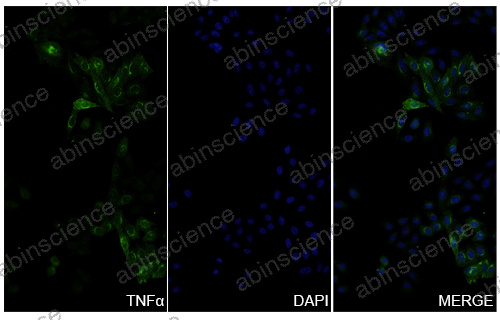
TNFα-overexpressing cells were fixed with 4% paraformaldehyde for 20 minutes, followed by blocking with 5% goat serum for 1 hour. The cells were then incubated with a primary antibody targeting TNF-alpha (HF879163) at a concentration of 20 ug/ml overnight at 4°C. Afterward, the cells were incubated with a secondary antibody, Goat Anti-Rabbit IgG (Alexa Fluor-488) at a concentration of 6.5 ug/ml for 50 minutes at room temperature. The localization of TNF-alpha was visualized (shown in green), and nuclear DNA was stained with DAPI (shown in blue).
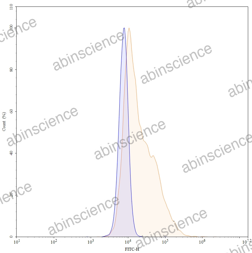
Flow-cytometry using anti-Human TNFa antibody. Overexpression cells were stained with an irrelevant antibody (Blue Histogram) or an anti-Human TNFa monoclonal antibody (Catalog HF879163, Yellow Histogram) at a concentration of 5 µg/ml for 30 mins at RT. After washing, bound antibody was detected using a Goat Anti-Rabbit IgG H&L Polyclonal Antibody, FITC (abinScience:TF690414) and cells analysed on a NovoCyte Flow Cytometer.
문의하기
+86-027-65523339
중국 우한시 심둔사로 666번지 C동, 우한, 430206


