In early October 2026, the fatal case of severe pneumonia in an anti‑plague research institute staff member in Irkutsk Oblast, Russia, prompted immediate public health interventions, including contact tracing, health monitoring, and transport sanitation protocols. Health screening along transit corridors, such as routes from Irkutsk to Moscow, brought pneumonic plague—a rare disease in modern times—back into public focus.
At the center of this incident is Yersinia pestis, the causative agent of plague. As a high‑consequence pathogen transmitted via respiratory droplets, Y. pestis remains a major target for global epidemiological surveillance due to its high virulence and rapid clinical progression. From a basic research perspective, its sophisticated virulence regulation, host adaptation strategies, and metabolic characteristics make it a classical model system in microbiology and infectious disease research.
The genus Yersinia comprises Gram‑negative bacilli belonging to the family Enterobacteriaceae. Although the genus name is most frequently associated with plague, Yersinia pestis is not the sole human pathogen in this group; Yersinia enterocolitica and Yersinia pseudotuberculosis are also prominent human pathogens. Despite close phylogenetic relationships, these three species exhibit distinct host adaptation strategies and clinical manifestations.
Y. pestis is the etiology of plague, maintained in nature through an enzootic cycle involving wild rodents and flea vectors. In contrast, Y. enterocolitica and Y. pseudotuberculosis primarily cause enteric infections. Y. enterocolitica presents as enterocolitis, terminal ileitis, and mesenteric lymphadenitis, whereas Y. pseudotuberculosis causes enteritis and, in certain geographical regions, systemic inflammatory disease. Both enteric species transmit predominantly through contaminated food, animal contact, or the fecal‑oral route—contrasting with the vector‑borne transmission cycle of Y. pestis.
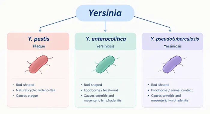
Figure 1. Major pathogenic species of the genus Yersinia.
This functional divergence demonstrates that while pathogenic Yersinia species share conserved virulence systems, their specific host ranges, transmission dynamics, and tissue tropisms are dictated by distinct genomic features and virulence regulation networks. For Y. pestis, the primary biological focus is not merely its taxonomic assignment within Yersinia, but how it adapts to the rodent–flea cycle and rapidly shifts its physiological state upon entry into mammalian hosts.
As a classic zoonotic pathogen, Y. pestis persists in natural foci primarily among wild rodents and their ectoparasitic fleas. Infected rodents function as reservoir hosts, while fleas act as biological vectors transmitting the bacterium within animal populations. Humans are accidental hosts, entering the transmission chain under specific ecological exposure conditions.
This transmission dynamic involves active physiological adaptation rather than passive mechanical transfer. Transitioning from ambient temperatures in the flea gut to the 37°C mammalian environment exposes Y. pestis to marked changes in temperature, nutrient availability, and immune pressures. To survive, the bacterium rapidly remodels its surface architecture, gene expression profile, and metabolic pathways. Multiple virulence systems in Y. pestis are thermosensitive; for instance, shift to host body temperature induces expression of the type III secretion system (T3SS) and alters lipopolysaccharide (LPS) structure.
Understanding this environmental transition is critical to deciphering Y. pestis pathogenesis. Within the host, the pathogen must overcome two main hurdles: evading rapid clearance by the innate immune system and acquiring essential nutrients for tissue replication. Vector transmission, immune evasion, tissue tropism, and metabolic maintenance thus operate not as isolated events, but as an integrated biological program driving host adaptation.
Following inoculation by an infected flea, early pathogenesis centers on bacterial survival in localized host tissue and subsequent entry into lymphatic or vascular systems, rather than immediate lung involvement. Differences in entry site and dissemination pathways dictate the resulting clinical form of plague.
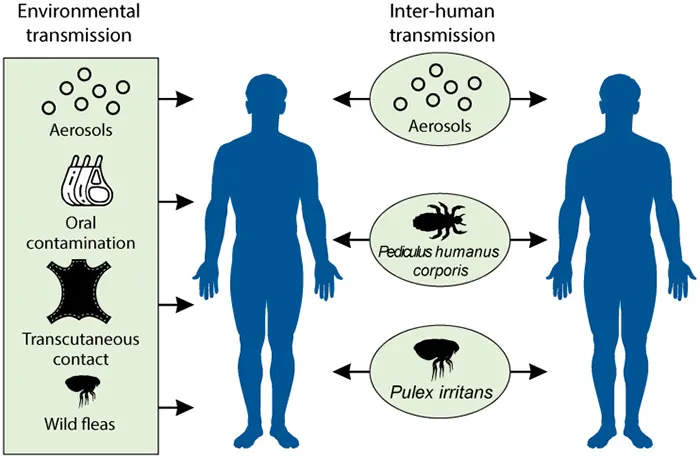
Figure 2. Enzootic cycle and human infection pathways of Yersinia pestis (DOI: 10.1128/CMR.00044-19)
Bubonic plague is the most common form, typically initiated by infected flea bites. Following cutaneous entry, Y. pestis drains via regional lymphatic channels to local lymph nodes, where it replicates and triggers intense localized inflammation. This produces characteristically enlarged, painful lymph nodes termed buboes. If the localized infection is not contained, bacteria can escape into the systemic circulation.
Septicemic plague develops when Y. pestis proliferates extensively in the bloodstream. It can manifest secondarily from unresolved bubonic plague or arise directly as primary septicemic plague without localized lymphadenitis. High‑grade bacteremia exposes the pathogen to systemic immune defenses while inducing severe hyperinflammation, tissue necrosis, and multi‑organ dysfunction.
Pneumonic plague exhibits distinct pathophysiological and epidemiological characteristics. It may develop secondarily via hematogenous seeding of the pulmonary parenchyma during bubonic or septicemic plague, or directly as primary pneumonic plague via inhalation of infectious aerosols. In primary cases, the bacterium bypasses skin and regional lymphatic barriers, directly colonizing the respiratory tract and alveolar spaces, leading to rapid disease progression.
Direct respiratory involvement underpins the heightened public health significance of pneumonic plague. Unlike vector‑dependent bubonic plague, pneumonic plague transmits directly from person to person via respiratory droplets. Pulmonary tissue acts simultaneously as a site of destructive bacterial replication and an amplification hub for airborne transmission, shifting both the clinical trajectory and ecological niche of the pathogen.
Pathogenesis in Y. pestis extends beyond simple intra‑host multiplication; it is governed by molecular interactions between bacterial surface structures, secretion systems, and host proteins. Capsular antigen fraction 1 (Caf1/F1) and plasminogen activator (Pla) protease represent two of the best‑characterized virulence factors.
The F1 capsular antigen, encoded by the caf1 gene, polymerizes to form a high‑molecular‑weight capsular polymer on the bacterial surface. F1 expression markedly reduces bacterial phagocytosis by host macrophages and neutrophils. Structural and functional analyses demonstrate that the F1 polymer physically inhibits phagocytic adhesion and sterically shields bacterial surface ligands from binding host phagocytic receptors. By constructing a physical surface barrier against innate immune destruction, F1 creates a permissive niche for extracellular bacterial survival during early infection. Given its high immunogenicity, F1 also serves as a key target in plague diagnostics, subunit vaccine design, and immunological assays.
While F1 facilitates physical immune evasion, Pla protease mediates enzymatic virulence and tissue invasion. Pla is an outer membrane omptin‑family protease with dual adhesin and invasin functions. A central biological role of Pla is activating host plasminogen into plasmin, which degrades fibrin networks and dysregulates hemostasis. Pla‑mediated fibrinolysis dismantles tissue barriers, enabling Y. pestis to disseminate rapidly from localized dermal sites into deeper tissues, lymphatics, and the circulation.
Pla function extends beyond simple extracellular matrix degradation. In pulmonary infection models, Pla cleaves host homeostatic and inflammatory regulatory proteins, such as plasminogen activator inhibitor‑1 (PAI‑1), thereby modulating immune cell recruitment, cytokine signaling, and vascular permeability. Thus, Pla actively reshapes the microenvironment rather than serving merely as an isolated digestive enzyme.
In addition to F1 and Pla, Y. pestis utilizes a highly conserved type III secretion system (T3SS) to translocate effector proteins, known as Yersinia outer proteins (Yops), directly into host cell cytoplasm. Yop effectors disrupt phagocytosis, actin cytoskeleton dynamics, and pro‑inflammatory signaling pathways, suppressing cellular immune responses. Thermal induction of T3SS expression at 37°C further highlights the pathogen's adaptive response to host entry.
Overall, Y. pestis virulence operates as a coordinated multi‑layered system: capsular polymers like F1 inhibit phagocytic uptake, proteases like Pla modulate proteolytic networks and tissue barriers, the T3SS/Yop array subverts host intracellular signaling, and auxiliary systems (such as iron acquisition and LPS modifications) sustain intra‑host metabolic survival. Synergies among these mechanisms dictate the complex pathogenic phenotype of Y. pestis.
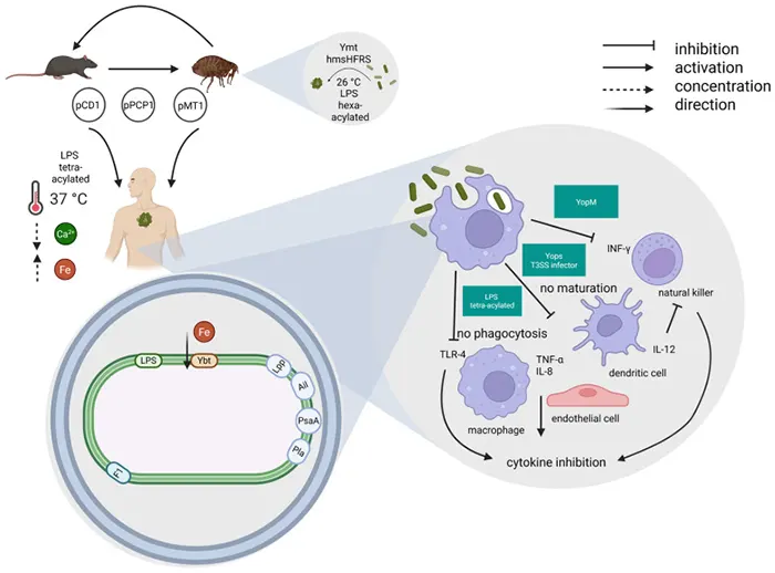
Figure 3. Primary virulence determinants of Yersinia pestis (DOI: 10.3390/biomedicines13102555)
While virulence factors mediate direct host–pathogen interactions, core bacterial metabolic pathways represent another critical axis in Y. pestis survival and targeted intervention.
Within host tissues, Y. pestis relies on continuous biosynthesis of nucleic acids, proteins, and cellular envelopes alongside immune evasion. Folate metabolism is essential to core bacterial viability. Unlike mammals, which absorb dietary folate, most bacteria synthesize folate de novo; hence, this pathway has long served as a validated antimicrobial drug target.
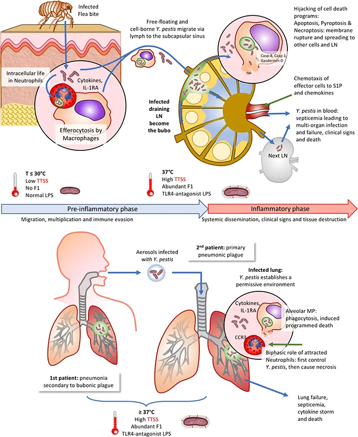
Figure 4. Evasion of innate immunity by Yersinia pestis (DOI: 10.1038/s41435-019-0065-0)
A key enzyme in this cascade is dihydropteroate synthase (DHPS, encoded by folP). In Y. pestis, DHPS catalyzes the condensation of 6‑hydroxymethyl‑7,8‑dihydropterin diphosphate with p‑aminobenzoic acid (PABA) to yield dihydropteroate, an essential precursor for downstream tetrahydrofolate biosynthesis. Structural biology studies of Y. pestis DHPS—including high‑resolution apo structures and inhibitor‑bound complexes—provide molecular models for elucidating catalytic mechanisms and rational drug design.
Reflecting on Y. pestis through the lens of recent public health events underscores the complex biology of this pathogen. Persisting within wild rodent–flea reservoirs, Y. pestis rapidly reprograms its surface structure, virulence factors, and metabolic networks upon mammalian invasion. Molecular players ranging from F1/Caf1 and Pla to the T3SS array and FolP distinctively coordinate immune escape, vascular dissemination, host cell signaling subversion, and metabolic sustenance.
From surface capsular antigens (F1) and host‑modifying proteases (Pla) to essential metabolic enzymes (DHPS/folP), the research targets of Y. pestis address distinct scientific questions in antigenicity, immune evasion, tissue dissemination, structural biology, and metabolic inhibition, together forming a structured framework for basic microbiological and translational research. Developing specialized recombinant proteins and antibodies against these targets serves purposes beyond simple diagnostic detection—these molecular tools enable precise functional assays, structural analyses, host‑factor interaction studies, and targeted screening platforms for novel therapeutics.
Y. pestis is thus both a historical pathogen of high public health relevance and a premier research model for vector‑borne transmission, host adaptation, tissue invasion, and essential bacterial physiology. Systematic investigation of these molecular targets deepens our understanding of microbial survival strategies while advancing structural biology, drug discovery, and diagnostic tool development.
To support research on Y. pestis core virulence factors (F1/Caf1, Pla, LcrV) and metabolic targets (DHPS/FolP), abinScience offers high‑purity recombinant proteins and specific antibodies. In addition, virulence‑associated proteins from other pathogenic Yersinia species such as Y. enterocolitica (ail, Invasin, YopH, YopB/YopD, OsmY) are important targets for host–pathogen interaction studies, and abinScience provides corresponding research tools.
| Catalog No. | Product Name |
|---|---|
| JN173012 | Yersinia pestis F1 capsule antigen/Caf1 Recombinant Protein (N‑His) |
| JN173022 | Yersinia pestis Plasminogen activator/Pla Recombinant Protein (N‑His) |
| JN173032 | Yersinia pestis DHPS/folP Recombinant Protein (N‑His) |
| JN853022 | Yersinia pestis LcrV Recombinant Protein (N‑His) |
| JN173013 | Anti‑Yersinia pestis F1 capsule antigen/Caf1 Recombinant Antibody (SAA3090) |
| JN173014 | Anti‑Yersinia pestis F1 capsule antigen/Caf1 Polyclonal Antibody |
| JN173024 | Anti‑Yersinia pestis Plasminogen activator/Pla Polyclonal Antibody |
| JN173034 | Anti‑Yersinia pestis DHPS/folP Polyclonal Antibody |
| JN173044 | Anti‑Yersinia pestis DHPS/folP Polyclonal Antibody |
| JN853013 | Anti‑Yersinia pestis LcrV Recombinant Antibody (SAA3091) |
| JN853014 | Anti‑Yersinia pestis LcrV Polyclonal Antibody |
| JN853023 | Anti‑Yersinia pestis LcrV Recombinant Antibody (SAA3092) |
| JN905012 | Yersinia enterocolitica ail/Attachment invasion locus Recombinant Protein (N‑His‑SUMO) |
| JN853012 | Yersinia enterocolitica LcrV/Virulence‑associated V antigen Recombinant Protein (N‑His) |
| JN026012 | Yersinia enterocolitica Invasin Recombinant Protein (N‑His‑SUMO) |
| JN934012 | Yersinia enterocolitica YopH Recombinant Protein (N‑His) |
| JN841012 | Yersinia enterocolitica YopD Recombinant Protein (N‑His) |
| JN885012 | Yersinia enterocolitica OsmY Recombinant Protein (N‑His‑SUMO) |
| JN885022 | Yersinia enterocolitica OsmY Recombinant Protein (C‑His) |
| JN174012 | Yersinia enterocolitica sctE/YopB Recombinant Protein (N‑His) |
| JN174014 | Anti‑Yersinia enterocolitica sctE/YopB Polyclonal Antibody |
Need Yersinia Research Reagents?
Browse our full collection of recombinant proteins and antibodies for Yersinia research.
+86-027-65523339
중국 우한시 심둔사로 666번지 C동, 우한, 430206


 中文
中文 English
 한국어
한국어 日本語
日本語 Español
 Français
 Русский