1. Recent WNV Activity in Europe Refocuses Attention on Viral Molecular Mechanisms
Recent activity surrounding West Nile virus (WNV) continues to draw significant attention. As of August 2026, 15 European countries have reported locally acquired WNV infections across 124 affected areas. For virology researchers, the significance of these periodic outbreaks extends beyond shifting case numbers—it brings this classic flavivirus back into the research spotlight.
WNV is by no means a newly discovered virus, and a robust body of foundational research exists. However, as molecular virology, structural biology, and immunology advance, numerous questions surrounding WNV warrant deeper investigation at the molecular level.
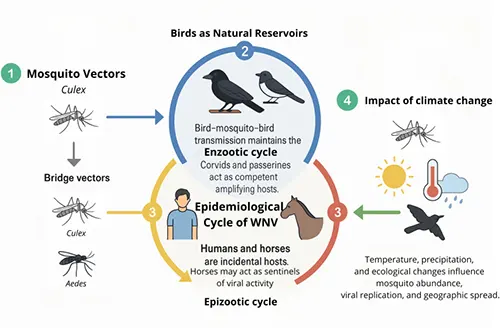
Figure 1. The epidemiological cycle of West Nile virus (DOI: 10.3389/fcimb.2025.1690827)
Consequently, understanding WNV requires moving beyond basic virus architecture or high‑level summaries of the infection cycle. The real scientific value lies in exploring how key viral molecules act in concert and interact with host cells and immune pathways—insights that ultimately clarify primary research targets and their scientific utility.
2. From Genome to Key Proteins: Molecular Composition of WNV
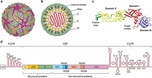
Figure 2. Structure and genome organization of West Nile virus (DOI: 10.1080/21505594.2021.1908740)
2.1 Single‑Stranded Positive‑Sense RNA Genome and Polyprotein Processing
WNV belongs to the genus Orthoflavivirus within the family Flaviviridae. Its genome consists of a single‑stranded positive‑sense RNA (+ssRNA) containing a 5′ untranslated region (UTR), a single large open reading frame (ORF), and a 3′ UTR. Unlike viruses that encode proteins via multiple independent ORFs, WNV translates its single ORF into a polyprotein precursor, which is subsequently cleaved by viral and host proteases to yield mature viral proteins.
These functional proteins fall into two main categories: structural and non‑structural. The structural proteins include the capsid (C), precursor membrane/membrane (prM/M), and envelope (E) proteins, which primarily drive viral particle assembly, maturation, and cell entry. The non‑structural proteins (NS1, NS2A, NS2B, NS3, NS4A, NS4B, and NS5) coordinate viral RNA replication, polyprotein processing, and host cellular microenvironment modulation.
This genomic organization offers a clear functional advantage: the virus maintains a single continuous coding sequence to generate diverse functional proteins via site‑specific cleavage. Rather than operating independently, these proteins form integrated functional modules that drive distinct stages of the viral life cycle.
2.2 Structural Proteins and Viral Particle Assembly
The C protein binds viral genomic RNA to form the nucleocapsid during particle assembly. The prM protein acts as a structural chaperone during viral maturation, stabilizing E protein conformation prior to proteolytic processing. The E protein is the principal envelope glycoprotein on the virion surface and mediates direct interactions with host cell receptors.
Composed of distinct structural domains, the E protein undergoes critical conformational rearrangements during entry. Upon endocytosis, the acidic environment of the endosome triggers E protein structural shifts, inducing fusion between the viral and endosomal membranes to release the viral genome into the cytoplasm. Because E protein directly mediates cell entry, its structural features constitute primary epitopes recognized by neutralizing antibodies.
Positioned at the intersection of viral entry and host immune recognition, the E protein remains a critical focal point in flavivirus structural biology and immunology.
2.3 NS1: A Key Protein Bridging Viral Replication and Host Modulation
Among the non‑structural proteins, NS1 occupies a unique role. Unlike replication proteins confined strictly to intracellular compartments, NS1 exists in multiple distinct forms: intracellular NS1 involved in genome replication, cell surface‑associated NS1, and a soluble secreted form (sNS1).
Intracellular NS1 associates with virus‑induced membrane structures to support replication complex (RC) assembly. Flavivirus replication does not occur freely in the cytosol; instead, it takes place within localized, ER‑derived membrane invaginations. NS1 collaborates with other non‑structural proteins to establish this microenvironment, optimizing conditions for viral RNA synthesis.
NS1's significance also stems from its extensive interactions with the host immune system. Studies demonstrate that WNV NS1 binds host complement regulatory factors, such as Factor H, attenuating complement activation; it also interferes with C4‑mediated complement cascades.
Furthermore, sNS1 is secreted into the extracellular space as a stable hexamer. Secreted NS1 exerts independent host‑modulatory functions and serves as an accessible circulating biomarker for research.
Thus, NS1 exemplifies a multifunctional target that bridges viral replication machinery, host immune evasion, and antigen detection research.
2.4 NS2–NS5: Essential Non‑Structural Components of the WNV Replication Machinery
Small transmembrane proteins NS2A and NS2B participate in membrane remodeling and cleavage event coordination. NS3 functions as a multifunctional enzyme: its N‑terminal domain pairs with NS2B to form the NS2B‑NS3 serine protease required for polyprotein processing, while its C‑terminal domain exhibits NTPase and RNA helicase activities crucial for RNA unwinding during replication. NS5, the largest non‑structural protein, harbors methyltransferase (MTase) and RNA‑dependent RNA polymerase (RdRp) activities, driving 5′ RNA capping and nascent RNA strand synthesis, respectively.
NS3 and NS5 act in tight functional synergy with NS1: NS1 facilitates replication complex assembly and membrane microenvironment formation, NS3 drives essential enzymatic processing and unwinding, and NS5 executes RNA synthesis. Together, they form the enzymatic core of the WNV replication machinery.
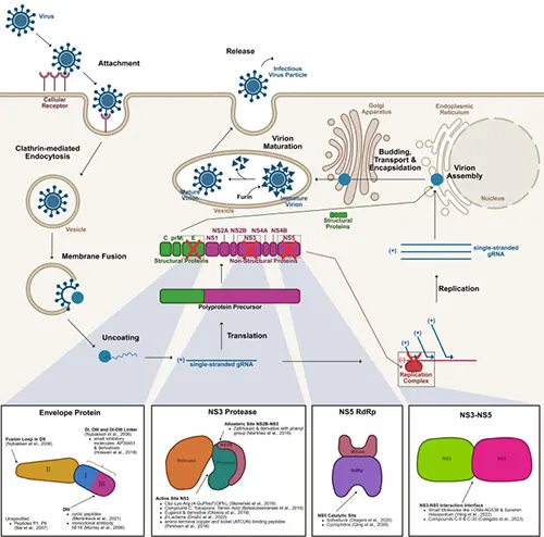
Figure 3. Life cycle and key research targets of West Nile virus (DOI: 10.3389/fcimb.2025.1568031)
3. The WNV Replication Cycle: From Entry to Virion Release
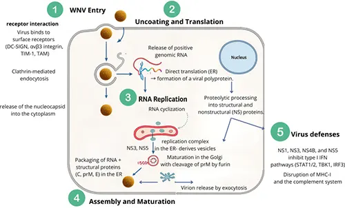
Figure 4. The replication cycle of WNV (DOI: 10.3389/fcimb.2025.1690827)
3.1 Viral Entry and Uncoating: E Protein‑Mediated Fusion
WNV entry is driven primarily by the E glycoprotein. Virions enter host cells via receptor‑mediated endocytosis. Low pH within mature endosomes triggers irreversible conformational changes in the E protein, driving fusion between the viral membrane and the endosomal membrane. Membrane fusion results in the release (uncoating) of viral genomic RNA into the host cytoplasm.
This entry pathway emphasizes that infection is not merely passive attachment, but a highly orchestrated process driven by structural transitions of the envelope protein.
3.2 Translation and Polyprotein Processing
Upon release into the cytosol, the +ssRNA genome functions directly as mRNA for translation. Without requiring prior negative‑strand intermediate synthesis, WNV utilizes host ribosomal machinery to synthesize the single polyprotein precursor.
Co‑ and post‑translational cleavage by viral and host proteases follows immediately. Specifically, the viral NS2B‑NS3 protease executes multiple site‑specific cleavages, releasing functional, mature viral proteins. Viral protein production is thus an integral early step of the replication cycle itself rather than an isolated post‑replication event.
3.3 Replication Complex Assembly and RNA Synthesis
WNV RNA replication occurs on specialized intracellular membranes. Viral infection induces extensive endoplasmic reticulum (ER) membrane remodeling, generating protective vesicular structures that host the replication complex. Non‑structural proteins including NS1, NS2A, NS3, NS4A, NS4B, and NS5 assemble within these membrane invaginations.
Within this sheltered niche, the RdRp synthesizes a negative‑sense RNA intermediate, which then serves as a template to generate abundant positive‑sense genomic RNA. Newly synthesized +ssRNA strands either re‑enter translation cycles or are packaged into nascent viral particles.
This localized replication strategy concentrates reaction components and shields double‑stranded RNA (dsRNA) intermediates from host cytosolic pattern recognition receptors (PRRs).
3.4 Virion Assembly, Maturation, and Release
Progenitor RNA associates with the C protein to form the nucleocapsid, which buds into the ER lumen acquiring host‑derived lipid membranes embedded with prM and E structural proteins. These immature, non‑infectious virions undergo transport through the trans‑Golgi network (TGN).
In the acidic TGN environment, host furin protease cleaves prM into mature M protein and a pr peptide, triggering structural reorganization of E proteins into homodimers. Mature infectious virions are subsequently released into the extracellular space via exocytosis to initiate new infection cycles.
Overall, the WNV life cycle functions as a continuous, highly coordinated cascade where entry, translation, cleavage, replication, and assembly are tightly coupled.
4. Host‑Pathogen Interactions: WNV Immune Evasion and Modulation
4.1 Innate Immune Sensing of WNV
Upon infection, host cells first detect viral genomic RNA and replication intermediates (such as dsRNA) rather than intact virions. Pattern recognition receptors (PRRs), including RIG‑I, MDA5, and specific TLRs, recognize these pathogen‑associated molecular patterns (PAMPs), triggering downstream signaling cascades that induce Type I interferons (IFN‑α/β) and pro‑inflammatory cytokines.
Type I IFNs induce hundreds of interferon‑stimulated genes (ISGs), establishing an antiviral state that represents the primary host defense against early WNV dissemination.
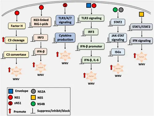
Figure 5. Immune evasion mechanisms of WNV (DOI: 10.3389/fmicb.2025.1711088)
4.2 Immune Evasion Mechanisms
WNV actively counters host innate defenses through multiple non‑structural proteins that target distinct signaling nodes in the IFN pathways. By attenuating IFN induction and downstream JAK‑STAT signaling, the virus maintains a cellular environment favorable for replication without causing premature host cell collapse.
NS1 plays a pivotal role in immune evasion. Beyond its cellular replication function, cell‑surface and secreted NS1 interact with host complement proteins to inhibit complement activation pathways, dampening local inflammatory cascades.
4.3 Adaptive Immune Responses and Neutralization
As infection progresses, adaptive immunity coordinates viral clearance. B cells generate target‑specific antibodies, while cytotoxic T lymphocytes eliminate infected host cells.
For antibody responses, functional efficacy depends not merely on binding affinity, but on targeted epitope specificity, stoichiometry, and structural accessibility for neutralizing virus entry. Thus, evaluating therapeutic or detection antibodies requires distinguishing binding capacity from functional neutralization efficacy.
4.4 Antibody‑Dependent Enhancement (ADE) in Flavivirus Biology
Antibody‑dependent enhancement (ADE) is a classic phenomenon observed in flavivirus immunology. When non‑neutralizing or sub‑neutralizing concentrations of antibodies bind viral particles, the resulting immune complexes can enter Fc‑receptor‑bearing immune cells (such as monocytes/macrophages), potentially increasing viral uptake and replication efficiency under specific experimental conditions.
While ADE has been observed in WNV experimental assays, outcomes vary significantly based on antibody concentration, epitope specificity, antibody subclass, and cell type. Consequently, ADE is best analyzed as a noteworthy immunological concept rather than an absolute mechanism during natural WNV pathology.
5. Key WNV Research Targets and Scientific Applications
Distinct WNV proteins serve specific applications across fundamental research, detection assay design, and therapeutic discovery:
- • NS1 Protein
NS1 is a premier target in WNV research due to its unique biomarker profile. As one of the few viral proteins secreted into extracellular fluid during early infection, sNS1 is detectable prior to seroconversion. Its high sequence conservation across WNV strains ensures robust target specificity. Functionally, NS1 serves as an essential tool for dissecting replication complex assembly, ER membrane remodeling, and complement evasion. Pragmatically, recombinant NS1 and high-affinity antibodies support acute-phase detection assay development, vector surveillance, and serological assay design.
- • E Protein
As the primary mediator of cell entry and major surface antigen, the E protein is essential for entry mechanism studies and neutralizing antibody screening. Its conformational dynamics are central to understanding membrane fusion events, while its immunogenic epitopes are key templates for vaccine design, therapeutic mAb discovery, and entry inhibitor screening.
- • NS3 and NS5 Proteins
NS3 and NS5 represent the core catalytic engine of WNV. Featuring distinct domain functions (NS3: protease/helicase; NS5: RdRp/methyltransferase), both proteins are highly conserved across flaviviruses. They represent primary targets for mechanistic studies on viral RNA replication and leading candidates for small‑molecule antiviral screening and enzyme inhibitor assays.
- • GP1 / Genome Polyprotein
The full‑length genome polyprotein encompasses the complete antigenic spectrum of all structural and non‑structural proteins. Compared to isolated recombinant domain antigens, the full polyprotein preserves native epitope representation across the entire viral proteome. It serves as an invaluable tool for broad‑spectrum antibody profiling, epitope mapping, cross‑reactivity studies across flaviviruses, and high‑throughput primary antibody screening.
These targets do not function in isolation. NS1, NS3, and NS5 collectively drive replication and host modulation; E protein mediates infection and humoral recognition; and the polyprotein captures the complete immunogenic landscape. Together, they form an integrated system for WNV research.
6. Conclusion
West Nile virus research spans complex interactions between viral proteins and host immune counter‑defenses. From E protein‑mediated cell entry to NS1‑driven replication and complement evasion, and NS3/NS5‑catalyzed genome synthesis, each component plays a coordinated role in the viral life cycle.
Deciphering protein function and domain structure is essential for advancing flavivirus virology, developing targeted detection tools, and designing effective antiviral therapeutics.
abinScience West Nile Virus Research Reagents
To support fundamental research and experimental needs for West Nile virus, we offer a comprehensive portfolio of scientific reagents covering key viral targets, including recombinant proteins, antibody reagents, ELISA kits, and antibody pair sets validated for ELISA, WB, IHC, and SPR applications.
1. Recombinant Proteins
| Product Name |
Cat. No. |
Primary Applications |
| West Nile Virus/WNV NS1 Recombinant Protein (N‑His) |
VK554012 |
ELISA, Immunogen Preparation, SDS‑PAGE, WB, Bioactivity Assays |
| West Nile Virus/WNV NS1 Recombinant Protein (C‑His) |
VK554011 |
ELISA, Immunogen Preparation, SDS‑PAGE, WB, Bioactivity Assays |
| West Nile Virus/WNV NS1 Recombinant Protein (C‑Fc) |
VK554021 |
ELISA, Immunogen Preparation, SDS‑PAGE, WB, Bioactivity Assays |
2. Antibody Reagents
| Product Name |
Cat. No. |
Primary Applications |
| Anti‑West Nile Virus/WNV NS1 Polyclonal Antibody |
VK554014 |
ELISA, IHC, WB |
| Anti‑West Nile virus/WNV GP1/Genome polyprotein Recombinant Antibody (CR4353) |
VK554013 |
ELISA, SPR, WB |
| Anti‑West Nile virus/WNV GP1/Genome polyprotein Recombinant Antibody (CR4374) |
VK554023 |
ELISA, SPR, WB |
| Anti‑West Nile virus/WNV GP1/Genome polyprotein Recombinant Antibody (CR4274) |
VK554033 |
ELISA, SPR, WB |
3. ELISA Kits
| Product Name |
Cat. No. |
| West Nile Virus/WNV NS1 Antigen ELISA Kit |
VK554018 |
| Anti‑West Nile Virus/WNV NS1 IgG ELISA Kit |
VK554028 |
| Anti‑West Nile Virus/WNV NS1 IgM ELISA Kit |
VK554038 |
| West Nile virus/WNV GP1/Genome Antigen ELISA Kit |
VK554048 |
4. Antibody Pair Sets
| Product Name |
Cat. No. |
| West Nile Virus/WNV NS1 Antibody Pair Set |
VK554908 |
| West Nile virus/WNV GP1/Genome Antibody Pair Set |
VK554918 |
References
- [1] Singh, P., Khatib, M. N., Ballal, S., Kaur, M., Nathiya, D., Sharma, S., Prasad, G. V. S., Sinha, A., Gaidhane, A. M., Mohapatra, P., Varma, A., Lakhanpal, S., Shabil, M., Bushi, G., Sah, S., & Abu Serhan, H. (2025). West Nile Virus in a changing climate: epidemiology, pathology, advances in diagnosis and treatment, vaccine designing and control strategies, emerging public health challenges ‑ a comprehensive review. Emerging Microbes & Infections, 14(1), 2437244. https://doi.org/10.1080/22221751.2024.2437244
- [2] Saiz, J. C., Martín‑Acebes, M. A., Blázquez, A. B., Escribano‑Romero, E., Poderoso, T., & Jiménez de Oya, N. (2021). Pathogenicity and virulence of West Nile virus revisited eight decades after its first isolation. Virulence, 12(1), 1145–1173. https://doi.org/10.1080/21505594.2021.1908740
- [3] Prieto‑Vega, S., Berzal‑Herranz, A., Garrido, J. J., Arias, A., Grande‑Pérez, A., Fernández‑Escamilla, A. M., & Montoya, M. (2025). West Nile virus unmasked: from gene variability to future challenges. Frontiers in Cellular and Infection Microbiology, 15, 1690827. https://doi.org/10.3389/fcimb.2025.1690827
- [4] Zhang, S., He, Y., Wu, Z., Wang, M., Jia, R., Zhu, D., Liu, M., Zhao, X., Yang, Q., Wu, Y., Zhang, S., Huang, J., Ou, X., Gao, Q., Sun, D., Zhang, L., Yu, Y., Chen, S., & Cheng, A. (2023). Secretory pathways and multiple functions of nonstructural protein 1 in flavivirus infection. Frontiers in Immunology, 14, 1205002. https://doi.org/10.3389/fimmu.2023.1205002
- [5] Kayesh, M. E. H., Kohara, M., & Tsukiyama‑Kohara, K. (2025). Innate immune sensing and vaccine strategies against West Nile virus: role of Toll‑like receptors and viral evasion mechanisms. Frontiers in Microbiology, 16, 1711088. https://doi.org/10.3389/fmicb.2025.1711088
- [6] Kocabiyik, D. Z., Álvarez, L. F., Durigon, E. L., & Wrenger, C. (2025). West Nile virus ‑ a re‑emerging global threat: recent advances in vaccines and drug discovery. Frontiers in Cellular and Infection Microbiology, 15, 1568031. https://doi.org/10.3389/fcimb.2025.1568031


 中文
中文 English
 한국어
한국어 日本語
日本語 Español
 Français
 Русский


