Release date:
2025-10-31 View count: 680
In multicolour flow cytometry data analysis, we are accustomed to discussing the intensity of positive signals, yet often overlook the definition of “negative”. The Isotype Control serves as a systematic reference to help us delineate this boundary—allowing us to observe the noise structure behind the signal and determine whether true expression is occurring.
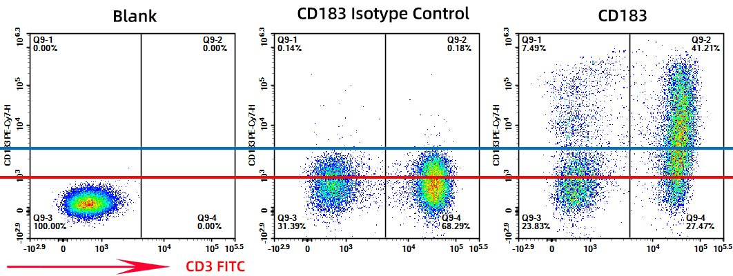
Figure 1. Comparison of gating using a blank (red line) vs. isotype control (blue line)
1. Starting from “Zero”: The Experimental Logic and Significance of Isotype Controls
Isotype control antibodies share the same host source, immunoglobulin subclass, and fluorochrome label as the target antibody, but lack specificity for the target antigen. They are not intended to “define the negative population” but rather provide a background baseline for the antibody-sample system.
In cases where the sample exhibits strong Fc receptor binding, non-specific interactions, or autofluorescence, the isotype control reveals the level of system noise, ensuring that the determination of “positive” is not based on subjective gating but rather on the inherent stability of the experimental system. More importantly, the isotype signal can act as a “magnifying glass” for potential system issues:
- Assessing residual Fc receptor binding: If signals are elevated in monocytes or B cells, this indicates inadequate Fc blocking.
- Identifying fluorescence dye aggregation or leakage: Some bright dyes (e.g., PE, PerCP-Cy5.5) show significant background variability.
- Verifying intracellular staining integrity: If background increases significantly post-fixation/permeabilization, the isotype control can highlight this change.
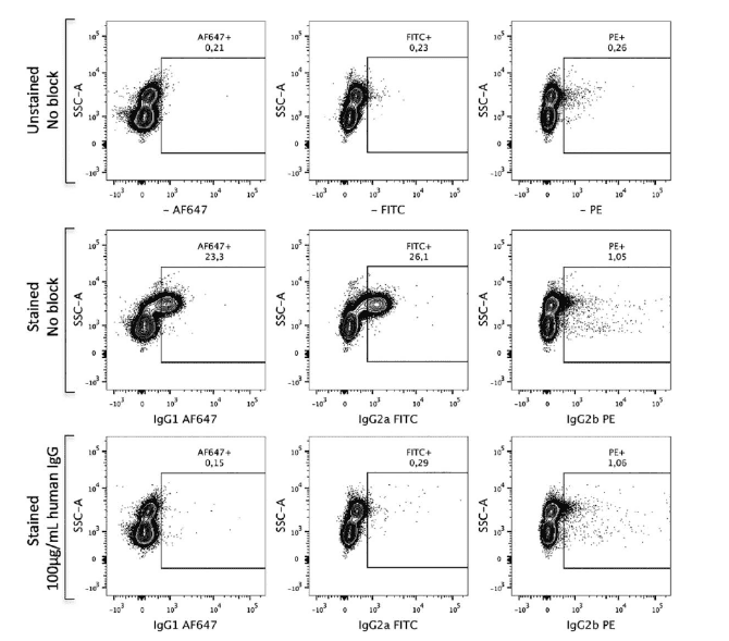
Figure 2. Staining results of CD45 under different treatments (Isotype control without Fc block, background elevated; Fc block reduces background)
2. Between Ideal and Reality: The Boundaries of Isotype Control Usage
In theory, as long as the isotype antibody matches the target antibody in species, subclass, fluorochrome ratio, and concentration, it should accurately reflect the background level. However, in practice, this is nearly impossible to achieve perfectly.Minor differences in fluorochrome-to-protein ratios, antibody purity, and batch variations can lead to signal drift. Therefore, while isotype controls are useful for reference, they should not be applied dogmatically.
When to Consider Adding an Isotype Control?
✅ Low/Medium-Low Expressing Antigens (difficult to set thresholds)
✅ FcR-High Expressing Cells (e.g., monocytes, macrophages, B cells, granulocytes)
✅ Intracellular Staining/Fixed Permeabilisation (background often increases)
✅ Samples that have been Stimulated/Cultured/Frozen-Thawed (increased system noise)
✅ New Panel Validation/New Batch of Antibodies (establish baseline reference)
When it May Not Be Necessary (if most or all conditions are met):
⭕ Clear Threshold Set by FMO with a large separation between positive and negative populations
⭕ Strong Positive Expression/Dual Peak Separation of Target Antigen
⭕ Dead Cell Exclusion Complete, Fc Block Adequate, Antibody Titration Optimised
⭕ Established Baseline and Quality Control Records Available
3. Enhancing Signal Credibility: Optimisation and Considerations in Practice
Even with isotype controls in place, experimental design should still ensure system background is appropriately controlled. Here are optimisation recommendations:
Validate and Titrate Antibodies: Excess antibody can cause negative peak shifts and broaden the baseline, interfering with threshold determination. Titrate to the optimal concentration to ensure clear signals.
Focus on Specific Binding Sources: If the isotype signal is high, prioritise using Fc blockers specific to the species of the sample (e.g., human IgG or specialised Fc block for human samples). Apply the block before adding the antibody. Blocking buffers containing immunoglobulins/serum can further reduce background.
Use Viability Dyes to Exclude Dead Cells: Dead cells exhibit strong non-specific staining, often leading to background “false positives”. Use amine-reactive viability dyes (e.g., Zombie dyes) to label dead cells before surface staining or sample loading.
Set Gating Thresholds Cautiously: The isotype control only reflects non-specific antibody binding and should not be used in isolation to define positive/negative populations. Spillover spreading (Spillover Spreading) should be evaluated using SSM/CSI indices in conjunction with single-stain and FMO controls.
Handling Samples with High Autofluorescence: For tissue, older, or myeloid samples, retain an unstained control and attempt autofluorescence correction in the channels/models. The isotype control only covers antibody-related background and cannot replace autofluorescence correction.
4. Isotype Control Selection and Application Example
Here is an example of selecting and applying an isotype control in a single-colour flow cytometry experiment to detect IL-17A-PE-Cy7 expression in human peripheral blood mononuclear cells (PBMCs):
1) Experimental Background: IL-17A is a low-expressing antigen, and PBMCs contain FcR-high expressing cells (monocytes, B cells). The experiment involves fixation/permeabilisation, necessitating an isotype control to assess non-specific binding. The instrument used was the BD FACS Canto II.
2) Isotype Control Selection: For IL-17A-PE-Cy7 (mouse IgG1 κ, PE-Cy7-labelled, concentration 1 µg/mL), select the isotype antibody from the same supplier and batch. The fluorochrome/antibody ratio should match, and the concentration should be adjusted to 1 µg/mL (titration to confirm equivalent background displacement).
3) Experimental Workflow: Take approximately 1×10⁶ PBMCs, first stain dead cells with Zombie Aqua (amine-reactive dye) (4°C, 15 minutes). After washing, block Fc receptors with human IgG (10 µg/mL) (4°C, 10 minutes). Add mouse IgG1 κ-PE-Cy7, incubate at 4°C for 30 minutes, wash, then fix and permeabilise with a reagent kit (eBioscience) using the same conditions as for fully stained samples. Use the same buffer, centrifugation, and PMT voltage (calibrated via CS&T QC), collecting at least 10,000 events.
4) Results and Interpretation: The MFI of the isotype control in the PE-Cy7 channel was around 200, close to the unstained sample MFI (150), indicating low background and effective Fc blocking. In contrast, the fully stained sample showed an IL-17A+ population with an MFI of 2,000, with clear separation between positive and negative populations (SI ≈ 20). If the isotype MFI was significantly higher than the unstained control (e.g., 500), this would warrant checking the Fc block, dead cell proportion, or antibody concentration. FMO controls were used to confirm the IL-17A threshold, and single-stain controls (if involving multicolour expansion) were used to generate the compensation matrix.
This example demonstrates how to select an isotype control for a low-expression antigen, emphasising the importance of strict matching (species, subclass, fluorochrome), titration consistency, Fc blocking, and dead cell exclusion, ensuring reliable background signals and providing a reference for threshold setting.
Isotype controls provide an important background reference in flow cytometry experiments. They reflect levels of non-specific binding and help researchers identify system noise, thereby improving the accuracy of signal interpretation. By flexibly assessing experimental goals and sample characteristics, and appropriately applying isotype controls, researchers can enhance data stability and reliability, providing more scientifically robust support for experimental results.
abinScience was founded in France and is focused on the development and production of high-quality life science reagents. Based in the innovative technology hub of Strasbourg, France, our vision is to "Empower Bioscience Discovery." Our flow cytometry antibody products cover common detection markers and are available in a wide range, catering to multi-species research needs. We offer stable and reliable support for scientific research. For more information on our flow cytometry antibodies, please visit:
Explore abinScience Flow Cytometry (FACS) Antibodies

 中文
中文 English
 한국어
한국어 日本語
日本語 Español
 Français
 Русский


