 中文
中文 English
| Catalog No. | HB104014 | ||||||||
|---|---|---|---|---|---|---|---|---|---|
| Species reactivity | Human, Macaca mulatta | ||||||||
| Applications | ELISA, IHC, WB, IF | ||||||||
| Host species | Rabbit | ||||||||
| Isotype | IgG | ||||||||
| Clonality | Polyclonal | ||||||||
| Immunogen | E. coli - derived recombinant Human CD326/EPCAM (Gln24-Lys265). | ||||||||
| Target | Epithelial glycoprotein 314, hEGP314, Major gastrointestinal tumor-associated protein GA733-2, EGP314, TACSTD1, Adenocarcinoma-associated antigen, TROP1, EPCAM, Epithelial glycoprotein, KS 1/4 antigen, Tumor-associated calcium signal transducer 1, M4S1, CD326, Ep-CAM, EGP, Epithelial cell adhesion molecule, M1S2, KSA, Cell surface glycoprotein Trop-1, GA733-2, Epithelial cell surface antigen, MIC18 | ||||||||
| Endotoxin level | Please contact with the lab for this information. | ||||||||
| Purification | Purified by antigen affinity column. | ||||||||
| Accession | P16422 | ||||||||
| Form | Liquid | ||||||||
| Storage buffer | 0.01M PBS, pH 7.4, 50% Glycerol, 0.05% Proclin 300. Please refer to the specific buffer information in the hardcopy of datasheet or the lot-specific COA. |
||||||||
| Product Usage Information |
| ||||||||
| Stability and Storage | Use a manual defrost freezer and avoid repeated freeze thaw cycles. Store at 2 to 8°C for frequent use. Store at -20 to -80°C for twelve months from the date of receipt. | ||||||||
| Note | For research use only. |
Hela cells were fixed with 4% paraformaldehyde for 20 minutes, followed by blocking with 5% goat serum for 1 hour. The cells were then incubated with a primary antibody targeting EpCAM (HB104014) at a concentration of 20.4 ug/ml overnight at 4°C. Afterward, the cells were incubated with a secondary antibody, Goat Anti-Rabbit IgG (Alexa Fluor-488) for 50 minutes at room temperature. The localization of EpCAM was visualized (shown in green), and nuclear DNA was stained with DAPI (shown in blue).
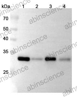
Western blot analysis was performed using anti-EPCAM polyclonal antibody at 1μg/ml on various samples.
Lane 1: HCT116 cell lysate
Lane 2: MCF7 cell lysate
Lane 3: HT-29 cell lysate
Lane 4: Mouse colon lysate


Contact us for custom quotes, bulk requests and any other issues.
Mail: support@abinScience.com
+86-027-65523339
Building C, No. 666, Shen Dun Si Lu, Wuhan, 430206, China


