What are Helper T Cells?
Helper T cells (Th cells) are one of the most important lymphocyte subpopulations in the immune system, primarily composed of CD4+ T cells. They play a crucial role in immune responses by acting as "commanders," coordinating the body's defence and immune homeostasis. This is achieved through the secretion of cytokines, aiding B cells in antibody production, enhancing macrophage-mediated pathogen clearance, and regulating the functions of other immune cells. Dysfunctions in Helper T cells are often linked to immune-related diseases such as infection intolerance, allergic reactions, autoimmune diseases, or tumour immune escape.
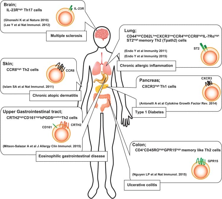
Figure 1. Helper T Cells and Immune Responses in Different Tissues (DOI: 10.1093/intimm/dxw006)
Differentiation of Helper T Cells
Naive CD4+ T cells are activated via their T cell receptor (TCR) in conjunction with co-stimulatory molecules like CD28, and through antigen-presenting cells (APCs) that express major histocompatibility complex (MHC) class II molecules, such as macrophages, dendritic cells, and B cells. Activation signals are transmitted through the recognition and binding of MHC II molecules presenting homologous antigen peptides on the surface. Upon complete activation, naive CD4+ T cells rapidly proliferate and differentiate into various subsets of Helper T cells, such as Th1, Th2 or Th17 (Figure 2).
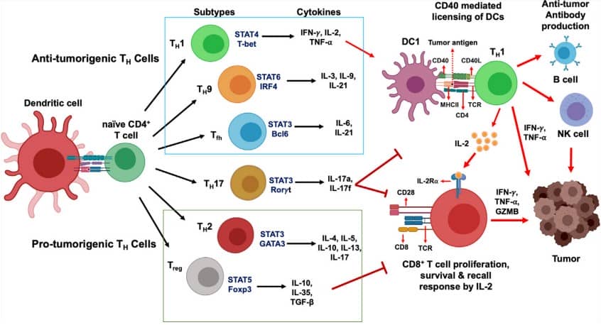
Figure 2. Development of CD4+ T Cells and Their Functional Subsets in Immunity (DOI: 10.3389/fimmu.2021.669474)
Helper T Cells and Disease
The balance of Helper T cell subsets is crucial for immune homeostasis, and imbalance often leads to disease development. For instance, an imbalance between Th1/Th2 cells is associated with susceptibility to allergies or infections, while excessive activation of Th17 cells is linked to autoimmune diseases such as rheumatoid arthritis and psoriasis. Defects in Treg function may result in graft rejection and failure of immune tolerance. In tumour immunotherapy, different Helper T cell subsets can provide important prognostic and therapeutic insights. Therefore, accurate and systematic detection of Helper T cell subsets is vital for both research and clinical applications.
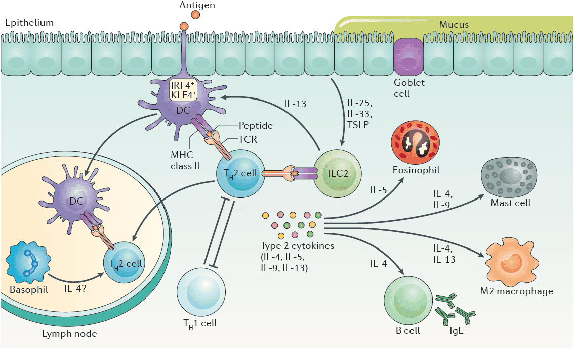
Figure 3. Th Cells and Type II Immune Responses (DOI: 10.1038/nri.2017.118)
Flow Cytometric Detection of Helper T Cells
Flow cytometry is a critical method for detecting Helper T cells and their subsets in both research and clinical settings. By analysing surface markers, secreted cytokines, and transcription factors, it allows for efficient identification and quantification of different subsets.
Table 1. Helper T Cell Subsets and Markers
|
Cell Subset
|
Surface Markers
|
Secreted Cytokines
|
Transcription Factors
|
|
Th1
|
CD4, CD183(CXCR3)*, CD195(CCR5), CD197(CCR7), CXCL9, CXCL10, CXCL11
|
IFN-γ*, LT-α, TNF, Perforin, Granzyme A, Granzyme B, Granulysin (Hu)
|
T-bet*, STAT1, STAT4, Runx3
|
|
Th2
|
CD4, CD194(CCR4), CD294(CRTH2)*, CD193(CCR3), CD198(CCR8), MDC, TCA3, TARC
|
IL-4*, IL-5, IL-6, IL-10, IL-13, IL-31
|
GATA3*, STAT5, STAT6, MAF, GFI-1, IRF4, c-Maf
|
|
Th9
|
CD4, CCL20, CD196(CCR6)
|
IL-9*, IL-10, CCL17 (TARC), CCL22 (MDC),TGF-β
|
GATA3 (early), SMADs, STAT6, PU.1, IRF4
|
|
Th17
|
CD4, CD161, CD194(CCR4), CD196(CCR6)*, IL23R, CCL4, CCL17, CCL22
|
IL-17A*, IL-17F, IL-21, IL-22, IL-24, IL-26 (Hu), TNF, CCL20 (MIP-3α)
|
RORγt*, RORα4, STAT3, Runx1, Batf, IRF4, c-Maf
|
|
Th22
|
CD4, CCR10, CD194(CCR4), CD196(CCR6), CD279 (PD-1)
|
IL-22*, TNF
|
AhR*
|
|
Tfh
|
CD4, CD185 (CXCR5)*, CD196(CCR6), CD278 (ICOS)*, CD279 (PD-1)*
|
IFN-γ, IL-4, IL-17A, IL-17F, IL-21*
|
Bcl-6* , MAF, STAT3
|
|
Treg
|
CD4, CD25*, CD127*-/low, CD152(CTLA4)
|
IL-10, IL-35, TGF-β
|
Foxp3*, Smad3, STAT5, AhR
|
*Note: * indicates characteristic markers of cells.
abinScience Recommended Flow Cytometry Antibodies for Helper T Cell Detection
|
Target
|
Clone
|
Species
|
Localization
|
|
CD3
|
OKT3
|
Human
|
Surface
|
|
CD4
|
SAA0002
|
Human
|
Surface
|
|
CD8
|
G10-1
|
Human
|
Surface
|
|
CD25
|
1H4
|
Human
|
Surface
|
|
CD127
|
N13B2-h3
|
Human
|
Surface
|
|
CD183
|
V3G6
|
Human
|
Surface (Chemokine receptor)
|
|
CD185
|
16D7
|
Human
|
Surface (Chemokine receptor)
|
|
CD193
|
5E8-G9-B4
|
Human
|
Surface (Chemokine receptor)
|
|
CD194
|
SAA0069
|
Human
|
Surface (Chemokine receptor)
|
|
CD195
|
PRO-140
|
Human
|
Surface (Chemokine receptor)
|
|
CD196
|
R612
|
Human
|
Surface (Chemokine receptor)
|
|
CD197
|
R707
|
Human
|
Surface (Chemokine receptor)
|
|
CD198
|
SAA1405
|
Human
|
Surface (Chemokine receptor)
|
|
CXCL9
|
SAA0465
|
Human
|
Secreted (Chemokines)
|
|
CXCL10
|
SAA0413
|
Human
|
Secreted (Chemokines)
|
|
CXCL11
|
SAA0466
|
Human
|
Secreted (Chemokines)
|
|
TCA3(CCL1)
|
SAA0526
|
Human
|
Secreted (Chemokines)
|
|
TARC(CCL17)
|
SAA0460
|
Human
|
Secreted (Chemokines)
|
|
IFNγ
|
SAA0414
|
Human
|
Secreted (Cytokines)
|
|
IL4
|
SAA0371
|
Human
|
Secreted (Cytokines)
|
|
IL9
|
SAA0377
|
Human
|
Secreted (Cytokines)
|
|
IL17A
|
SAA0386
|
Human
|
Secreted (Cytokines)
|
|
IL22
|
SAA0391
|
Human
|
Secreted (Cytokines)
|
|
GATA3
|
SAA0103
|
Human
|
Transcription Factor
|
|
FOXP3
|
1A210
|
Human
|
Transcription Factor
|
|
CD3
|
145-2C11
|
Mouse
|
Surface
|
|
CD4
|
GK1.5
|
Mouse
|
Surface
|
|
CD8
|
53-6.72
|
Mouse
|
Surface
|
|
CD25
|
PC61/PC61.5.3
|
Mouse
|
Surface
|
|
IFNγ
|
XMG1.2
|
Mouse
|
Secreted (Cytokines)
|
|
IL4
|
11B11
|
Mouse
|
Secreted (Cytokines)
|
|
IL17A
|
BZN035
|
Mouse
|
Secreted (Cytokines)
|
About Us
abinScience was founded in France and is focused on the development and production of high-quality life science reagents. Based in the innovative technology hub of Strasbourg, France, our vision is to "Empower Bioscience Discovery." Our flow cytometry antibody products cover common detection markers and are available in a wide range, catering to multi-species research needs. We offer stable and reliable support for scientific research. For more information on our flow cytometry antibodies, please visit: www.abinscience.com
References
[1] Basu A, Ramamoorthi G, Albert G, Gallen C, Beyer A, Snyder C, Koski G, Disis ML, Czerniecki BJ, Kodumudi K. Differentiation and Regulation of TH Cells: A Balancing Act for Cancer Immunotherapy. Front Immunol. 2021 May 3;12:669474.
[2] Walker JA, McKenzie ANJ. TH2 cell development and function. Nat Rev Immunol. 2018 Feb;18(2):121-133.
[3] Luckheeram RV, Zhou R, Verma AD, Xia B. CD4⁺T cells: differentiation and functions. Clin Dev Immunol. 2012;2012:925135.
[4] Nakayama T, Hirahara K, Onodera A, Endo Y, Hosokawa H, Shinoda K, Tumes DJ, Okamoto Y. Th2 Cells in Health and Disease. Annu Rev Immunol. 2017 Apr 26;35:53-84.
[5] Mills KHG. IL-17 and IL-17-producing cells in protection versus pathology. Nat Rev Immunol. 2023 Jan;23(1):38-54.
[6] Geng J, Yu S, Zhao H, Sun X, Li X, Wang P, Xiong X, Hong L, Xie C, Gao J, Shi Y, Peng J, Johnson RL, Xiao N, Lu L, Han J, Zhou D, Chen L. The transcriptional coactivator TAZ regulates reciprocal differentiation of TH17 cells and Treg cells. Nat Immunol. 2017 Jul;18(7):800-812.

 中文
中文 English


