In June, multiple groundbreaking studies supported by abinScience, focusing on antibody and protein research, were published in top-tier journals such as Aging Cell and Scientific Reports. These studies cover critical areas, including the pro-inflammatory mechanism of the key Alzheimer's disease protein Galectin-9 in Aβ aggregation and the adhesion mechanism of small molecule pathogens to host cell cytoskeleton proteins. Some of these findings were made possible by abinScience's high-purity recombinant proteins and antibodies, enabling significant breakthroughs in understanding Alzheimer's disease mechanisms and screening targets for infectious diseases. In this literature highlight, we review these advancements from leading laboratories, emphasizing the application of protein research in neurodegenerative diseases and infection control.
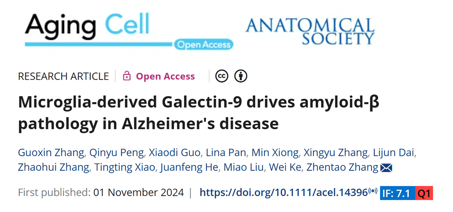
Title: Microglia-derived Galectin-9 drives amyloid-β pathology in Alzheimer's disease
Journal: Aging Cell
Impact Factor: 7.1
Author Affiliation: Department of Neurology, Renmin Hospital of Wuhan University

This study investigates the role of microglia-derived Galectin-9 (Gal-9) in Alzheimer's disease (AD) and its association with amyloid-β (Aβ) pathology. Microglia in AD induce neuroinflammation and neurodegeneration by releasing pro-inflammatory cytokines. While Galectin family proteins (e.g., Gal-3) are known to regulate microglial activation and immune responses, the specific role of Gal-9 remains unclear. The researchers explored whether Gal-9 contributes to AD's Aβ pathology and its underlying mechanisms. Using immunostaining, Western blot, and ELISA, they found that Gal-9 is significantly overexpressed in the brain tissue and cerebrospinal fluid (CSF) of AD patients and APP/PS1 mice, co-localizing with Aβ plaques. Further experiments showed that the interaction between Gal-9 and Aβ fibers (Gal-9-Aβ) enhances microglial inflammatory responses and neuronal toxicity, leading to more severe neuronal apoptosis and motor dysfunction. In APP/PS1 mice with Gal-9 knockout (Gal-9 KO), Aβ deposition, neuroinflammation, memory deficits, and synaptic dysfunction were reduced, suggesting that Gal-9 plays a critical role in driving AD pathology. The findings indicate that Gal-9 could be a novel therapeutic target for AD.
abinScience provided Gal-9 recombinant protein (Catalog No. HT173011), used in vitro to study Gal-9's effect on Aβ aggregation. The protein was co-incubated with Aβ42, and ThT fluorescence assays and electron microscopy confirmed that Gal-9 significantly accelerates Aβ42 aggregation, alters its fibril morphology, and enhances its neurotoxicity and seeding activity. Additionally, a neutralizing anti-Gal-9 antibody was used to validate the specificity of this effect, demonstrating that it blocks Gal-9's promotion of Aβ aggregation.
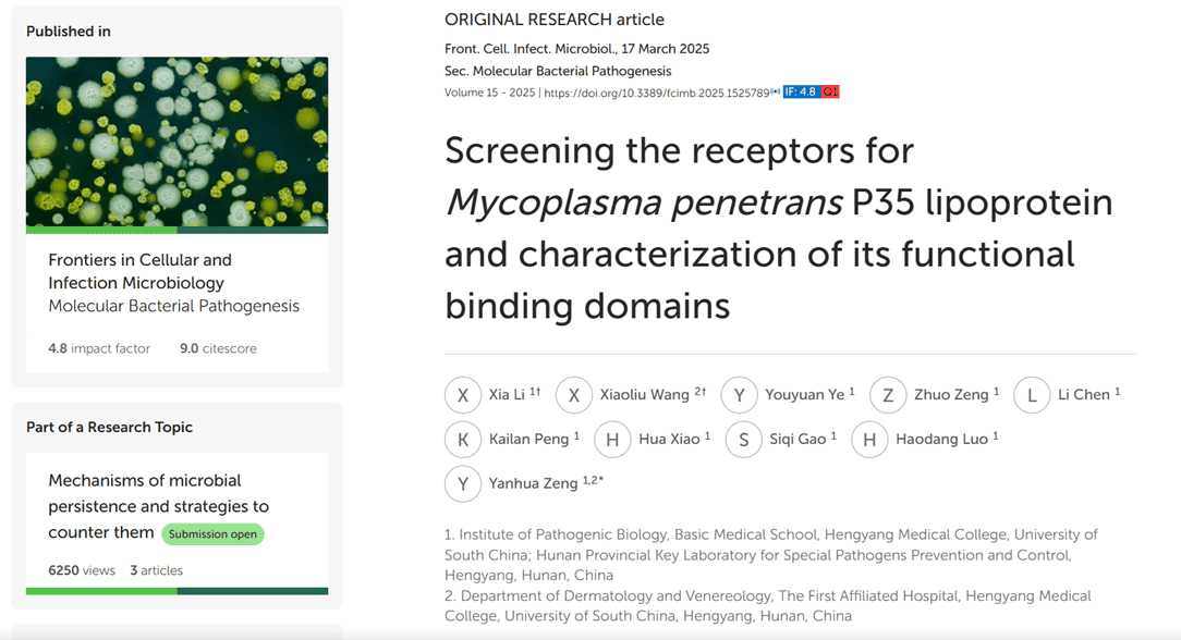
Title: Screening the receptors for Mycoplasma penetrans P35 lipoprotein and characterization of its functional binding domains
Journal: Frontiers in Cellular and Infection Microbiology
Impact Factor: 4.8
Author Affiliation: Institute of Pathogenic Biology, School of Basic Medical Sciences, Hengyang Medical College, University of South China

This study focuses on the mechanism by which the human pathogen Mycoplasma penetrans mediates host cell adhesion through its surface lipoprotein P35. P35 is a major surface immunogen and adhesion protein of M. penetrans, but its specific interactions with host cell receptors remain unclear. Using modified virus overlay protein binding assays (VOPBA), far-western blot, and siRNA knockdown, the researchers identified the host cell membrane protein ACTG1 (γ-actin) as a key receptor for P35. They further pinpointed amino acid residues 35–42 and 179–186 of P35 as the functional domains for ACTG1 binding. Disrupting ACTG1 via antibody blocking, protein pretreatment, or siRNA knockdown significantly reduced the adhesion of rP35 and M. penetrans to SV-HUC-1 cells, confirming ACTG1's critical role in adhesion. This study not only reveals the functional binding sites of P35 for the first time but also provides a theoretical basis for developing anti-infection strategies targeting the P35-ACTG1 interaction.
abinScience provided recombinant human ACTG1 protein (Catalog No. HX058012), which was used in far-western blot experiments to confirm the specific binding between rP35 and ACTG1, showing a clear band at 35 kDa, with no binding signals for other proteins (e.g., KRT8). In ELISA assays, abinScience's ACTG1 protein served as a coated antigen, successfully detecting binding activity with rP35 and its key synthetic peptides (e.g., P35-4), further supporting ACTG1 as the specific receptor for P35.
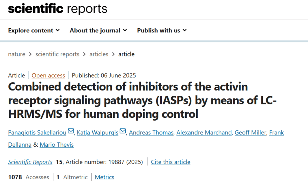
Title: Combined detection of inhibitors of the activin receptor signaling pathways (IASPs) by means of LC-HRMS/MS for human doping control
Journal: Scientific Reports
Impact Factor: 3.8
Author Affiliation: Institute of Biochemistry, German Sport University Cologne

This study focuses on detecting emerging "inhibitors of the activin receptor signaling pathways (IASPs)" in the field of anti-doping. These drugs, including various therapeutic antibodies and fusion proteins, can promote muscle growth or enhance erythropoiesis, making them potential performance-enhancing substances banned by the World Anti-Doping Agency (WADA). To address the challenges of IASPs' diversity, complex structures, and limited detection methods, the researchers developed and validated a multiplex detection method based on LC-HRMS/MS, capable of identifying nine IASPs in serum, plasma, and urine samples. Combining (immuno)affinity purification, trypsin digestion, and high-resolution mass spectrometry, the method achieved a sensitivity of 10–50 ng/mL and successfully detected long-term residues of Luspatercept and Sotatercept in real samples, providing an efficient and feasible solution for routine IASP anti-doping screening.
abinScience provided key reference standards, including Sotatercept (Catalog No. HY428026), Elritercept (Catalog No. HB949026), Luspatercept (Catalog No. HF869016), Ramatercept (Catalog No. HT343066), and Apitegromab (Catalog No. HT343056). These standards were used to develop, optimize, and validate the new detection method. abinScience's high-quality Luspatercept standard was repeatedly used for sensitivity validation, target peptide selection, and mass spectrometry signal confirmation, ensuring the method’s accuracy and reliability. Compared to Luspatercept materials from other suppliers, abinScience's product demonstrated stable and consistent properties, helping resolve prior controversies about Luspatercept's target-binding capacity and supporting the scientific validity of the proposed affinity purification and multi-target identification strategy.
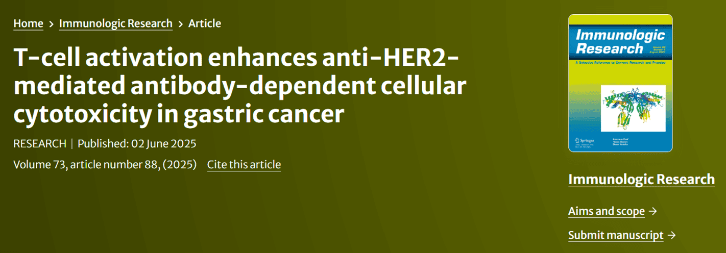
Title: T-cell activation enhances anti-HER2-mediated antibody-dependent cellular cytotoxicity in gastric cancer
Journal: Immunologic Research
Impact Factor: 3.3
Author Affiliation: Department of Gastrointestinal Surgery, Zhongshan Hospital

This study addresses resistance to anti-HER2 monoclonal antibodies (e.g., Trastuzumab, TRA) in HER2-positive gastric cancer patients. The researchers found that a lack of T cells in the tumor microenvironment is a previously underrecognized mechanism of resistance to anti-HER2 therapy. Analysis of clinical samples revealed that the infiltration level of T cells, particularly CD4+ T cells, is closely associated with patients' response to anti-HER2 therapy and survival. In vitro experiments and organoid models further confirmed that CD4+ T cells enhance NK cell function and antibody-dependent cellular cytotoxicity (ADCC). The study also demonstrated that activating T cells with T-cell agonists or HER2×CD3 bispecific antibodies enhances NK cell anti-tumor effects, thereby improving the efficacy of anti-HER2 monoclonal antibodies.
abinScience provided a critical reagent—the anti-HER2×CD3 bispecific antibody Ertumaxomab (Catalog No. HY286086)—used to activate T cells and enhance immune responses in the tumor microenvironment. The researchers found that Ertumaxomab effectively induces T cells to secrete IL-2, a cytokine essential for maintaining NK cell activity and promoting ADCC. Combining Ertumaxomab with TRA significantly increased NK cell killing of HER2-positive gastric cancer cells and enhanced CD16 re-expression, demonstrating the importance of T-cell assistance in TRA's anti-tumor effects.
Literature Citation Reward Program
To express gratitude for the support and trust of researchers, abinScience is launching a Literature Citation Reward Program! Whether you're a pioneer in life sciences or a dedicated laboratory researcher, you can apply for generous rewards by publishing articles in SCI journals using abinScience's full range of products.

 中文
中文 English


