Health authorities in the Democratic Republic of Congo (DRC) have declared a new outbreak of the Ebola virus, which has claimed the lives of 15 people since the end of August, according to the health minister's statement on Thursday, September 08, 2025, at 04:10 PM +08. The Ebola virus, first identified in 1976 near the Ebola River in what is now the DRC, emerged during two simultaneous outbreaks—one in the DRC and the other in South Sudan. Named after the river where it was initially detected, the virus belongs to the Filoviridae family and is known for causing severe, often fatal hemorrhagic fever in humans and primates. Since its discovery, the DRC has experienced multiple outbreaks, with this latest incident marking the 16th occurrence in the country, highlighting the ongoing challenge of managing this deadly disease in the region.
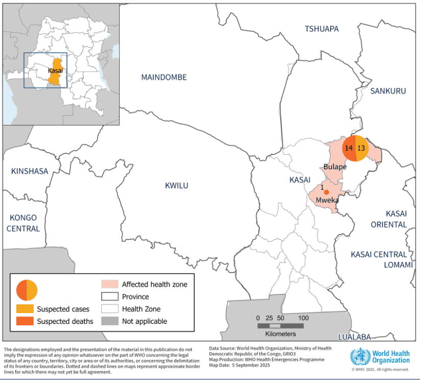
Fig.1 Map of suspected cases and deaths of Ebola virus disease by health zone, as of 4 September 2025(WHO)
Ebola Virus: Pathogenesis, Genomic Structure, and Challenges in Prevention and Treatment
As a member of the Filoviridae family, the Ebola virus (EBOV) is a single-stranded, negative-sense RNA virus known for causing severe hemorrhagic fever with historically reported fatality rates up to 90%. Its pathogenesis involves immune evasion, vascular leakage, and multi-organ failure, driven by key viral proteins. The genome encodes seven structural proteins: NP (nucleoprotein), VP35 (polymerase cofactor), VP40 (matrix protein), GP (glycoprotein), VP30 (transcription activator), VP24 (nucleocapsid component), and L (RNA-dependent RNA polymerase).
Key pathogenic factors include GP for host cell entry and immune modulation, VP24 and VP35 for interferon antagonism, and NP for genome encapsulation. These are highlighted in the table below for their critical roles in viral replication and host interaction.
| Protein |
Function |
Role in Pathogenesis |
Application | Validated in | Recommended Pairing |
| GP (Glycoprotein) |
Host cell attachment and entry |
Triggers immune dysregulation and vascular damage |
Vaccine target, antibody development | WB, ELISA | Neutralizing antibodies, pseudovirus systems |
| VP35 |
Polymerase cofactor, immune evasion |
Suppresses interferon signaling |
Drug target for broad antivirals | SPR, Flow | NP, polymerase inhibitors |
| VP24 |
Nucleocapsid maturation, immune suppression |
Inhibits STAT1 signaling |
Assembly inhibitors, diagnostics | WB, ELISA | Immune modulation assays |
| NP (Nucleoprotein) |
Genome encapsulation and nucleocapsid assembly |
Forms helical nucleocapsid; essential for replication |
Structural studies, antiviral screening | IF, Co-IP | VP35, subcellular fractionation |
| VP40 |
Matrix protein, budding |
Virion assembly and release |
Virus-like particle studies | IF, budding assays | VLP production kits |
| VP30 |
Transcription activation |
Regulates viral gene expression |
Replication cycle analysis | RT-PCR | Transcription factor studies |
| L |
RNA polymerase |
Genome replication and transcription |
Polymerase inhibitor development | Enzymatic assays | Polymerase substrates |
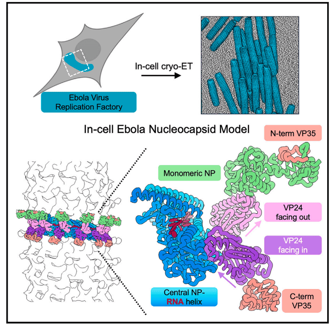
Fig.2 Ebola virus nucleocapsid assembly revealed by in situ cryo-electron tomography(Cell 187:5587–5603.)
Challenges in prevention and treatment include the virus's high lethality, rapid mutation potential, and outbreaks in resource-limited areas. Host factors such as the NPC1 receptor, which serves as an intracellular receptor for Ebola virus entry, and TIM-1, which enhances viral attachment, play crucial roles in pathogenesis; therapeutic strategies targeting NPC1–GP interactions remain exploratory. Vaccine development faces hurdles like ensuring broad protection against multiple strains and long-term immunity. Current vaccines, such as Ervebo (rVSV-ZEBOV-GP), have shown efficacy in ring vaccination strategies, reducing EVD rates by over 95% post-day 10, as evidenced in the 2018-2020 DRC outbreak. However, broader therapeutics targeting conserved interfaces (e.g., NP-VP35) are needed for pan-filovirus coverage.
Key Insights from Recent Studies and abinScience's Supporting Products
From August 2018 to January 2020, ring vaccination established 1,853 rings and vaccinated 265,183 individuals in the eastern DRC. EVD incidence fell abruptly around day 10; among participants disease-free at day 10, EVD onset during days 10–29 was 0.16 per 1,000. No vaccine safety concerns were identified. This approach proved operationally efficient and dose-sparing in insecure contexts.
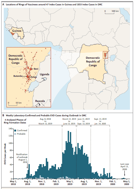
Fig.3 Ring Vaccination Sites in Guinea (2015) and Democratic Republic of Congo (2018–2020).(N Engl J Med 391:2327–2336.)
Note: Observational ring-vaccination evidence coexisted with other public-health interventions; effects should be interpreted in context.
Recent studies provide groundbreaking insights: The NEJM study on DRC's ring vaccination with rVSV-ZEBOV-GP demonstrates its role in curtailing outbreaks. The Cell study uses in situ cryo-ET to reveal intracellular nucleocapsid assembly, uncovering a third NP layer tethered by VP35, offering a structural target for antivirals based on conserved NP–VP35 interfaces. These findings underscore the need for high-quality reagents in research.
Vaccine platforms vary; the rVSV-ZEBOV-GP uses a vesicular stomatitis virus vector for rapid immunity, ideal for acute outbreak control, while adenovirus-based vaccines like Ad26.ZEBOV/MVA-BN-Filo offer durable responses through a prime-boost regimen, suited for stable population immunization.
About abinScience
abinScience offers premium recombinant proteins and antibodies for ZEBOV research, enabling structural, functional, and therapeutic studies. Our products are designed for scientists and distributors seeking reliable tools to accelerate discoveries.
For Research Use Only. Not for use in diagnostic procedures.
Our reagents support diverse lab workflows, such as neutralization assays with GP antibodies, co-immunoprecipitation with NP/VP35 pairs, and pseudovirus systems for viral entry studies, enhancing translational Ebola research.
abinScience Ebola Products
| Type |
Catalog No. |
Product Name |
Protein
|
VK623011 |
Recombinant ZEBOV GP/GP1, 2 Protein, C-His |
| VK623021 |
Recombinant ZEBOV GP1 Protein, C-His |
| VK623031 |
Recombinant ZEBOV GP1 Protein, C-Fc |
| VK623012 |
Recombinant ZEBOV GP1 Protein, N-His |
| VK661012 |
Recombinant ZEBOV VP24 Protein, N-His-SUMO |
| VK095012 |
Recombinant ZEBOV VP40 Protein, N-His-SUMO |
| VK783012 |
Recombinant ZEBOV VP40 Protein, N-His-SUMO |
| VK661022 |
Recombinant ZEBOV VP35 Protein, N-His |
| VK095022 |
Recombinant ZEBOV VP24 Protein, N-His |
| VK783022 |
Recombinant ZEBOV VP40 Protein, N-His |
| Type |
Catalog No. |
Product Name |
Antibody
|
VK623106 |
Research Grade Anti-REBOV/SEBOV/TAFV/ZEBOV GP/Envelope glycoprotein Antibody (ADI-15878) |
| VK623010 |
InVivoMAb Anti-ZEBOV GP/Envelope glycoprotein Antibody (Iv0195) |
| VK623020 |
InVivoMAb Anti-ZEBOV GP/Envelope glycoprotein Antibody (Iv0196) |
| VK623030 |
InVivoMAb Anti-ZEBOV GP/Envelope glycoprotein Antibody (Iv0197) |
| VK623040 |
InVivoMAb Anti-ZEBOV GP/Envelope glycoprotein Antibody (6D8) |
| VK623050 |
InVivoMAb Anti-SEBOV/ZEBOV GP/Envelope glycoprotein Antibody (Iv0198) |
| VK518010 |
InVivoMAb Anti-REBOV/SEBOV/TAFV/ZEBOV NP/Nucleoprotein Antibody (MJ20) |
| VK623023 |
Anti-ZEBOV GP/Envelope glycoprotein Antibody (mAb100) |
| VK623043 |
Anti-ZEBOV GP/Envelope glycoprotein Antibody (GPE118) |
| VK518023 |
Anti-ZEBOV NP/Nucleoprotein Antibody (KZ51) |
| VK661013 |
Anti-ZEBOV VP35/Polymerase cofactor VP35 Antibody (F9) |
| VK783013 |
Anti-ZEBOV VP40/Matrix protein VP40 Antibody (DSTL094) |
| VK623053 |
Anti-ZEBOV GP/Envelope glycoprotein Neutralization Antibody (KZ52) |
| VK661014 |
Anti-ZEBOV VP35 Polyclonal Antibody |
| VK095014 |
Anti-ZEBOV VP24 Polyclonal Antibody |
| VK783014 |
Anti-ZEBOV VP40 Polyclonal Antibody |
| VK623014 |
Anti-ZEBOV GP1 Polyclonal Antibody |
| VK623086 |
Research Grade Anti-ZEBOV GP/Glycoprotein (ANP-015) |
| VK623096 |
Research Grade Anti-ZEBOV GP/Glycoprotein (Zmapp) |
| VK623013 |
Anti-Zaire ebolavirus/ZEBOV GP/GP1, 2 Nanobody (SAA1248) |
For Research Use Only. Not for use in diagnostic procedures.
References
- Muyembe, J.-J., et al. (2024). Ebola Outbreak Response in the DRC with rVSV-ZEBOV-GP Ring Vaccination. N Engl J Med 391:2327–2336. doi:10.1056/NEJMoa1904387
- Watanabe, R., et al. (2024). Intracellular Ebola virus nucleocapsid assembly revealed by in situ cryo-electron tomography. Cell 187:5587–5603. Published Sept 17, 2024; accepted Aug 21, 2024. doi:10.1016/j.cell.2024.08.044
Distribution Opportunities

 中文
中文 English
 한국어
한국어 日本語
日本語 Español
 Français
 Русский


