Release date:
2025-11-17 View count: 738
When performing flow cytometry on peripheral blood or tissue single-cell suspensions, red blood cells (RBCs) are often "invisible troublemakers." Their abundance, small size, lack of nuclei, and absence of target immune epitopes make them prone to masking white blood cell signals, increasing background noise, interfering with gating, and even leading to misinterpretations when staining or performing absolute count measurements. Therefore, removing RBCs is the first step toward obtaining clean and reproducible flow cytometry results. The commonly used method for RBC removal in laboratories is the use of Red Blood Cell Lysis Buffer (also known as ACK Lysis Buffer).
1. Standard Recipe and Lysis Mechanism of Red Blood Cell Lysis Buffer
Table 1: 1× ACK Lysis Buffer
|
Reagent
|
Quantity (for 500 mL)
|
Final Concentration
|
|
NH4Cl
|
4.16 g
|
150 mM
|
|
KHCO3
|
0.5 g
|
10 mM
|
|
Na2EDTA
|
0.02 g
|
0.1 mM
|
Prepare the buffer by dissolving 4.16 g of NH₄Cl, 0.5 g of KHCO₃, and 0.02 g of Na2EDTA in 100 mL of deionized water. Adjust the pH to 7.2, then bring the volume to 500 mL with deionized water. Store at 4°C and bring to room temperature before use.
The core component of the RBC lysis buffer is ammonium chloride. During lysis, ammonium ions (NH₄⁺) cannot pass through the cell membrane, while other ions (such as chloride ions, Cl⁻) can pass freely. This selective permeability creates a difference in ionic concentration across the cell membrane, leading to an osmotic pressure difference. As a result, water diffuses into the RBCs due to this osmotic pressure difference, causing the RBCs to swell and eventually lyse. In contrast, nucleated cells, due to differences in their membrane structure and composition, are more resistant to this osmotic shock and remain intact, enabling efficient separation of RBCs from nucleated cells.
2. Instructions for Use
2.1 Blood Samples
Take 100 µL of fresh anticoagulated blood in a centrifuge tube and add 1:10-1:20 of RBC lysis buffer. Mix thoroughly.
Incubate at room temperature or 4°C for 5-10 minutes until the blood sample becomes transparent.
Centrifuge at 300-500×g for 5 minutes and carefully discard the supernatant using a pipette. (If RBC lysis is incomplete, repeat steps 1 and 2; usually, a small amount of residual RBCs does not significantly affect subsequent experiments.)
Wash by adding 2-5 mL of PBS, HBSS, saline, or serum-free medium to resuspend the pellet. Centrifuge at 300-500×g for 2-3 minutes and discard the supernatant.
Resuspend the cells in an appropriate solution as required by subsequent experiments.
2.2 Tissue Samples
Fresh tissue is digested with trypsin or collagenase to obtain a single-cell suspension, then centrifuge and discard the supernatant.
Add RBC lysis buffer to the cell pellet at a ratio of 1:3-1:10. Gently pipette to mix and incubate for 3-5 minutes. (Specific time should be adjusted based on lysis efficiency.)
Add 5-10 times the volume of PBS, HBSS, saline, or serum-free medium to terminate the lysis.
Centrifuge at 300-500×g for 5 minutes and discard the supernatant.
Resuspend the cells in an appropriate solution for subsequent experiments.
3. Common Issues with RBC Lysis
3.1 How to Assess Lysis Efficiency
Direct Observation: After complete lysis, the solution becomes clear and transparent, as shown in Figure 1.
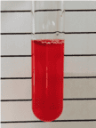
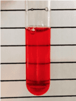
Figure 1. Incomplete RBC Lysis (left) vs. Complete RBC Lysis (right)
Machine Detection: Samples with complete lysis show clear cell populations upon analysis, as demonstrated in Figure 2.
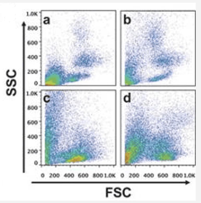
Figure 2. Complete RBC Lysis (a,b) vs. Incomplete RBC Lysis (c,d) Results (Images sourced from the internet)
3.2 Incomplete RBC Lysis
|
Issue
|
Possible Cause
|
Solution
|
|
Incorrect Lysis Buffer
|
• Incorrect concentration (e.g., 10× stock not diluted)
• Improper storage causing inefficacy
• pH outside required range
|
• Ensure 1× working concentration
• Store and use within the effective period
• Accurately adjust pH to 7.2-7.4 when preparing
|
|
Improper Technique
|
• Inadequate or excessive lysis time
• Incomplete mixing after adding lysis buffer
• Insufficient washing steps
• Incorrect centrifuge speed
|
• Optimise lysis time based on sample type
• Gently mix immediately after adding lysis buffer
• Ensure adequate washing volume and steps
• Use 300-500×g centrifuge speed
|
|
Sample-Specific Issues
|
• Poor anticoagulation
• Blood from special disease models or drug treatments
• Mismatch between sample and lysis buffer
• Species differences (e.g., murine RBCs)
|
• Use fresh, properly anticoagulated samples
• Optimise lysis conditions for special samples
• Choose a lysis buffer compatible with the sample
|
3.3 Why Are Mouse & Rat RBCs Harder to Lyse?
|
Possible Cause
|
Solution
|
|
Tougher RBC Membrane: Mouse RBCs have membranes rich in cholesterol and sphingolipids, making them more robust and harder to rupture.
|
Increase lysis buffer volume and extend lysis time.
|
|
Smaller RBC Size & Thicker Membrane: Mouse RBCs are smaller and have a thicker membrane compared to human RBCs, making them more difficult to lyse.
|
Perform multiple rounds of lysis but be cautious not to over-lyse, as this may compromise leukocyte integrity.
|
|
Stronger Antioxidant Defense: Mouse RBCs have a higher resistance to oxidative stress from the lysis buffer, which can hinder lysis.
|
Conduct multiple lysis steps with appropriate care to prevent over-lysis.
|
About Us
As a strategic venture of AtaGenix (established 2011), abinScience was founded in 2023 to deliver premium life science reagents that accelerate discovery. Our flow cytometry antibody products cover commonly used detection markers, with a wide variety to meet the research needs of multiple species (Human, Mouse, Rat, Dog, Hamster, Monkey, etc.). We provide stable and reliable support for scientific research. For more information on abinScience flow cytometry antibodies, please click:
abinScience Flow Cytometry (FACS) Antibodies

 中文
中文 English
 한국어
한국어 日本語
日本語 Español
 Français
 Русский


