Small-diameter vascular grafts (inner diameter <6 mm) remain one of the most persistent unmet needs in cardiovascular surgery. Autologous vessel harvesting is limited by donor-site morbidity, while existing synthetic materials such as ePTFE continue to suffer from chronic inflammation, calcification, and intimal hyperplasia — leaving clinicians without an ideal small-caliber graft solution. Erythrocyte membrane vesicles (EMVs) have attracted considerable interest in nanomedicine due to their inherent biocompatibility and immune-evasion properties. Yet this "don't eat me" paradigm exploits only the passive immunological features of the red blood cell membrane, leaving its active immunomodulatory potential largely untapped.
Phosphatidylserine (PS) is a conserved apoptotic signaling lipid that is normally confined to the inner leaflet of the plasma membrane. When externalized to the outer leaflet, it serves as an "eat me" signal that triggers macrophage recognition and efferocytosis. A research team at Huazhong University of Science and Technology, publishing in Nature Communications, has now introduced a fundamentally new approach: engineering PS-externalized erythrocyte membrane vesicles (PS-EMVs) via a phosphatidylserine externalization method (PEM), functionalizing small-diameter vascular grafts with these vesicles to actively engage the efferocytic pathway and drive immune-mediated vascular tissue regeneration.
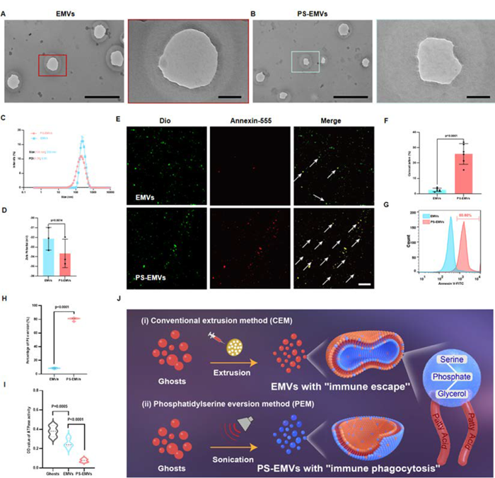
Fabrication and Characterization of PS-EMVs
Red blood cells isolated and purified from rabbit whole blood were processed using the PEM approach — a combination of hypotonic treatment and ultrasonication — to generate PS-EMVs. Transmission electron microscopy (TEM) revealed a well-defined spherical nanovesicle morphology with a mean diameter of approximately 134 nm, notably smaller than the ~230 nm EMVs produced by conventional extrusion (CEM). Zeta potentials were comparable between the two preparations, both carrying a negative surface charge. Fluorescence co-localization analysis confirmed that DiI signal co-localized with Annexin V-FITC-labeled PS at a significantly higher rate in PS-EMVs than in EMVs. Flow cytometry further demonstrated that approximately 80.60% of PS-EMVs had achieved PS externalization, far exceeding the efficiency of CEM-derived EMVs.
The team additionally validated PS exposure using the Anti-Phosphatidylserine Reference Antibody (Bavituximab, RUO) supplied by abinScience (Cat. No. YP862016), achieving high-sensitivity quantification that was fully consistent with Annexin V-based detection and further confirmed the efficiency of PS externalization. Mechanistically, ATPase activity assays showed that PEM treatment substantially reduced the activity of flippase ATP11, the enzyme responsible for maintaining membrane phospholipid asymmetry — establishing this as the central mechanism driving forced PS externalization.
Immunomodulatory Effects of PS-EMVs on Pro-inflammatory Macrophages
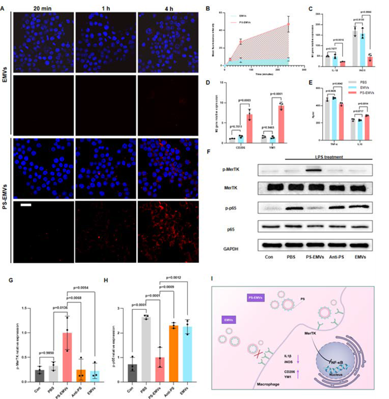
Having established the structural identity of PS-EMVs, the researchers moved to validate their immunomodulatory function in RAW264.7 macrophage cultures. DiI-labeled PS-EMVs and EMVs were introduced separately into macrophage cultures; fluorescence microscopy revealed a time-dependent increase in PS-EMV uptake from 20 minutes through 4 hours, consistently exceeding that of EMVs and confirming that PS externalization substantially enhances efferocytic uptake.
In LPS-induced M1 macrophages, PS-EMV treatment significantly downregulated the M1 marker genes iNOS and IL-1β while upregulating the M2 markers CD206 and YM1. ELISA measurements correspondingly showed decreased secretion of the pro-inflammatory cytokine TNF-α and increased release of the anti-inflammatory cytokine IL-10. Western blot analysis further demonstrated that PS-EMVs activated MerTK phosphorylation and suppressed the NF-κB pathway (evidenced by reduced p65 phosphorylation). Critically, blocking PS with an anti-PS antibody abolished all of these effects, confirming that the PS–MerTK axis is the central signaling node governing this anti-inflammatory response.
Engineering PS-EMV-Functionalized Small-Diameter Vascular Grafts
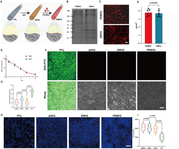
Building on the cell-level findings, the team next integrated PS-EMVs into an implantable device platform. Small-diameter tubular scaffolds were fabricated from polycaprolactone (PCL) by electrospinning, then sequentially coated with a positively charged polydopamine (pDA) layer, followed by electrostatic adsorption of negatively charged PS-EMVs, yielding the final PS-EMV-functionalized vascular graft (PEMVG). SDS-PAGE protein profiling showed that the protein composition of PEMVG was highly consistent with that of EMV-functionalized grafts (EMVG).
Fluorescence labeling confirmed uniform distribution of PS-EMVs across the graft surface, with protein quantification indicating an adsorption density of approximately 3.7 μg/cm². After 10 days of simulated in vitro circulation, about 10.91% of PS-EMVs were retained on the graft surface, suggesting a capacity for sustained signal release. All four graft formulations (PCL, pDAG, EMVG, and PEMVG) exhibited burst pressures well above the physiological blood pressure range (90–120 mmHg). The pDA coating also markedly reduced non-specific BSA protein adsorption and promoted endothelial cell adhesion.
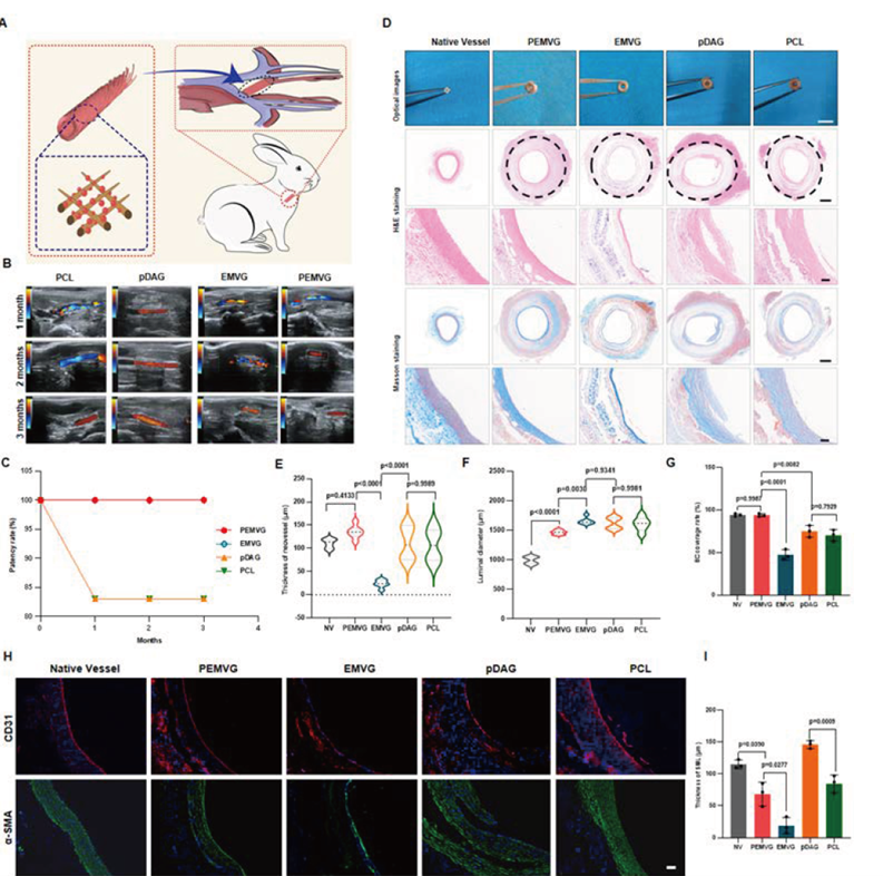
In Vivo Evaluation in a Rabbit Carotid Artery Replacement Model
The four graft formulations were evaluated over three months in a rabbit carotid artery replacement model. Doppler ultrasound follow-up showed that both the PEMVG and EMVG groups maintained a patency rate of 100%, significantly outperforming pDAG (83.3%) and PCL (83.3%). H&E and Masson's trichrome staining revealed that PEMVG neointima was dense, continuous, and uniform in thickness — closely resembling native vessels — whereas the other three groups displayed irregular, loosely organized intimal structures.
Immunofluorescence staining demonstrated markedly superior CD31-positive endothelial cell coverage in the PEMVG group, with α-SMA-positive smooth muscle cell layer thickness most closely approximating that of native arteries. Supplementary analyses further showed that PEMVG significantly reduced M1 macrophage density, increased the proportion of M2 macrophages, and exhibited the strongest resistance to calcification among all groups.
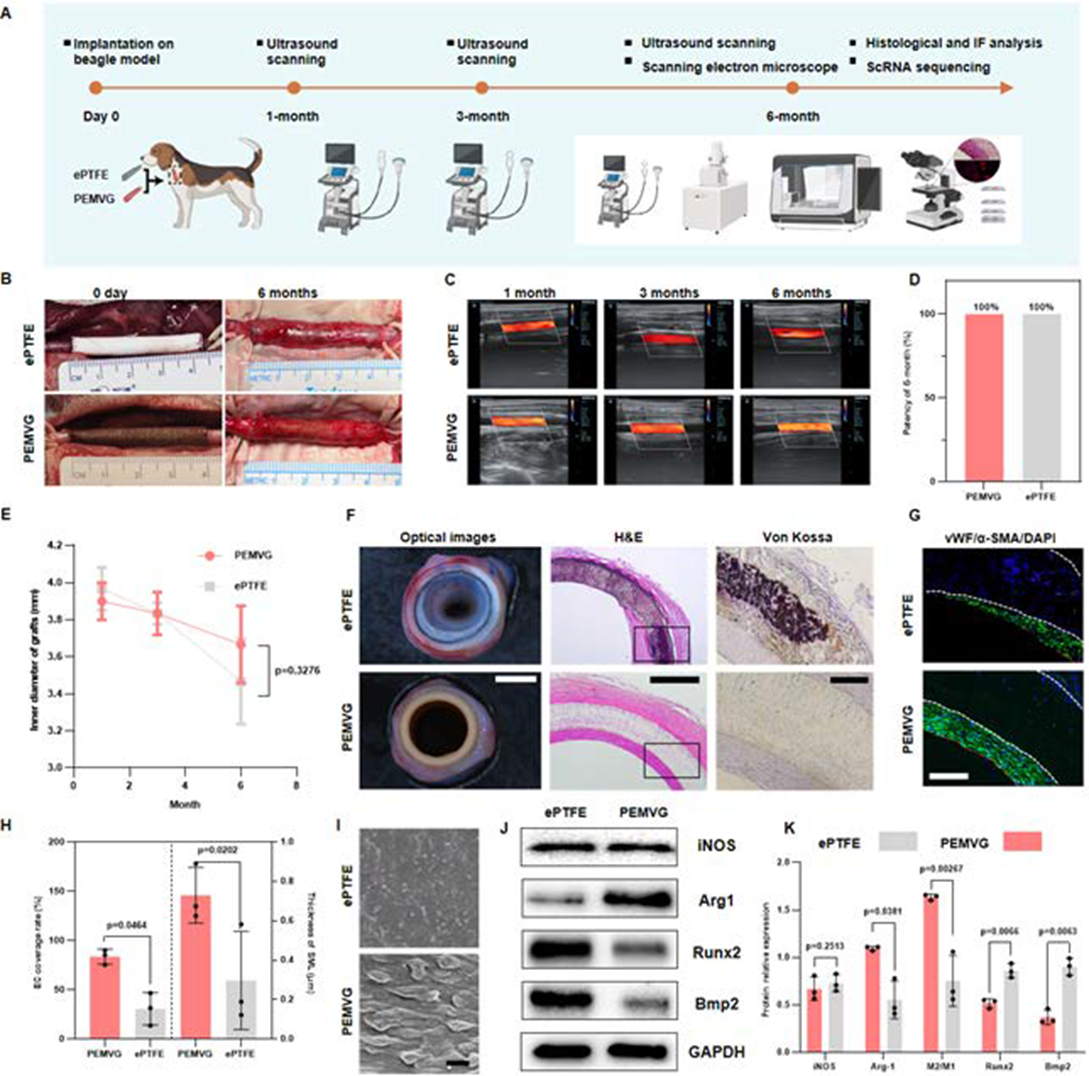
Pre-clinical Translational Evaluation in a Canine Carotid Artery Model
To assess translational potential, the researchers established a beagle carotid artery replacement model, benchmarking PEMVG against a commercial ePTFE graft over six months. Doppler ultrasound showed 100% patency in both groups at the six-month endpoint, with no significant difference in luminal diameter changes. However, detailed post-explant analysis revealed clear divergence between the two: H&E and Von Kossa staining showed that PEMVG lumens were thrombus-free with uniform, non-calcified neointima, whereas ePTFE grafts exhibited prominent calcific deposits.
Co-immunofluorescence staining for vWF and α-SMA confirmed that PEMVG achieved superior endothelial coverage and more physiological smooth muscle layer thickness compared to ePTFE. Scanning electron microscopy (SEM) revealed a complete endothelial monolayer on the PEMVG luminal surface, while ePTFE surfaces showed conspicuous platelet adhesion. Western blot quantification indicated a higher M2/M1 macrophage ratio and lower expression of the osteogenic proteins Runx2 and BMP2 in the PEMVG group.
Single-Cell RNA Sequencing Reveals the Efferocytic Regulatory Mechanism
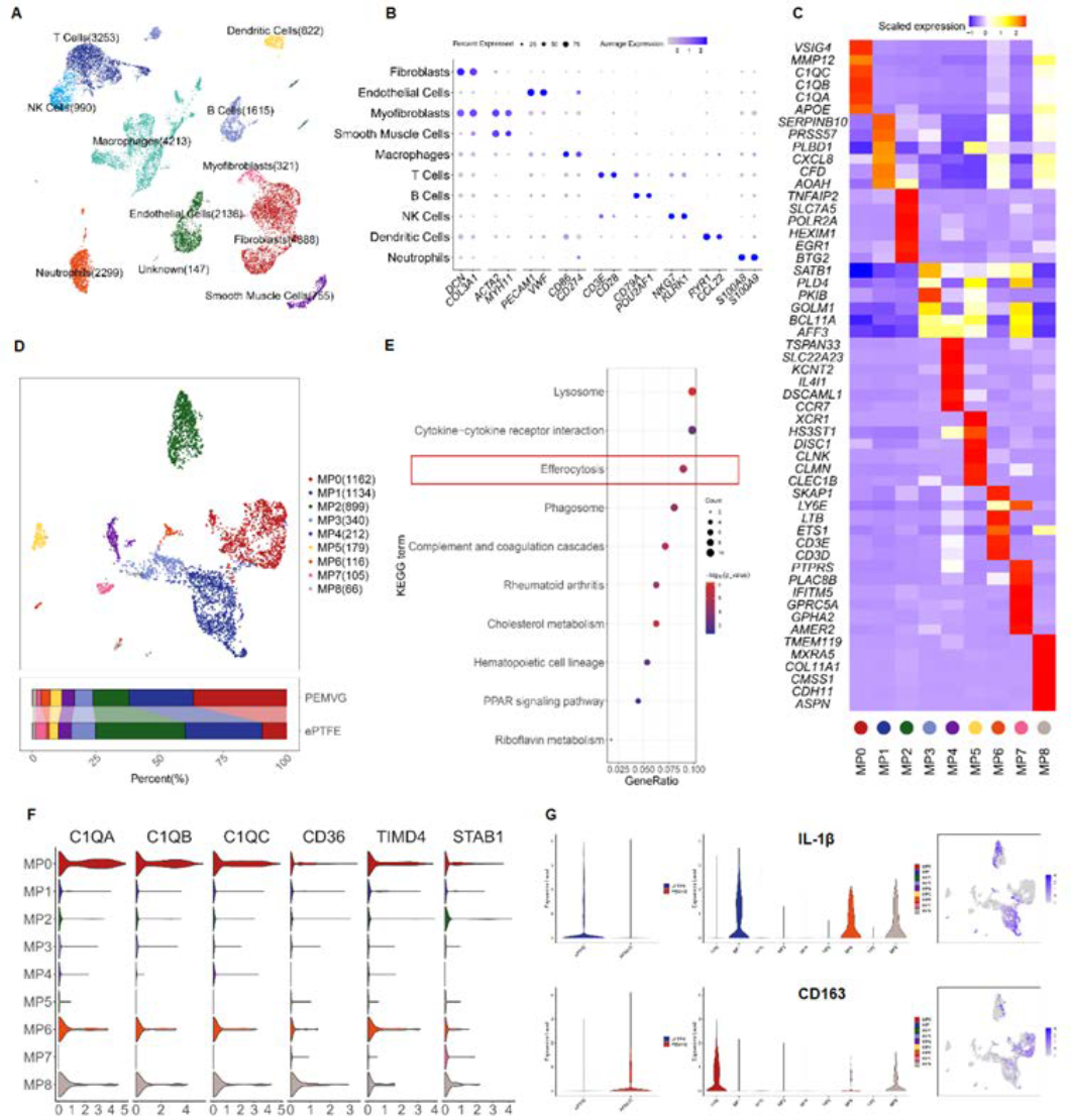
To dissect the molecular basis of PEMVG's immunomodulatory effects, the team performed single-cell transcriptomic sequencing (scRNA-seq) on tissue harvested from explanted canine grafts. Integrative analysis yielded 20,739 high-quality single-cell transcriptomes spanning 11 cell types, including endothelial cells, smooth muscle cells, T cells, B cells, and macrophages. Macrophage subpopulation analysis identified nine distinct clusters (MP0–MP8). Compared to the ePTFE group, the proportion of the MP0 subpopulation was significantly elevated in PEMVG tissue.
KEGG pathway enrichment analysis showed that MP0 was highly enriched for lysosomal, efferocytic, phagosomal, and cholesterol metabolic pathways, with efferocytosis ranking among the top 10 enriched terms. Expression of canonical marker genes — C1QA, C1QB, C1QC, CD36, TIMD4, and STAB1 — confirmed MP0 as a macrophage subset with robust efferocytic activity. Further analysis revealed significant downregulation of IL-1β and upregulation of CD163 within MP0, consistent with a strongly anti-inflammatory, pro-regenerative phenotype. Overlapping expression of LIPA, CD36, and CTSS across the efferocytosis–phagosome–lysosome axis indicated that MP0 orchestrates a complete, sequential efferocytic cascade.
Conclusion
Through an engineering-driven approach, this study achieved active externalisation of phosphatidylserine on the erythrocyte membrane, generating a biointerface with potent efferocytosis-inducing capability. By actively reshaping the local immune microenvironment, PEMVG successfully addressed the twin challenges of thrombosis and calcification that have long plagued small-diameter vascular grafts — marking a paradigm shift from passive "immune shielding" to active "immune-instructed regeneration". This strategy provides a rigorous mechanistic foundation and compelling pre-clinical evidence for the rational design of next-generation cardiovascular biomaterials.
Supported by abinScience
abinScience provided the PS-specific antibody Anti-Phosphatidylserine Reference Antibody (Bavituximab, RUO) (Cat. No. YP862016) for this study. The antibody was used in flow cytometry to quantitatively assess the level of phosphatidylserine exposure on the PS-EMV surface, enabling high-sensitivity verification of PS externalization efficiency and providing critical technical support for downstream immunoprecipitation and protein interaction analyses.
Looking for Tools to Support PS Externalization, Efferocytosis, or Vascular Regeneration Research?
abinScience offers a curated portfolio of antibodies and recombinant proteins targeting phosphatidylserine, macrophage polarization, and immune regulation — validated for flow cytometry, Western blot, immunofluorescence, and ELISA applications.
Email: info@abinscience.com | Tel: +86-27-65523339

 中文
中文 English
 한국어
한국어 日本語
日本語 Español
 Français
 Русский