 中文
中文 English
 한국어
한국어 日本語
日本語 Español
 Français
 Русский
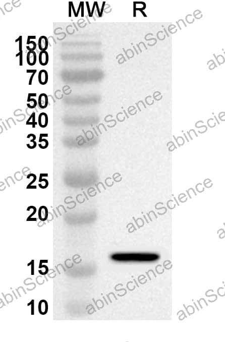
| Catalog No. | VK564014 | ||||||||
|---|---|---|---|---|---|---|---|---|---|
| Species reactivity | Porcine epidemic diarrhea virus (strain CV777) (PEDV) | ||||||||
| Applications | ELISA, IHC, WB | ||||||||
| Host species | Rabbit | ||||||||
| Isotype | IgG | ||||||||
| Clonality | Polyclonal | ||||||||
| Immunogen | E. coli - derived recombinant PEDV Spike glycoprotein (RBD) (Phe617-Val745). | ||||||||
| Target | Spike glycoprotein, S glycoprotein, E2, Peplomer protein, S | ||||||||
| Purification | Purified by antigen affinity column. | ||||||||
| Accession | Q91AV1 | ||||||||
| RRID | Anti-PEDV Spike glycoprotein (RBD) Polyclonal Antibody (abinScience Cat# VK564014, RRID:AB_3723714) |
||||||||
| Form | Liquid | ||||||||
| Storage buffer | 0.01M PBS, pH 7.4, 50% Glycerol, 0.05% Proclin 300. Please refer to the specific buffer information in the hardcopy of datasheet or the lot-specific COA. |
||||||||
| Product Usage Information |
| ||||||||
| Stability and Storage | Use a manual defrost freezer and avoid repeated freeze thaw cycles. Store at 2 to 8°C for frequent use. Store at -20 to -80°C for twelve months from the date of receipt. | ||||||||
| Background | Spike glycoprotein (RBD) is a ~151 kDa protein. S1 region attaches the virion to the cell membrane by interacting with host ANPEP/aminopeptidase N, initiating the infection. Binding to the receptor probably induces conformational changes in the S glycoprotein unmasking the fusion peptide of S2 region and activating membranes fusion. S2 region belongs to the class I viral fusion protein. Under the current model, the protein has at least 3 conformational states: pre-fusion native state, pre-hairpin intermediate state, and post-fusion hairpin state. During viral and target cell membrane fusion, the coiled coil regions (heptad repeats) regions assume a trimer-of-hairpins structure, positioning the fusion peptide in close proximity to the C-terminal region of the ectodomain. RBD is the therapeutic target of casirivimab/imdevimab (REGEN-COV). | ||||||||
| Note | For research use only. |
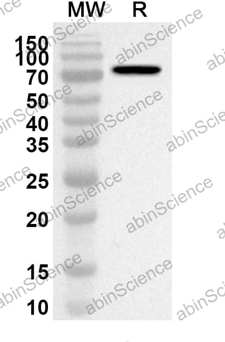
Western blot analysis was performed using anti-PEDV Spike glycoprotein (RBD) polyclonal antibody at 1ug/mL on recombinant PEDV Spike glycoprotein (RBD) (Catalog No. VK564062).


Contact us for custom quotes, bulk requests and any other issues.
Mail: support@abinScience.com
+86-27-65523339
Building C, No. 666, Shen Dun Si Lu, Wuhan, 430206, China


