 中文
中文 English
 한국어
한국어 日本語
日本語 Español
 Français
 Русский

| Catalog No. | HY546317 |
|---|---|
| Description |
Anti-Human CD66e/CEA/CEACAM5 Antibody (RG7813), FITC [RG7813] (HY546317) is a human monoclonal antibody detecting CEACAM5 in FCM. Suitable for Human.
Highlights
|
| Species reactivity | Human |
| Applications | FCM |
| Host species | Human |
| Isotype | IgG1, kappa |
| Clone ID | RG7813 |
| Conjugation | FITC |
| Clonality | Monoclonal |
| Target | Meconium antigen 100, CEACAM5, CEA, CD66e, Carcinoembryonic antigen-related cell adhesion molecule 5, Carcinoembryonic antigen |
| Endotoxin level | Please contact with the lab for this information. |
| Accession | P06731 |
| Form | Liquid |
| Storage buffer | 0.01M PBS, pH 7.4, 0.2% BSA, 0.05% Proclin 300. Please refer to the specific buffer information in the hardcopy of datasheet or the lot-specific COA. |
| Stability and Storage | Store at 4°C for 12 months. Protect from light. Do not freeze. |
| Background | Cell adhesion molecule CEACAM5 (CD66e) is a ~76 kDa protein. Cell surface glycoprotein that plays a role in cell adhesion, intracellular signaling and tumor progression. Mediates homophilic and heterophilic cell adhesion with other carcinoembryonic antigen-related cell adhesion molecules, such as CEACAM6. Plays a role as an oncogene by promoting tumor progression; induces resistance to anoikis of colorectal carcinoma cells. 1. Taheri, M. et al. (2000) The Journal of biological chemistry 275, 26935-43. PMID: 10864933 2. Ordoñez, C. et al. (2000) Cancer research 60, 3419-24. PMID: 10910050 3. Oikawa, S. et al. (1989) Biochemical and biophysical research communications 164, 39-45. PMID: 2803308 |
| Note | For flow cytometric staining, the suggested use of this reagent is 0.5 µg per million cells in 100 µL volume. It is recommended that the reagent be titrated for optimal performance for each application. For research use only. |
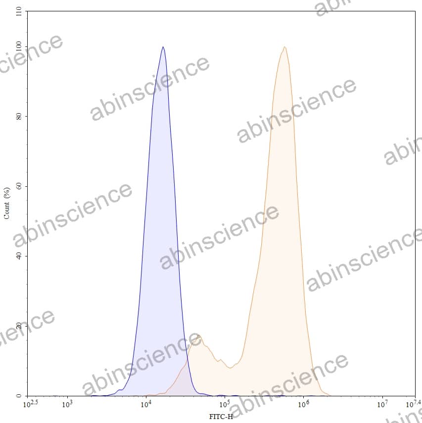
Flow-cytometry using FITC anti-human CD66e antibody. HPAC cells were stained with an irrelevant antibody (Blue Histogram) or an FITC anti-human CD66e monoclonal antibody (Catalog HY546317, Yellow Histogram) at a concentration of 5 µg/ml for 30 mins at RT. After washing, and cells analysed on a NovoCyte Flow Cytometer.
Contact us for custom quotes, bulk requests and any other issues.
Mail: support@abinScience.com
+86-27-65523339
Building C, No. 666, Shen Dun Si Lu, Wuhan, 430206, China


