 中文
中文 English
 한국어
한국어 日本語
日本語 Español
 Français
 Русский

| Catalog No. | HB984016 |
|---|---|
| Description |
Anti-Human CD9 (AT14-012) (HB984016) is a research-grade recombinant antibody targeting CD9. Produced in mammalian cells with native-like glycosylation.
Highlights
|
| Species reactivity | Human |
| Applications | ELISA, Bioactivity: FACS, Functional assay, Research in vivo |
| Host species | Human |
| Isotype | IgG1-kappa |
| Clone ID | AT14-012 |
| Expression system | Mammalian cells |
| Clonality | Monoclonal |
| Target | Motility-related protein, Leukocyte antigen MIC3, TSPAN29, Cell growth-inhibiting gene 2 protein, CD9, MIC3, p24, Tspan-29, MRP-1, 5H9 antigen, Tetraspanin-29, CD9 antigen |
| Endotoxin level | Please contact the lab for this information. |
| Purity | >95% purity as determined by SDS-PAGE. |
| Purification | Protein A/G purified from cell culture supernatant. |
| Accession | P21926 |
| Form | Liquid |
| Storage buffer | 0.01M PBS pH 7.4 Please refer to the specific buffer information in the hardcopy of datasheet or the lot-specific COA. |
| Stability and Storage | Use a manual defrost freezer and avoid repeated freeze thaw cycles. Store at 2 to 8°C for frequent use. Store at -20 to -80°C for twelve months from the date of receipt. |
| Alternate Names | AT14-012, AT14012, AT14012 |
| Background | CD9 antigen is a ~25 kDa protein. Integral membrane protein associated with integrins, which regulates different processes, such as sperm-egg fusion, platelet activation and aggregation, and cell adhesion. Present at the cell surface of oocytes and plays a key role in sperm-egg fusion, possibly by organizing multiprotein complexes and the morphology of the membrane required for the fusion. In myoblasts, associates with CD81 and PTGFRN and inhibits myotube fusion during muscle regeneration. In macrophages, associates with CD81 and beta-1 and beta-2 integrins, and prevents macrophage fusion into multinucleated giant cells specialized in ingesting complement-opsonized large particles. Also prevents the fusion between mononuclear cell progenitors into osteoclasts in charge of bone resorption. 1. Higginbottom, A. et al. (2003) Biochemical and biophysical research communications 311, 208-14. PMID: 14575715 2. Nakazawa, Y. et al. (2008) Blood 112, 1730-9. PMID: 18541721 3. Ikeyama, S. et al. (1993) The Journal of experimental medicine 177, 1231-7. PMID: 8478605 4. Takeda, Y. et al. (2003) The Journal of cell biology 161, 945-56. PMID: 12796480 5. Masellis-Smith, A. et al. (1994) Journal of immunology (Baltimore, Md. : 1950) 152, 2768-77. PMID: 7511626 |
| Note | For research use only. Not suitable for clinical or therapeutic use. |
Detects Recombinant Human CD9 Protein, C-His (Cat No.: HB984021) in indirect ELISA.
SDS-PAGE for Research Grade Anti-Human CD9 (AT14-012).
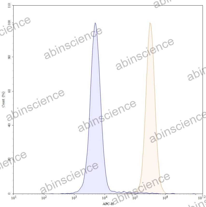
Flow-cytometry using APC anti-human CD9 antibody. HCT-116 cells were stained with an irrelevant antibody (Blue Histogram) or an APC anti-human CD9 monoclonal antibody (Catalog HB984016, Yellow Histogram) at a concentration of 5 µg/ml for 30 mins at RT. After washing, and cells analysed on a NovoCyte Flow Cytometer.
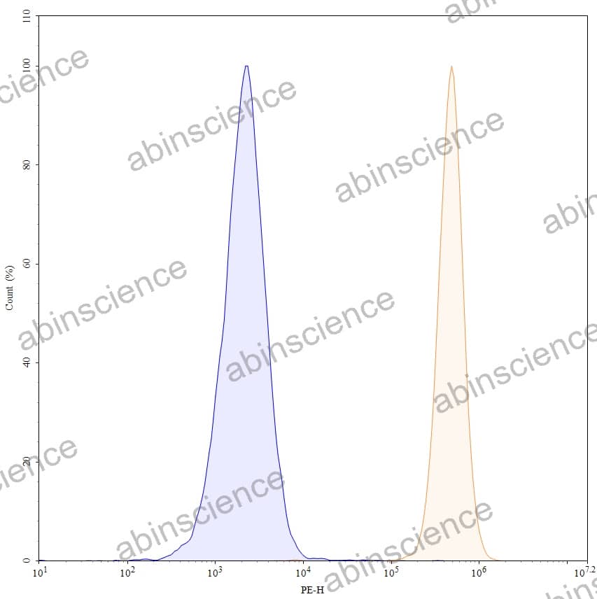
Flow-cytometry using PE anti-human CD9 antibody. HCT-116 cells were stained with an irrelevant antibody (Blue Histogram) or an PE anti-human CD9 monoclonal antibody (Catalog HB984016, Yellow Histogram) at a concentration of 5 µg/ml for 30 mins at RT. After washing, and cells analysed on a NovoCyte Flow Cytometer.
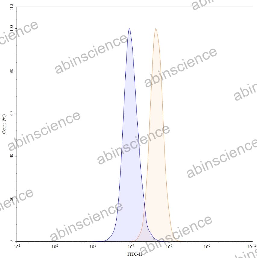
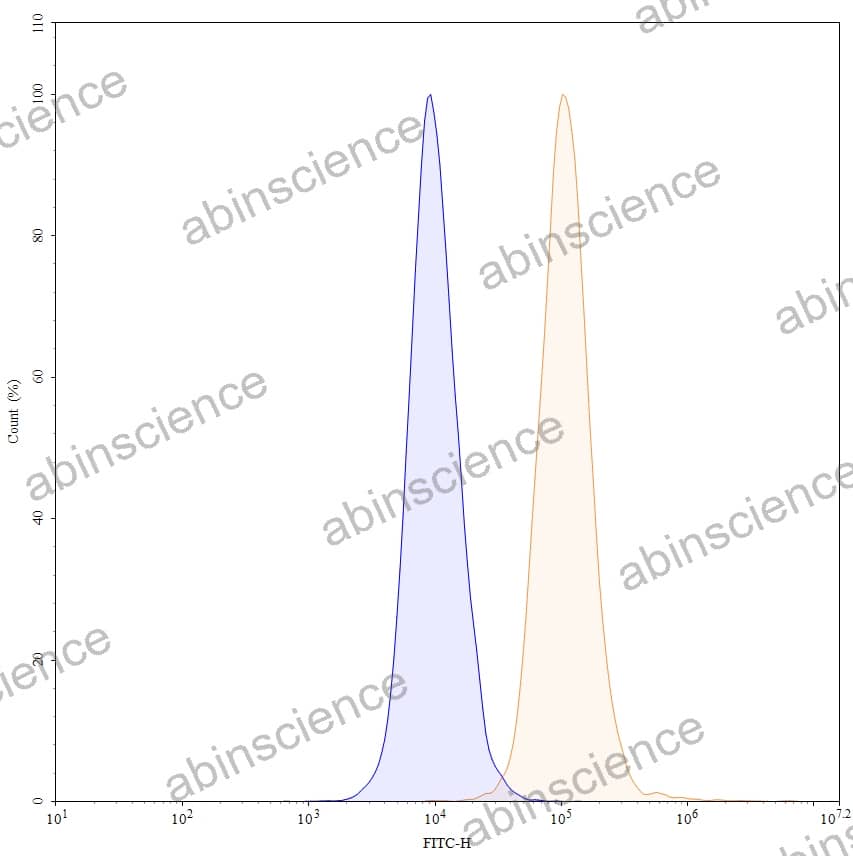
Flow-cytometry using FITC anti-human CD9 antibody. HCT-116 cells were stained with an irrelevant antibody (Blue Histogram) or an FITC anti-human CD9 monoclonal antibody (Catalog HB984016, Yellow Histogram) at a concentration of 5 µg/ml for 30 mins at RT. After washing, and cells analysed on a NovoCyte Flow Cytometer.


Contact us for custom quotes, bulk requests and any other issues.
Mail: support@abinScience.com
+86-27-87433958
Building C, No. 666, Shen Dun Si Lu, Wuhan, 430206, China


