Marburg Virus (MARV) Research Reagents
Marburg virus (MARV) is a filamentous, enveloped, negative-sense single-stranded RNA virus of the family Filoviridae, causing severe hemorrhagic fever with case fatality rates of 25–90% in humans. The virion contains four major structural proteins: glycoprotein (GP, primary attachment and membrane fusion), nucleoprotein (NP, genome packaging), viral protein 35 (VP35, immune antagonist and transcription factor), and VP40 (matrix protein essential for virion assembly). MARV-specific antibodies, recombinant antigens, and diagnostic assays enable rapid outbreak detection, vaccine development, therapeutic antibody discovery, post-exposure prophylaxis, and seroprevalence monitoring in endemic and emerging regions.
Research Use Only (RUO)Not intended for diagnostic or therapeutic procedures.
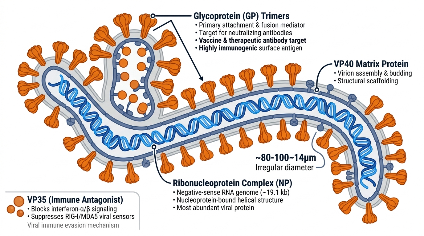
Fig. 1 MARV structure. Glycoprotein (GP) forms characteristic spike-like trimeric clusters on the lipid envelope; VP40 provides matrix scaffolding beneath the membrane layer. Filamentous morphology (branched, looped structures) and pleomorphic particles distinguish MARV from spherical or rod-shaped viruses. Nucleoprotein (NP) complexes the negative-sense RNA genome (~19.1 kb).
abinScience provides recombinant antigens (full-length and domain fragments), monoclonal and polyclonal detection antibodies, and sandwich ELISA kits for MARV research, diagnostics, vaccine development, and therapeutic antibody screening. Products are manufactured by AtaGenix Laboratories under ISO 9001 & ISO 13485. Contact us for strain-specific reagents, validated antibody pairs, and diagnostic assay optimization.
Key Research Targets
Glycoprotein (GP) — Primary Immunogen & Vaccine Target — GP is the major surface antigen and sole target for neutralizing antibodies, mediating viral attachment to host receptors and membrane fusion. GP is highly immunogenic and the primary focus for vaccine development and therapeutic antibody discovery. Monoclonal antibodies targeting GP enable sandwich ELISA assay development, serological diagnosis, rapid lateral flow testing, and viral neutralization assays. Recombinant GP (full-length and mucin-like domain fragments) support vaccine immunogenicity assessment, therapeutic antibody screening, and diagnostic assay optimization.
→ Browse GP products
Nucleoprotein (NP) — Most Abundant Viral Antigen — NP is the most abundant viral protein and serves as the primary serological marker for MARV infection. Antibodies against NP enable rapid detection of infected individuals through ELISA and lateral flow assays. Monoclonal anti-NP antibodies provide high specificity for MARV diagnosis and discrimination from other filoviruses (Ebola, Bundibugyo, Taï Forest). Recombinant NP antigens support serological surveillance, vaccine potency assessment, and convalescent serum characterization.
→ Browse NP products
VP35 & VP40 (Immune Antagonists & Structural Proteins) — VP35 functions as a potent antagonist of innate immunity, blocking interferon-α/β signaling and suppressing viral RNA sensors (RIG-I, MDA5). VP40 provides structural scaffolding and is essential for virion assembly and budding. Antibodies against VP35 and VP40 enable mechanistic studies of viral pathogenesis, host immune evasion strategies, and identification of antiviral drug targets. Support functional studies of immune antagonism unique to filoviruses and comparative virology.
→ Browse VP Protein products
Recommended Applications
Rapid Diagnostic Assay Development & Outbreak Response — GP and NP-specific monoclonal antibodies enable sandwich ELISA, lateral flow immunochromatography, and multiplex immunoassay development for point-of-care diagnosis during hemorrhagic fever outbreak response. Essential for rapid case identification, isolation, and contact tracing in endemic regions.
Vaccine Development & Immunogenicity Assessment — Recombinant GP and NP antigens serve as vaccine immunogens, benchmarks, or quality control standards. Monoclonal antibody panels enable quantification of serological responses, assessment of strain-specific protection, potency testing of vaccine candidates, and DIVA assay development (Differentiate Infected from Vaccinated Animals).
Therapeutic Antibody Discovery & Post-Exposure Prophylaxis — GP-targeting monoclonal antibodies enable high-throughput screening of neutralizing antibody libraries and human convalescent serum repositories. Support identification of therapeutic candidates for post-exposure prophylaxis (within 14 days of exposure) and treatment of acute hemorrhagic fever infections.
Seroprevalence & Epidemiological Surveillance — GP and NP-specific ELISA kits enable large-scale serological surveys in bat reservoirs (fruit bats, insectivorous bats), non-human primates, and human populations. Critical for understanding MARV ecology, zoonotic transmission patterns, geographical risk, and burden of human exposure in endemic regions.
How to Select the Right Product
Step 1: Identify your research objective — Vaccine development requiring immunogenic antigens? Rapid diagnostic assay for outbreak response? Therapeutic antibody discovery from screening libraries? Serological surveillance for epidemiological mapping?
Step 2: Select antigen type — Choose full-length GP or NP for broad reactivity, vaccine immunogenicity assessment, and serological assays; domain fragments (GP1/GP2 subunits, mucin-like domain, nucleoprotein core domain) for epitope-specific diagnostics, therapeutic antibody screening, and reduced background in multiplex assays.
Step 3: Choose antibody format — Use monoclonal pairs for sandwich ELISA with high specificity, strain discrimination, and assay manufacturability; polyclonal for Western blot, immunofluorescence, immunoprecipitation, and capture assays requiring broad viral variant recognition.
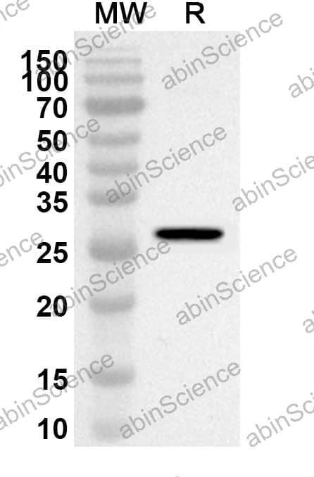
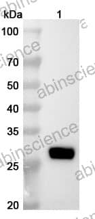
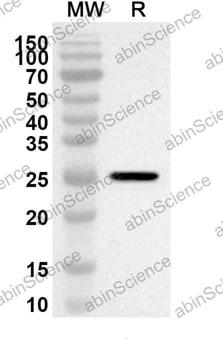
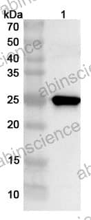
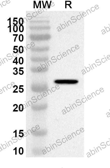
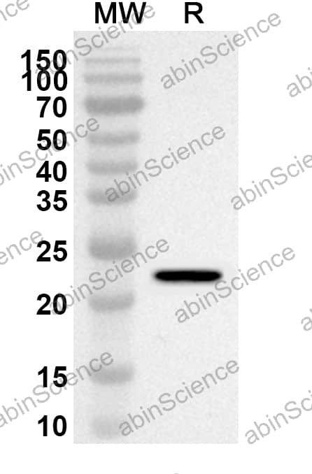
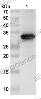
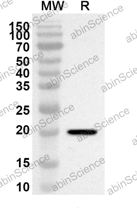
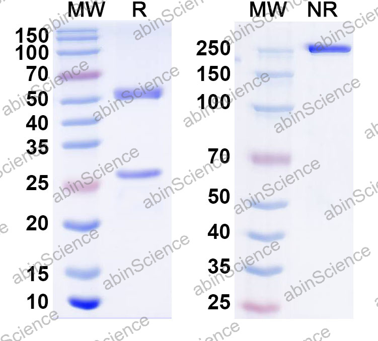
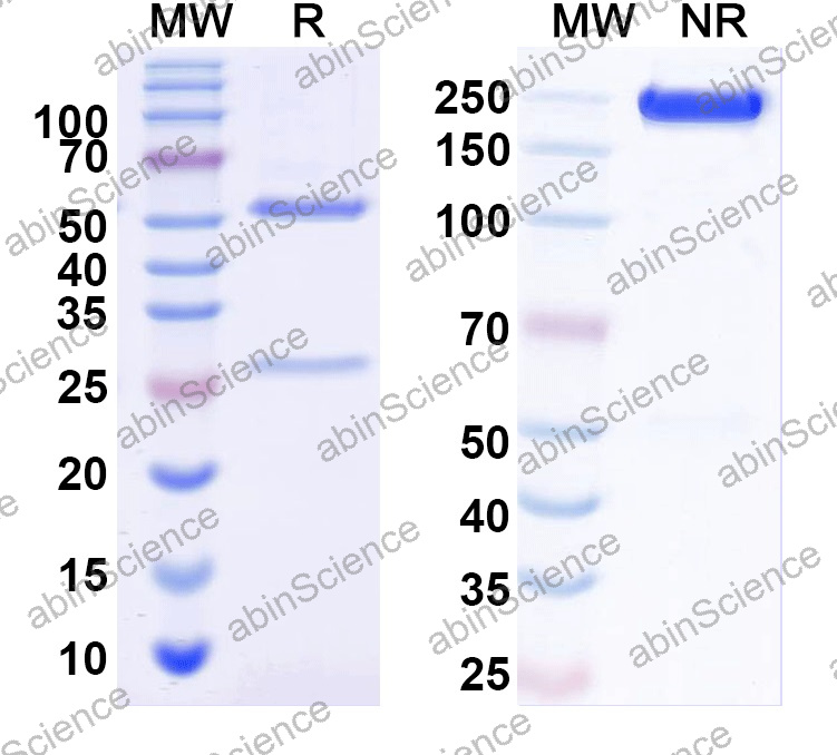
Step 4: Specify quality parameters — Request endotoxin testing (≤0.1 EU/μg for cell-based assays), purity verification (≥90% by SDS-PAGE), lot-specific Certificate of Analysis (CoA), and application-specific recommendations. Request pre-validated antibody pairs for sandwich ELISA if available.
Documents & QC Data
Download product datasheets, Certificate of Analysis (CoA), SDS-PAGE verification data, endotoxin testing reports, and application notes for each MARV reagent. All documents are lot-specific and include purity verification, endotoxin levels, recommended usage concentrations for ELISA/WB/IF, and shelf-life validation data.
FAQs
What reagent types are available for MARV research?
We offer recombinant antigens (full-length and domain fragments: GP, GP1/GP2, GP mucin-like domain, NP, VP35, VP40), monoclonal & polyclonal detection antibodies, and sandwich ELISA kits for quantitative measurement and large-scale serological surveys.
Are your MARV antigens suitable for diagnostic kit development?
Yes. All antigens are produced under ISO 9001/13485 quality systems with verified low endotoxin (≤0.1 EU/μg), high purity (≥90% by SDS-PAGE), and documented batch-to-batch consistency for assay manufacturing and regulatory compliance.
What expression systems do you use for MARV proteins?
HEK293/CHO systems for glycoproteins (GP, post-translational modifications required); E. coli for nucleocapsid proteins and simpler structural antigens (NP, VP35 core regions). Custom expression available for specialized applications.
How do I choose between monoclonal and polyclonal antibodies?
Monoclonal antibodies provide high epitope specificity, excellent for sandwich ELISA pairs, strain discrimination, and therapeutic screening. Polyclonal antibodies offer broader recognition across MARV variants and species, ideal for Western blot, immunofluorescence, and capture assays.
Do you offer pre-validated antibody pairs for sandwich ELISA?
Yes — we provide characterized capture/detection antibody pairs with recommended concentrations and coat/block conditions for MARV sandwich ELISA development. Contact us for pairing optimization, custom combinations, and volume pricing for diagnostic kit manufacturing and high-throughput screening.
References
1. Feldmann H, Geisbert TW. Ebola haemorrhagic fever. Lancet. 2011;377(9768):849-862. doi:10.1016/S0140-6736(10)60927-6
2. Kuhn JH, Becker S, Ebihara H, et al. Proposal for a revised taxonomy of the family Filoviridae: Classification, names of taxa and place of Marburg-like viruses. Arch Virol. 2010;155(12):2061-2067. doi:10.1007/s00705-010-0814-x
3. Towner JS, Sealy TK, Khristova ML, et al. Newly discovered Marburg virus associated with multiple deaths in Uganda. J Virol. 2008;82(11):5377-5388. doi:10.1128/JVI.02639-07
4. Volchkov VE, Becker S, Volchkova VA, et al. GP mRNA of Ebola virus is edited by the viral polymerase and by cellular editing endonucleases. Proc Natl Acad Sci USA. 1995;92(23):10957-10961. doi:10.1073/pnas.92.23.10957
5. Geisbert TW, Hensley LE. Catalytic properties of nanoparticles influence their suitability as vaccine adjuvants and immunological agents. Acc Chem Res. 2013;46(10):2441-2450. doi:10.1021/ar300135y
All products manufactured by AtaGenix Laboratories under ISO 9001 & ISO 13485. Contact us for custom antibody development, strain-specific reagent panels, validated antibody pairs, and specialized filovirus research services.


 中文
中文 English
 한국어
한국어 日本語
日本語 Español
 Français
 Русский