Background & Research Objectives
OMIP-023 is designed to systematically dissect the major subsets and activation status of human peripheral blood leukocytes in a single tube via 10-color flow cytometry. It systematically addresses the limitations of conventional multi-tube assays—excessive sample consumption, low detection efficiency, and fragmented data integration—making it ideal for both basic research and clinical settings where comprehensive immune insights are needed with limited sample volumes.
Panel Design: Multiparametric Reagent Reutilization
The core innovation of OMIP-023 lies in fluorescence channel multiplexing to maximize information density per experiment. CD8, CD14, and CD19 are co-labeled in the FITC channel, with distinct cell populations differentiated by scatter properties and other lineage markers. Meanwhile, CD16 and CD56 share the PE-Cy7 channel, enabling simultaneous identification of neutrophils, NK cells, NKT cells, and monocyte subsets. This design allows for systematic characterization of complex leukocyte networks in one single assay.
Sample Type & Optimized Experimental Workflow
Optimized for fresh EDTA-anticoagulated peripheral blood and umbilical cord blood, OMIP-023 adopts a whole-blood protocol post-red blood cell lysis—eliminating the interference of cell separation on immune cell phenotype and activation status. Antibody titration optimization and extended incubation time reduce reagent usage while ensuring stable staining, laying the foundation for reliable detection of low-expression activation markers.
The power of OMIP-023 stems from its streamlined yet robust antibody-fluorochrome combination. By leveraging innovative multiplexing strategies (e.g., simultaneous detection of CD8, CD14, and CD19 in the FITC channel), it integrates 13 key antibodies into a 10-color system, enabling in-depth and systematic analysis of all major peripheral blood leukocyte subsets and their functional subpopulations in a single run.
1. OMIP-023 Panel
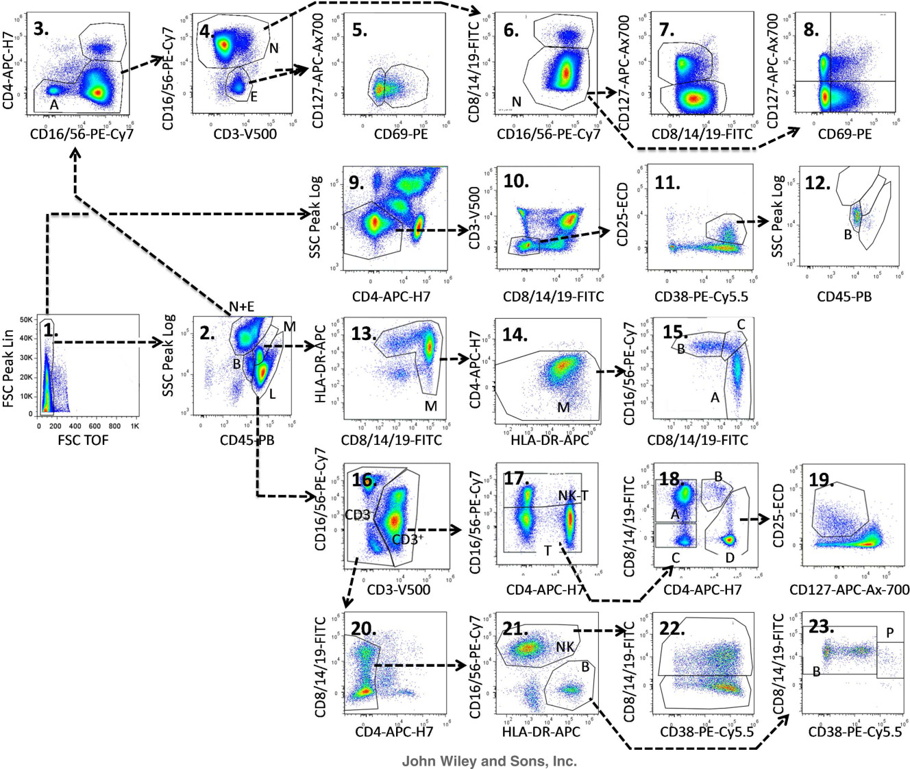
2. Gate Logic and Experimental Results
1
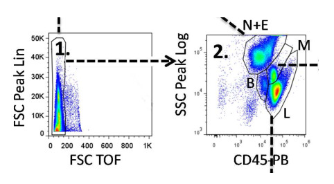
Figures 1-2: Basic Gating
Figure 1: Debris exclusion.
Figure 2: Gating via CD45/SSC to identify lymphocytes (L), monocytes (M), basophils (B), and neutrophils + eosinophils (N+E).
2
Figures 3-8: Analysis of Neutrophils (N) and Eosinophils (E) (N+E population in Figure 2)
Figure 3: Exclusion of CD4+ signals.
Figure 4: CD16++ for neutrophils; CD16- for eosinophils.
Figure 5: CD69/CD127 analysis for eosinophil activation status.
Figure 6: Exclusion of CD8+ signals to isolate pure neutrophils.
Figures 7-8: Assessment of neutrophil activation status.
3
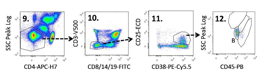
Figures 9-12: Basophil Analysis (Direct analysis, not the B population in Figure 2)
Figure 9: Exclusion of CD4+ cells and selection of SSC-low populations.
Figure 10: Exclusion of CD8+ T cells, CD14+ monocytes, CD19+ B cells, and CD3+ T cells.
Figure 11: CD38hiCD25+ to define basophils.
Figure 12: CD45/SSC confirmation (consistent with basophil localization in Figure 2).
4
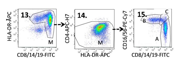
Figures 13-15: Monocyte Subset Analysis (M population in Figure 2)
Figure 13: Exclusion of CD8-HLA-DR- populations.
Figure 14: Exclusion of CD4+ populations.
Figure 15: CD14/CD16 to distinguish monocyte subsets—classical (A: CD14++, CD16-), non-classical (B: CD14+, CD16++), and intermediate (C: CD14++, CD16+).
5
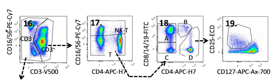
Figures 16-19: Lymphocyte and Subset Analysis (L population in Figure 2)
Figure 16: Gating of CD3+ T cells.
Figure 17: Identification of NKT cells and T cells.
Figure 18: Differentiation of CD4+ T cells and CD8+ T cells.
Figure 19: CD25+CD127- to gate regulatory T cells (Tregs).
6
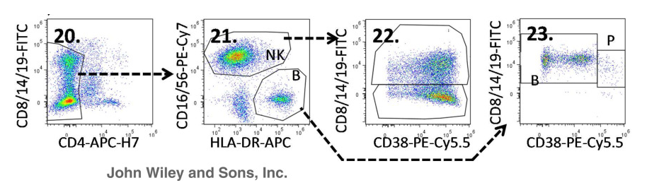
Figures 20-23: Non-T Cell Subset Analysis (CD3- population in Figure 16)
Figure 20: Exclusion of CD4+ signals.
Figure 21: NK cells (CD16+CD56+) and plasma cells (CD16/CD56-HLA-DR++).
Figure 22: NK cell subdivision into CD8+ NK and CD8- NK subsets.
Figure 23: Plasma cell subdivision into CD38-high plasma cells and CD38-variant B cells.
3. Panel Interpretation
1. Simplified Multiparametric Immunophenotyping
OMIP-023 enables detailed phenotyping of major leukocyte subsets (T cells, B cells, monocytes, NK cells) in one experiment. By combining lineage markers (CD3, CD8, CD4, CD19) with activation markers (CD25, CD127, HLA-DR), it assesses both cell composition and activation status—reducing the need for multi-tube assays, saving time, and minimizing sample usage. This is particularly valuable for studies involving pediatric samples or rare diseases with limited sample availability.
2. Innovative Fluorochrome and Marker Combination
Unlike traditional panels relying on single markers per channel, OMIP-023 innovatively uses multiple markers in a single fluorescence channel to boost throughput without compromising specificity. For example, CD14, CD19, and CD8 are co-labeled in the same channel, enabling clear differentiation of monocytes, B cells, and T cells. This strategy maximizes channel utility, allowing analysis of more immune cell types with limited samples.
3. Tailored Gating Strategy
OMIP-023’s gating logic leverages scatter and fluorescence signals to precisely distinguish distinct cell subsets—including rare or hard-to-detect populations like Tregs and NKT cells. It also incorporates specialized gating for granulocytes (eosinophils, neutrophils, basophils), which are often challenging to differentiate in standard flow cytometry assays.
4. Applicable Research Directions
Dynamic monitoring of immunotherapy efficacyt Tumor immune microenvironment analysis Autoimmune disease research Immune signature characterization of inflammatory diseases Hematopoietic and immune development research
5. Conclusion
OMIP-023 effectively overcomes the core challenges of traditional leukocyte phenotyping—high sample demand, low detection efficiency, and incomplete data coverage—through innovative fluorescence channel multiplexing, hierarchical marker design, and refined gating logic. This single-tube assay enables simultaneous detection of leukocyte subsets and their activation status, balancing efficiency, cost-effectiveness, and precision. It meets the standardization needs of clinical diagnosis while satisfying the demand for in-depth immune cell analysis in basic research, providing robust technical support for mechanistic studies, diagnostic marker screening, and therapeutic development of immune-related diseases. OMIP-023 drives immune analysis toward standardization, efficiency, and refinement.
Get OMIP-023 Compatible Flow Cytometry Antibodies
abinScience provides validated flow cytometry antibodies covering key targets in this panel, supporting your leukocyte subsets research
References
[1] Bocsi J, Melzer S, Dähnert I, Tárnok A. OMIP-023: 10-color, 13 antibody panel for in-depth phenotyping of human peripheral blood leukocytes. Cytometry A. 2014 Sep;85(9):781-4.

About Us
As a strategic venture of AtaGenix (established 2011), abinScience was founded in 2023 to deliver premium life science reagents that accelerate discovery. Our flow cytometry antibody products cover commonly used detection markers, with a wide variety to meet the research needs of multiple species (Human/Mouse/Rat/Dog/Hamster/Monkey, etc.). We provide stable and reliable support for scientific research.
Explore abinScience Flow Cytometry Antibodies


 中文
中文 English
 한국어
한국어 日本語
日本語 Español
 Français
 Русский