Challenges in Analyzing Antigen-Specific T Cell Function
In the study of infections, vaccines, or tumor immunology, antigen-specific T cells are typically present at very low frequencies and exhibit high functional heterogeneity. Conventional single-cytokine assays often fail to capture the co-expression patterns of cytokines comprehensively, thereby limiting an accurate assessment of T cell polyfunctionality and the quality of immune responses.
Systematic Integration of Multilayered Functional Markers
OMIP-014 integrates seven functional markers—core cytokines (IFN‑γ, IL‑2, TNF‑α) and extended markers (CD107a, CD40L)—into a 12‑color intracellular staining panel. It supports simultaneous multifunctional profiling of both CD4+ and CD8+ T cells, extending beyond Th1/Th2 cytokines to include degranulation and B‑cell helper markers for broader immune characterization.
Validated and Standardized Workflow
OMIP-014 features a fully optimized and standardized workflow covering antibody titration, stimulation, staining, and analysis. Key refinements in CD40L staining and MIP‑1β detection enhance signal‑to‑noise and reproducibility. The protocol is validated for reliable use in GCLP‑compliant clinical sample testing.
Accurate characterization of antigen-specific T cell polyfunctionality is critical for evaluating immune protection efficacy and disease progression. It plays a vital role in vaccine assessment, infection immunology, and immunotherapy monitoring. OMIP-014 is a comprehensively optimized and validated 12-color intracellular cytokine staining panel, designed specifically for profiling multifunctional antigen-specific CD4+ and CD8+ T cells in human peripheral blood mononuclear cells (PBMCs). Through its layered marker combination and stringent operational procedures, it enables high-specificity, reproducible analysis of low-frequency T cell responses, providing researchers with a reliable tool for multifunctional T cell characterization.
1. OMIP-014 Panel
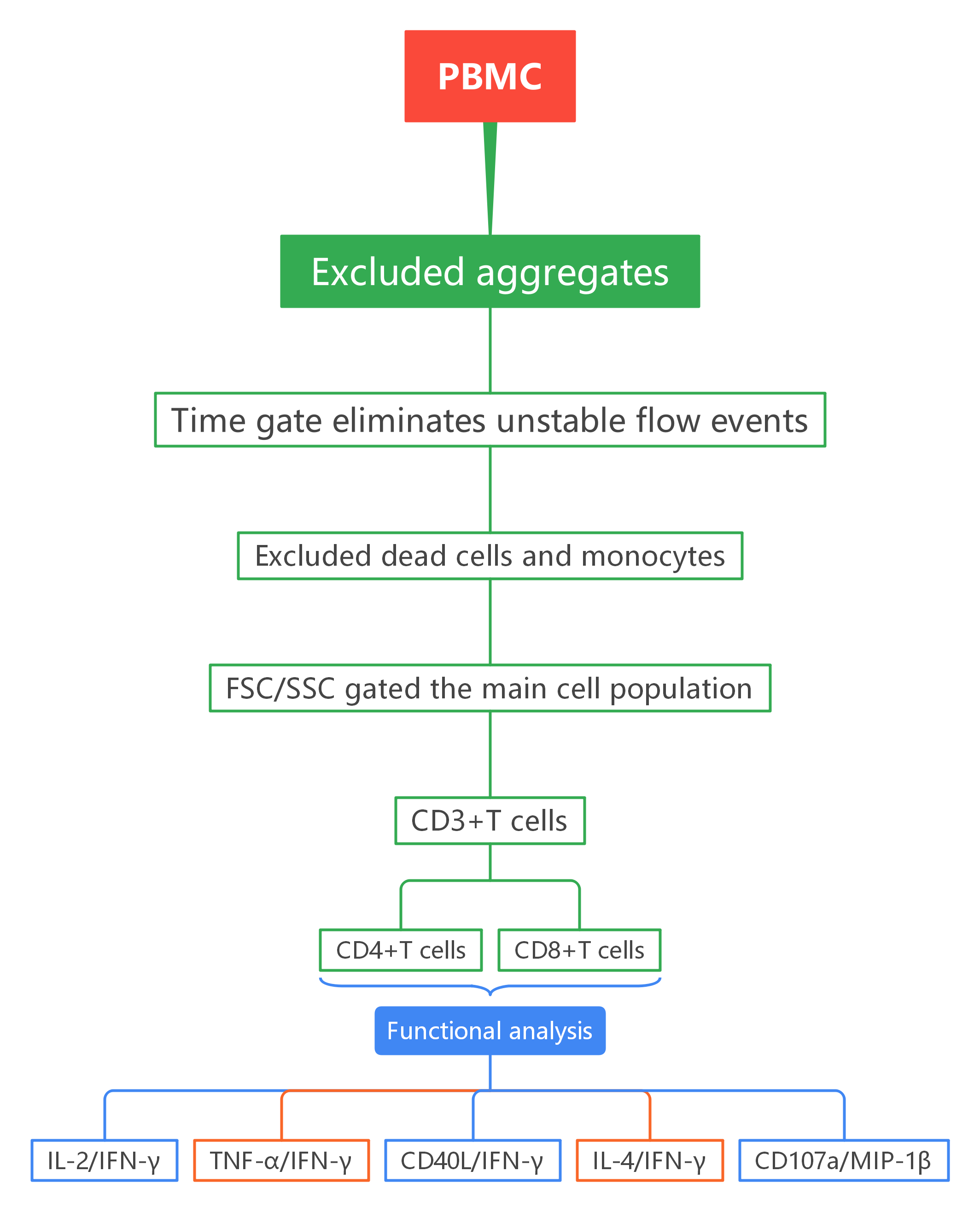
2. Gating Logic
1
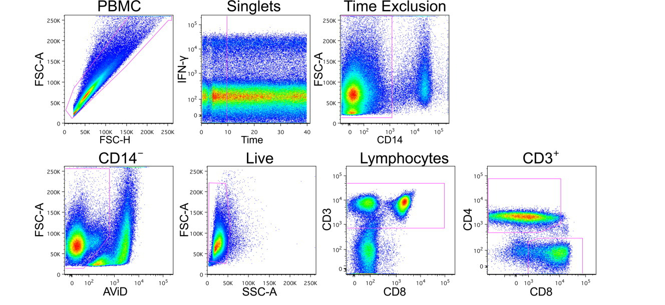
Live T-Cell Isolation
PBMCs,excluded aggregates, unstable flow events, dead cells and monocytes.
2
T Cell Subsetting
Within live lymphocytes, CD3+ T cells were gated, followed by separation into CD4+ and CD8+ subsets.
3
Independent Gating for Functional Markers
Each functional marker (e.g., IFN-γ, IL-2, TNF-α) was gated independently with a high threshold within CD4+ and CD8+ T cell populations to suppress background signal.
4
Boolean Combination Analysis
Based on the independent gates, Boolean logic was applied to identify cell subsets co-expressing different functional markers, enabling systematic dissection of T cell polyfunctionality.
3. Experimental Results
1). PBMC, excluded aggregates, unstable flow events, dead cells and monocytes, and the main cell population were gated. CD4+ and CD8+ T cells were subsequently gated based on CD3, CD4, and CD8 expression.
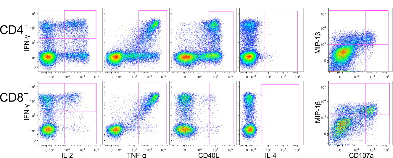
2). Within CD4+ and CD8+ T cells, different functional populations were identified using IFN-γ, IL-2, TNF-α, IL-4, MIP-1β, CD40L (CD154), and CD107a.
Key Analytical Features:
① Independent gating combined with Boolean analysis: Each functional marker is first gated independently (IL-2+, IFN-γ+, TNF-α+, etc.) without considering co-expression, establishing an objective and unbiased definition. Boolean gating then systematically identifies all possible polyfunctional subsets. This ensures standardization and reproducibility.
② High-threshold gating to suppress background noise: Functional gates are set with relatively high thresholds. This approach intentionally sacrifices some sensitivity but greatly enhances specificity, ensuring that low-frequency signals detected in antigen-stimulated samples are genuine and not due to background noise.
4. Panel Interpretation
4.1 Clear biological rationale:
IFN-γ, IL-2, and TNF-α are defined as "core cytokines" with the highest priority, ensuring robust detection of central T cell effector functions. MIP-1β, CD107a, IL-4, and CD40L serve as "functional expansion modules" for deeper characterization of subset properties (e.g., cytotoxicity, B cell help, Th1/Th2 polarity). This hierarchical design allows researchers to reliably assess core functions while flexibly exploring more complex biological questions.
4.2 Optimized experimental workflow
CD40L is stained intracellularly rather than via surface staining during co-culture, as surface staining is incompatible with Brefeldin A. Storing cells overnight at 4°C after stimulation—instead of freezing—significantly improves the signal-to-noise ratio for MIP-1β staining.
4.3 Quantitative use of staining index
The staining index is applied as a quantitative metric to determine optimal antibody titration concentrations. By quantifying the separation between positive and negative populations, it precisely identifies the point of highest signal-to-noise ratio, effectively preventing background fluorescence increase due to antibody excess and ensuring high data quality from the start.
5. Applications
Vaccine immunogenicity evaluation T cell response monitoring in infectious diseases Functional analysis of tumor antigen-specific T cells Mechanism studies in autoimmune diseases Immunotherapy efficacy assessment Mucosal and tissue immune response analysis Clinical sample testing in GCLP environments
6. Conclusion
In summary, OMIP-014 represents a highly refined detection panel that transforms the complex task of T cell polyfunctional analysis into a standardized and reliable workflow. Through its pre-optimized antibody combination, tailored procedures for critical steps (such as CD40L staining and MIP-1β detection), and a specificity-focused data analysis strategy, it provides researchers with an "out-of-the-box" toolkit.
Get OMIP-014 Compatible Flow Cytometry Antibodies
abinScience provides validated flow cytometry antibodies covering key targets in this panel, supporting your Antigen-Specific T Cell Function research
References
[1] De Rosa SC, Carter DK, McElrath MJ. OMIP-014: validated multifunctional characterization of antigen-specific human T cells by intracellular cytokine staining. Cytometry A. 2012 Dec;81(12):1019-21.

About Us
As a strategic venture of AtaGenix (established 2011), abinScience was founded in 2023 to deliver premium life science reagents that accelerate discovery. Our flow cytometry antibody products cover commonly used detection markers, with a wide variety to meet the research needs of multiple species (Human/Mouse/Rat/Dog/Hamster/Monkey, etc.). We provide stable and reliable support for scientific research.
Explore abinScience Flow Cytometry Antibodies

 中文
中文 English
 한국어
한국어 日本語
日本語 Español
 Français
 Русский