In flow cytometry, non-specific staining constitutes a primary confounding factor compromising data accuracy, with Fc receptor-mediated non-specific antibody binding serving as the most prevalent cause. As a crucial procedural step in flow cytometry, Fc receptor blocking substantially mitigates non-specific binding, thereby optimizing the signal-to-noise ratio and bolstering the reliability of experimental outcomes.
I. Biological Foundations of Fc Receptors and Antibodies
To elucidate the function of Fc receptor blocking, it is imperative to first delineate the structural properties of antibodies, the characteristics of Fc receptors, and their mechanisms of interaction.
1. Functional Domains of Antibodies
A canonical IgG antibody is composed of two heavy chains and two light chains covalently linked via disulfide bonds, configuring a symmetrical "Y"-shaped architecture. Its functional domains are bifurcated into two core regions:
● Fab Region (Fragment Antigen-Binding): Comprising the variable domains of both the heavy and light chains (VH/VL), this region is responsible for the specific recognition and binding of target antigen epitopes, functioning as the central domain for target-specific detection in flow cytometry.
● Fc Region (Fragment Crystallizable): Constituted by the constant domains of the heavy chains (CH2/CH3), this region is independent of antigen recognition. Its primary role is to mediate downstream immune effector functions, serving concomitantly as the recognition and binding locus for Fc receptors
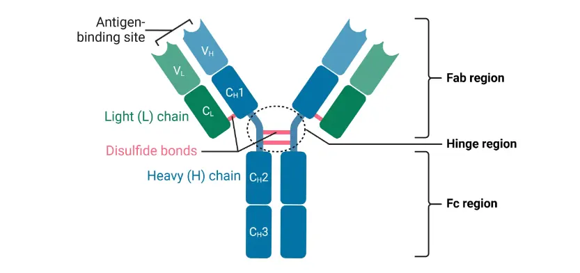
Figure 1. Antibody Structure
2. Classification and Expression of Fc Receptor Families
Fc receptors (FcRs) constitute a class of transmembrane glycoproteins broadly expressed on the surfaces of immune cells. Their principal physiological function involves the specific recognition of the antibody Fc region, thereby bridging humoral and cellular immunity. They mediate critical immune effector functions, including the phagocytic clearance of antigen-antibody complexes, the transduction of activation or inhibitory signals within immune cells, and antibody-dependent cellular cytotoxicity (ADCC).
Categorized by the specific antibody isotype (immunoglobulin subclass) they recognize, Fc receptors are classified into five primary families:
● Fcγ Receptor Family (FcγR): Recognizes and binds IgG antibodies. This family is the most critical in immunological assays, exhibiting the highest correlation with non-specific binding, and corresponds to the most frequently utilized antibody isotype in flow cytometry and immunoassays.
● Fcε Receptor Family (FcεR): Recognizes and binds IgE antibodies. It is primarily subdivided into the high-affinity FcεRI and the low-affinity FcεRII (CD23), acting as the central receptors in mediating Type I hypersensitivity reactions.
● Fcα Receptor Family (FcαR): Recognizes and binds IgA antibodies. The core subtype is FcαRI (CD89), which predominantly mediates effector functions associated with mucosal immunity.
● Fcα/μ Receptor (Fcα/μR): Capable of recognizing both IgA and IgM antibodies, this receptor plays a significant role in intestinal mucosal immunity and B cell activation.
● Fcμ Receptor (FcμR): Specifically recognizes and binds IgM antibodies. It is predominantly expressed on the surfaces of B cells and macrophages, participating in IgM-mediated immunomodulation.
Among all Fc receptor families, the FcγR family serves as the primary catalyst for non-specific binding in antibody-based detection assays. This phenomenon occurs because the overwhelming majority of research-grade detection antibodies—including flow cytometry antibodies, primary and secondary antibodies for immunohistochemistry (IHC), and ELISA detection antibodies—are of the IgG isotype. Consequently, their Fc regions can be directly recognized and bound by cell-surface FcγRs, a process entirely independent of the antigen specificity inherent to the Fab region.
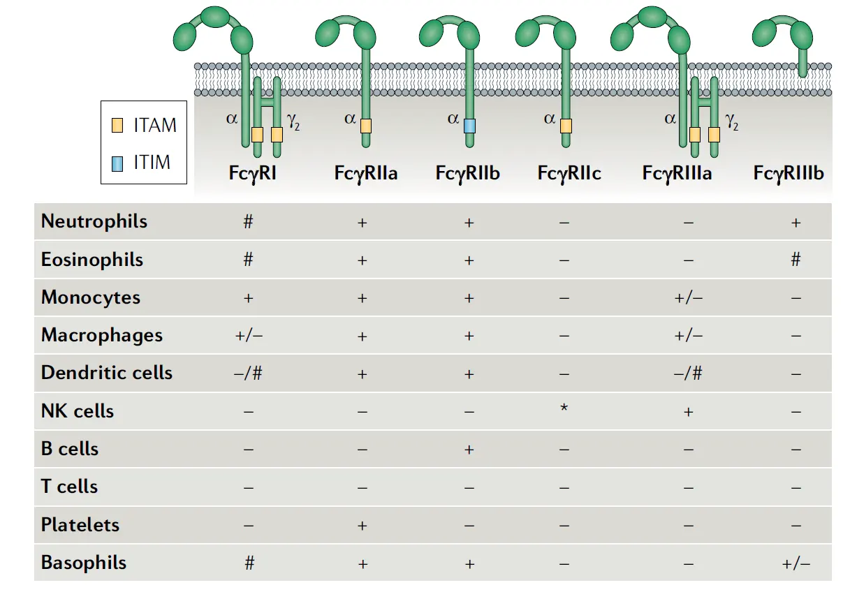
Figure 2. Major Types of Fcγ Receptors and Their Cellular Distribution (+, constitutive expression; –, no expression; #, inducible expression)
II. The Rationale for Fc Receptor Blocking
Under physiological conditions, the binding of Fc receptors to the antibody Fc region is a fundamental mechanism utilized by the immune system for pathogen clearance; however, in the context of flow cytometry assays, this interaction introduces a significant source of experimental artifact.
1. Mechanisms of Fc Receptor-Mediated Non-Specific Binding
In cells expressing Fc receptors, the antibody's Fc region can be recognized and bound by surface Fc receptors irrespective of the antigen specificity of the Fab region (Figure 3). This binding occurs independently of specific antigen-antibody recognition, resulting in the non-specific adherence of antibodies to the cell surface and the subsequent generation of extraneous signals.
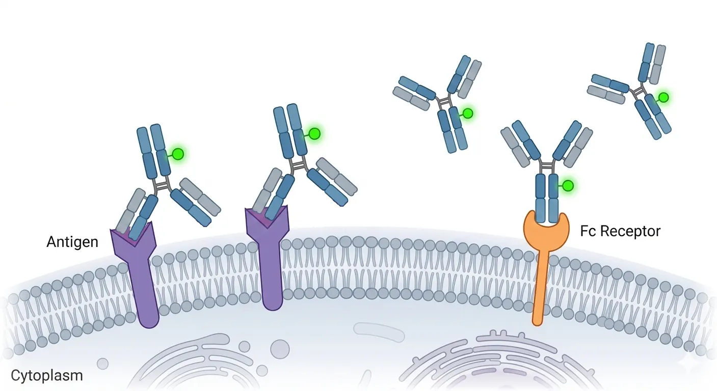
Figure 3. Specific Antigen Binding versus Fc Receptor-Mediated Non-Specific Bindin
2. Experimental Consequences of Omitting Fc Blocking
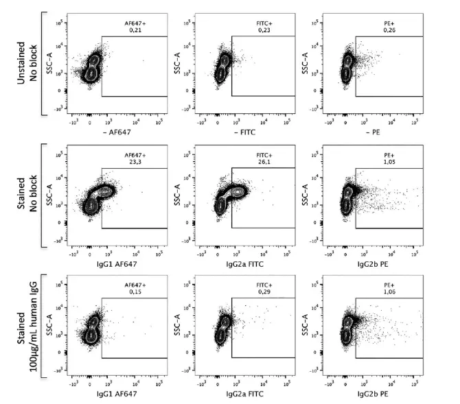
Figure 4. Comparative Analysis of Results With and Without Fc Receptor Blocking
Such non-specific binding exerts multiple deleterious effects on experimental outcomes:
● False-Positive Staining: As the most direct consequence, cells lacking target antigen expression may be erroneously classified as positive due to Fc receptor-mediated binding of the antibody's Fc region, thereby confounding phenotypic characterization (as illustrated in Figure 4).
● Elevated Background Fluorescence and Diminished Signal-to-Noise Ratio: Non-specific binding markedly amplifies overarching background signals (Figure 5), precipitating a reduction in the signal-to-noise ratio. Consequently, authentic signals from low-abundance antigens are masked by background noise, precluding effective detection.
● Obfuscated Cell Populations: In the context of immune cell subset analysis, non-specific background noise blurs the demarcations between distinct cell subpopulations. This prevents precise population delineation and subsequently impedes rigorous gating and quantitative analysis.
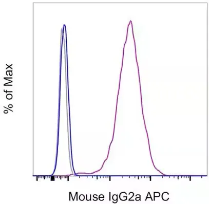
Figure 5. Cells lacking Fc receptor blocking exhibit pronounced false-positive signals (purple line), whereas the implementation of Fc receptor blocking effectively abolishes these signals (blue line). The gray line designates the autofluorescence of unblocked and unstained cells.
III. Distinguishing and Strategizing Between Isotype Controls, Live/dead Staining, and Fc Blocking live/dead
While Fc receptor blocking is a principal intervention for eliminating non-specific antibody binding, researchers in applied laboratory settings occasionally conflate its utility with that of isotype controls and live/dead staining, or incorrectly postulate that the three are interchangeable. Such cognitive discrepancies not only induce critical deficiencies in experimental design but also facilitate the misinterpretation of data.
1. Core Functions and Methodological Roles
| Experimental Component |
Primary Function |
Mechanism of Action |
Methodological Role |
| FcR Blocking |
Eliminates Fc receptor-mediated non-specific binding. |
Employs blocking reagents to pre-saturate cell surface Fc receptors, precluding the binding of Fc regions on subsequently applied staining antibodies. |
Proactive pre-treatment intervention; eradicates interference at its origin. |
| Isotype Control |
Evaluates the baseline level of non-specific binding. |
Utilizes an antibody matched in isotype and fluorochrome to the detection antibody, but devoid of target specificity, to assess the aggregate background of the assay system. |
Experimental control; facilitates gating and background evaluation but lacks the capacity to eliminate interference. |
| live/dead staining |
Differentiates live from dead cells to exclude dead-cell-derived artifacts. |
Leverages fluorochromes impermeable to intact membranes to label dead cells, enabling their exclusion during analysis. |
Dead cells inherently possess an elevated propensity for non-specific adsorption, substantially confounding results. |
2. Application Strategies in Experimental Design
These three components are not mutually exclusive; rather, their concurrent application represents the gold standard for acquiring precise data in the majority of immune cell-oriented flow cytometry assays:
● Pre-treatment Phase: Initially execute Fc receptor blocking to proactively negate Fc receptor-mediated non-specific binding, thereby attenuating background interference at its source.
● Staining Phase: Introduce live/dead dyes to label dead cells, facilitating their exclusion during downstream analysis. The non-specific adsorption capacity of dead cells substantially exceeds that of viable cells, constituting another major origin of background noise.
● Control Implementation: Concurrently incorporate isotype controls to aid in assessing non-specific background levels. This is particularly critical when interrogating low-expression antigens or rare cellular subsets, where isotype controls facilitate accurate positive/negative gating.
IV. Frequently Asked Questions (FAQ)
1. Which experiment necessitate Fc receptor blocking?
Fc receptor-mediated non-specific antibody binding is not a confounding factor exclusive to flow cytometry; it represents a ubiquitous source of central background interference across all immunological assays predicated on specific antibody recognition. Whenever an assay system simultaneously incorporates biological samples expressing Fc receptors (e.g., intact cells, tissue sections) and antibody-based detection reagents, a potential risk for Fc receptor-mediated non-specific binding exists, thereby mandating the consideration of Fc receptor blocking.。
2. Which cell types require Fc receptor blocking?
The primary criterion for ascertaining the necessity of Fc receptor blocking is the determination of whether the target cells express Fc receptors. The subsequent table systematically delineates the primary categories and cellular distributions of Fc receptors across human leukocytes:
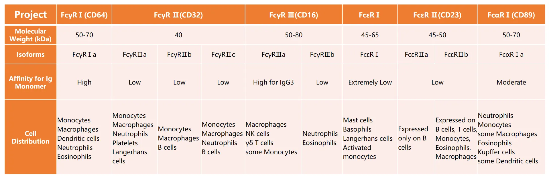
Figure 6. Comprehensive Summary of Fc Receptor Categories and Their Distribution on Human Leukocytes
As clearly evidenced in Figure 6, Fc receptors are not strictly confined to myeloid lineages; rather, they are ubiquitously distributed across virtually all leukocyte subpopulations. From monocytes/macrophages and neutrophils (which natively exhibit high FcγR expression) to B lymphocytes, natural killer (NK) cells, and even T lymphocytes (which manifest low baseline expression but robustly upregulate Fc receptors upon activation), varying typologies and expression levels of Fc receptors are prevalent. Furthermore, under inflammatory microenvironments or cellular activation states, non-immune cells—including epithelial cells, endothelial cells, and fibroblasts—can inducibly express Fc receptors, thereby exacerbating the risk of non-specific binding.
Consequently, to stringently abrogate Fc receptor-mediated non-specific antibody binding and safeguard the accuracy, stability, and reproducibility of experimental data, the integration of an Fc receptor blocking step is advocated, irrespective of the specific cellular composition of the sample.
Conclusion
Fc receptor-mediated non-specific binding constitutes a significant confounding artifact that compromises data fidelity in flow cytometry and a diverse array of antibody-based immunological assays. Fc receptor blocking is the definitive procedure to eradicate this interference,, optimize assay signal-to-noise ratios, and elevate result reproducibility, establishing it as an indispensable element of rigorous quality control in immunological research.
About Us
As a strategic venture of AtaGenix (established 2011), abinScience specializes in the development and production of high-quality life science reagents, and is committed to providing researchers with high-performance, highly reliable Flow Cytometry Antibodies, we offer:
1. Stringently validated flow cytometry antibodies including CD3, CD4, CD8 ensuring high sensitivity and minimal background noise;
2. A comprehensive portfolio of immunology research tools for multiple species, including Human, Mouse, Rat, Dog, Hamster, etc.;
3. Expert technical support and panel design consultation to help you accurately resolve complex immune cell populations.
For more information on abinScience flow cytometry antibodies, please visit:
abinScience Flow Cytometry Antibodies


 中文
中文 English
 한국어
한국어 日本語
日本語 Español
 Français
 Русский