
 中文
中文 English
 한국어
한국어 日本語
日本語 Español
 Français
 Русский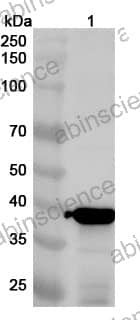
| Catalog No. | VK583014 | ||||||||
|---|---|---|---|---|---|---|---|---|---|
| Description |
Anti-PIV5 F/Fusion glycoprotein F0 Polyclonal Antibody (VK583014) is a rabbit polyclonal antibody detecting Fusion glycoprotein F0 in ELISA, IHC, WB.
Highlights
|
||||||||
| Species reactivity | Parainfluenza virus 5 (PIV5) | ||||||||
| Applications | ELISA, IHC, WB | ||||||||
| Host species | Rabbit | ||||||||
| Clonality | Polyclonal | ||||||||
| Isotype | IgG | ||||||||
| Immunogen | E. coli - derived recombinant Parainfluenza virus 5 (PIV5) F/Fusion glycoprotein F0 (Ala126-Ser487). | ||||||||
| Target | Fusion glycoprotein F0, Fusion glycoprotein F2, Fusion glycoprotein F1, F, Parainfluenza virus 5 (strain W3) (PIV5) (Simian virus 5) | ||||||||
| Purification | Purified by antigen affinity column. | ||||||||
| Accession | P04849 | ||||||||
| Form | Liquid | ||||||||
| Storage buffer | 0.01M PBS, pH 7.4, 50% Glycerol, 0.05% Proclin 300. Please refer to the specific buffer information in the hardcopy of datasheet or the lot-specific COA. |
||||||||
| Product Usage Information |
| ||||||||
| Stability and Storage | Use a manual defrost freezer and avoid repeated freeze thaw cycles. Store at 2 to 8°C for frequent use. Store at -20 to -80°C for twelve months from the date of receipt. | ||||||||
| Background | Fusion glycoprotein F0 is a ~56 kDa protein. Class I viral fusion protein. Under the current model, the protein has at least 3 conformational states: pre-fusion native state, pre-hairpin intermediate state, and post-fusion hairpin state. During viral and plasma cell membrane fusion, the heptad repeat (HR) regions assume a trimer-of-hairpins structure, positioning the fusion peptide in close proximity to the C-terminal region of the ectodomain. The formation of this structure appears to drive apposition and subsequent fusion of viral and plasma cell membranes. Directs fusion of viral and cellular membranes leading to delivery of the nucleocapsid into the cytoplasm. | ||||||||
| Note | For research use only. |
Western blot analysis was performed using anti-F-PIV5 polyclonal antibody at 1ug/mL on various samples.
Lane 1: recombinant PIV5 F/Fusion glycoprotein F0 (Catalog No: VK583012)
Contact us for custom quotes, bulk requests and any other issues.
Mail: [email protected]
+86-27-65523339
Building C, No. 666, Shen Dun Si Lu, Wuhan, 430206, China


