Acute ischemic stroke (AIS) accounts for approximately 76% of all stroke cases and remains a leading cause of long-term disability and death worldwide. While mechanical thrombectomy and intravenous thrombolysis can restore blood flow, around 40% of patients still experience residual disability after successful recanalization. The underlying culprit is cerebral ischemia-reperfusion injury (CIRI) — a condition driven by a self-amplifying cycle of oxidative stress and neuroinflammation in which excess reactive oxygen species (ROS) directly damage cells while triggering inflammatory cascades, ultimately causing irreversible neuronal loss. Multi-target agents with both antioxidant and anti-inflammatory activity hold particular therapeutic promise, yet conventional drug delivery struggles to cross the blood-brain barrier (BBB) and accumulate at the ischemic site — a bottleneck that has long remained unsolved.
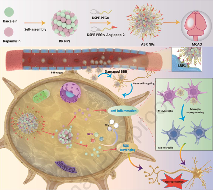
In recent years, carrier-free molecular self-assembled nanomedicines have attracted growing attention for their exceptionally high drug-loading capacity (exceeding 70 wt%) without the need for additional excipients, making them a viable platform for multi-target synergistic delivery. A research team from Army Medical University, publishing in Materials Today Bio, co-assembled a natural flavonoid from the traditional Chinese medicine herb Scutellaria baicalensis with an established immunosuppressant into a single nanoparticle platform. By further decorating the surface with a targeting ligand, they enabled the nanoparticles to traverse the BBB and deliver simultaneous antioxidant and anti-inflammatory neuroprotection.
~77% w/wTotal drug loading — far exceeding conventional nanocarriers (typically <20%)
37.88% → 7.84%Cerebral infarct volume in MCAO mice: saline group vs. ABR NPs group
18.69× → 2.59×Intracellular ROS fluorescence intensity in OGD/R cells: before vs. after ABR NPs treatment
01Assembly of Baicalein–Rapamycin Self-Assembled Nanoparticles
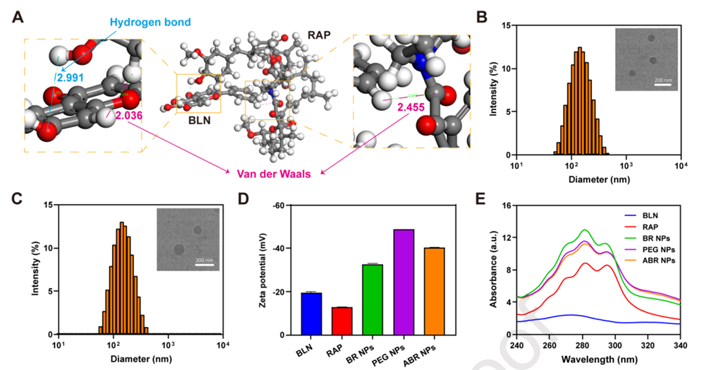
The study began with two pharmacologically distinct molecules: baicalein (BLN), a natural flavonoid free-radical scavenger extracted from the Chinese medicinal herb Scutellaria baicalensis, and rapamycin (RAP), a well-established mTOR inhibitor and immunosuppressant. Researchers systematically evaluated self-assembly at various molar ratios and identified 1:1 as the optimal formulation (particle size 134 nm, PDI 0.19 — the best across all tested ratios), designated BR NPs. Molecular dynamics simulations confirmed that BLN and RAP form stable nanoaggregates through hydrogen bonding and van der Waals interactions, while TEM imaging revealed a regular spherical morphology. The nanoparticles were subsequently surface-modified with DSPE-PEG2k-Angiopep-2 to yield ABR NPs (particle size 138.5 nm, zeta potential −40.53 mV, Figure 1C–D) — endowed with the ability to cross the BBB and actively target neurons. Total drug loading reached approximately 77% w/w, substantially higher than that of conventional nanocarriers (typically <20%).
02PEGylation Is Essential for Colloidal Stability in Physiological Media
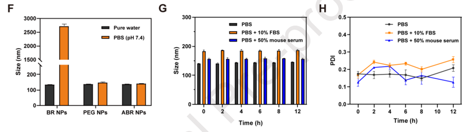
Unmodified BR NPs exhibited significant aggregation within 30 minutes of incubation in PBS, whereas PEG NPs and ABR NPs both maintained stable size distributions under the same conditions — demonstrating that PEGylation is critical for preserving colloidal stability in physiological saline. ABR NPs were further incubated at 37°C for 12 hours in PBS, PBS containing 10% FBS, and PBS containing 50% mouse serum; neither particle size nor PDI showed significant fluctuation across any condition, indicating that the platform can maintain its structural integrity under conditions that more closely approximate the in vivo environment.
03Angiopep-2 Modification Enhances Cellular Uptake Efficiency
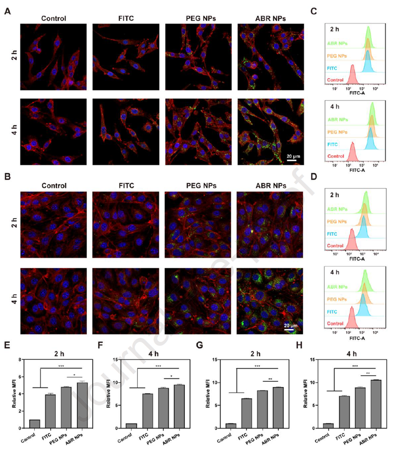
Researchers first established a safe concentration window for ABR NPs in three cell lines — PC12 (neuron-like cells), BV2 (microglia), and bEnd.3 (cerebrovascular endothelial cells) — identifying 10 μM as the uniform working concentration for subsequent experiments. At this concentration, confocal laser scanning microscopy (CLSM) and flow cytometry (FCM) revealed that ABR NPs produced markedly stronger fluorescence uptake signals in both PC12 and bEnd.3 cells compared with untargeted PEG NPs, at both 2- and 4-hour time points — indicating that Angiopep-2 modification genuinely enhances nanoparticle internalization by these two cell types. Mechanistic studies showed that uptake was substantially reduced at 4°C, confirming that the internalization process is energy-dependent — consistent with receptor-mediated endocytosis. The specific receptor responsible was ultimately identified using an LRP1 inhibitor supplied by abinScience.
04Dual Validation via In Vitro BBB Model and In Vivo Imaging
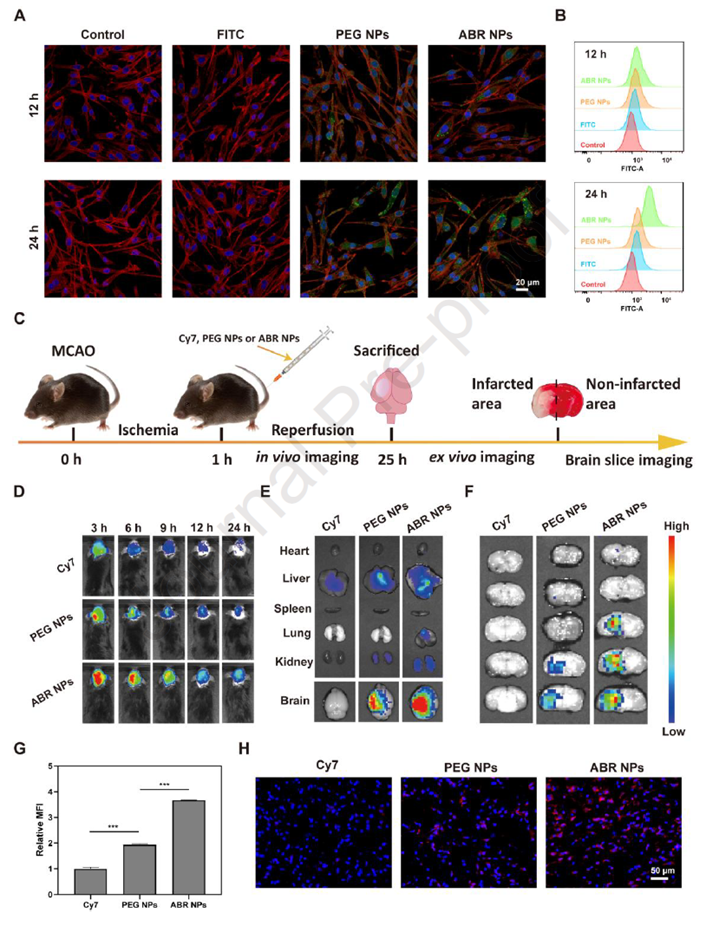
Researchers constructed an in vitro blood-brain barrier model using a bEnd.3 monolayer, with PC12 cells placed in the lower chamber to simulate brain parenchyma. At both 12 and 24 hours, the fluorescence intensity transported to the lower chamber and internalized by PC12 cells was significantly higher in the ABR NPs group than in the free FITC or PEG NPs groups. In a more pathophysiologically relevant setting — the middle cerebral artery occlusion (MCAO) mouse model — real-time brain fluorescence distribution was monitored at 3, 6, 9, 12, and 24 hours via an IVIS in vivo imaging system. ABR NPs consistently produced significantly stronger brain signals than PEG NPs throughout the observation period, while free Cy7 was rapidly cleared from systemic circulation. Ex vivo brain slice imaging further demonstrated that ABR NPs predominantly accumulated in the ischemic hemisphere, with minimal signal detected contralaterally; accumulation intensity was also markedly greater than that of PEG NPs. Confocal imaging of cryosectioned brain tissue corroborated this targeted distribution. Taken together, these findings establish that Angiopep-2 modification confers active BBB-crossing capability and ischemic lesion-targeting, rather than relying solely on the passive retention afforded by PEGylation.
05Antioxidant Efficacy Validated in Cell-Free Systems and at the Cellular Level
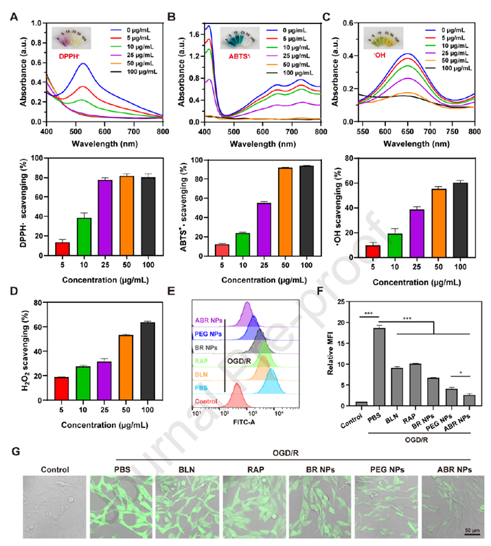
In cell-free chemical assays, ABR NPs demonstrated dose-dependent scavenging activity against DPPH radicals, ABTS radicals, hydrogen peroxide, and hydroxyl radicals. At the cellular level, researchers employed an oxygen-glucose deprivation/reoxygenation (OGD/R) model to simulate ischemia-reperfusion, using the DCFH-DA fluorescent probe to quantify intracellular ROS levels in PC12 cells. Flow cytometry showed that OGD/R treatment elevated intracellular ROS fluorescence intensity to 18.69-fold that of controls — confirming successful model induction — while ABR NPs treatment reduced this figure to 2.59-fold, outperforming PEG NPs (4.18-fold) and non-targeted BR NPs (6.76-fold). CLSM observations were fully consistent with the flow cytometry data. These results demonstrate that ABR NPs not only possess chemical antioxidant activity but can also efficiently neutralize excess ROS generated by ischemia-reperfusion at the intracellular level. Biosafety evaluation confirmed a hemolysis rate well below the 5% safety threshold, and a 30-day in vivo toxicity study revealed no abnormalities in blood counts, hepatic or renal function, or histology of major organs — indicating that the nanoplatform carries a favorable safety profile.
06Therapeutic Efficacy in the MCAO Model
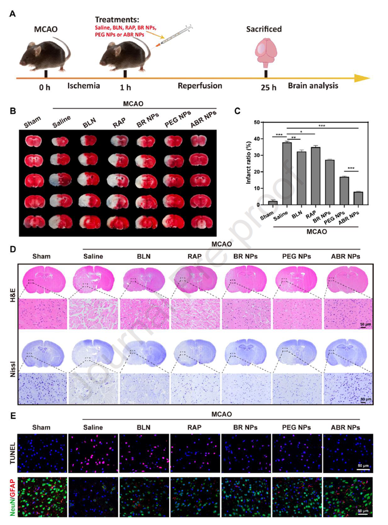
At the cellular level, the late apoptosis rate of OGD/R-challenged PC12 cells dropped from 21.2% in controls to 13.1% following ABR NPs treatment. More strikingly, ABR NPs demonstrated the most potent neuroprotective effect in the MCAO mouse model. At 24 hours after intravenous administration post-reperfusion, TTC staining revealed a cerebral infarct volume of 37.88% in the saline group, which was dramatically reduced to 7.84% in the ABR NPs group — significantly outperforming PEG NPs (16.87%), BR NPs (27.14%), and all other control groups. Zea-Longa neurological deficit scores also showed marked functional improvement in ABR NPs-treated mice. Histopathological analyses provided further corroboration: H&E staining showed largely preserved neuronal architecture in the ABR NPs group; Nissl staining revealed substantially reduced disorganization and morphological disruption; TUNEL staining demonstrated a marked decrease in apoptotic cell numbers; and NeuN staining confirmed a higher density of surviving neurons compared with all other treatment groups.
07Dual-Pathway Mechanism: ROS Scavenging and Microglial M1/M2 Polarization Modulation
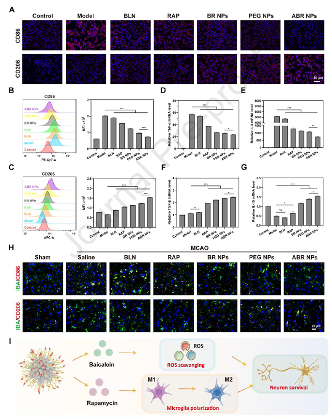
To elucidate the anti-inflammatory mechanism, researchers established an LPS-induced BV2 microglial inflammation model and confirmed that RAP-containing formulations exert anti-inflammatory effects by suppressing mTOR phosphorylation. After ABR NPs treatment, pro-inflammatory markers (CD86, TNF-α, IL-6) were downregulated while anti-inflammatory markers (CD206, IL-10, TGF-β) were upregulated; consistent in vivo results confirmed that ABR NPs promote the transition of microglia from the pro-inflammatory M1 phenotype to the anti-inflammatory M2 phenotype. Taken together, ABR NPs achieve synergistic neuroprotection against ischemia-reperfusion injury through a dual-pathway mechanism — baicalein-mediated ROS scavenging combined with rapamycin-driven microglial polarization modulation — delivered to the ischemic lesion via Angiopep-2-mediated BBB transcytosis.
Experimental Support from abinScience
In the experiments designed to elucidate the targeting mechanism of ABR NPs, the research team used an LRP1 (Low-Density Lipoprotein Receptor-Related Protein 1) inhibitor (Cat. No. HB705022) supplied by abinScience. LRP1 is the endogenous receptor for Angiopep-2, highly expressed on the surface of cerebrovascular endothelial cells and neurons, and serves as the key molecular target mediating Angiopep-2-directed transcytosis of nanoparticles across the BBB. PC12 and bEnd.3 cells were pre-treated with the abinScience LRP1 inhibitor prior to co-incubation with FITC-labeled nanoparticles, and uptake efficiency was evaluated by CLSM and FCM. The results showed that the LRP1 inhibitor had no appreciable effect on the uptake of free FITC or PEG NPs, but significantly suppressed internalization of ABR NPs — reducing it to levels comparable with PEG NPs. This confirms that LRP1 is the key mediator of ABR NPs internalization by neurons and cerebrovascular endothelial cells, providing functional validation that Angiopep-2 modification confers active targeting capability.
Reference
- Xianru Bi, Meng He, Wenxuan Cao, Li Xiang, Dong Liu, Liangbo Sun, Jiqin Lian, Xia Li, Lu Lu. Molecularly self-assembled nanoplatforms for targeted ROS-scavenging and neuroinflammation alleviation in acute ischemic stroke. Materials Today Bio, Volume 38, 2026, 103247, ISSN 2590-0064. https://doi.org/10.1016/j.mtbio.2026.103247
Looking for Research Tools for BBB Delivery or Ischemia-Reperfusion Injury Studies?
abinScience offers LRP1-related inhibitors and antibodies, ROS detection reagents (DPPH, ABTS, DCFH-DA, and more), and antibodies against microglial M1/M2 polarization markers (CD86, CD206, TNF-α, IL-6, IL-10, TGF-β) — supporting a wide range of experimental applications including cellular uptake assays, BBB transcytosis studies, and established ischemia-reperfusion models such as OGD/R and MCAO.
Email: [email protected] | Tel: +86-27-65523339


 中文
中文 English
 한국어
한국어 日本語
日本語 Español
 Français
 Русский