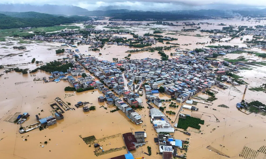
According to CCTV News, this year's Typhoon No. 10, Maysak, brought historic, widespread and sustained heavy rainfall to Guangxi. The Liulan Reservoir and Yunbiao Reservoir in Hengzhou, Nanning, along with the Liuwang Reservoir in Binyang County, experienced overtopping and breach incidents in quick succession, flooding multiple downstream villages and towns. As of now, 63 counties and districts across 14 prefecture-level cities in the region have been affected, with 6 deaths and 11 people missing — including 4 deaths and 8 missing in Hengzhou alone — and 130,000 people have been urgently evacuated and relocated.
Floodwaters surge in — and eventually recede. But for affected residents and rescue workers, the real health risks often begin only after the water goes down. Prolonged water immersion, sewage overflow, mosquito proliferation, and damaged sanitation infrastructure make the post-flood period a high-risk window for a range of infectious diseases. In this article, we take a research-oriented look at several infectious diseases that commonly surge after flooding, covering their pathogenic mechanisms, research frontiers, and key molecular targets.
Cholera
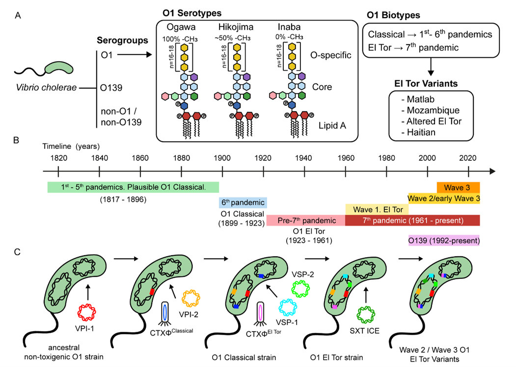
Cholera is caused by oral ingestion of Vibrio cholerae from contaminated water or food, and is one of the most typical "waterborne diseases" following flood disasters. It can cause profuse watery diarrhea and dehydration, with a relatively high case fatality rate among severe patients who do not receive timely treatment. After entering the small intestine, V. cholerae attaches to and colonizes the intestinal wall, releasing its core virulence factor — cholera toxin. This toxin binds to specific receptors on the surface of intestinal epithelial cells, enters the cells, and disrupts normal cell signaling, causing intestinal cells to secrete massive amounts of water and electrolytes — the direct cause of cholera's characteristic profuse watery diarrhea.
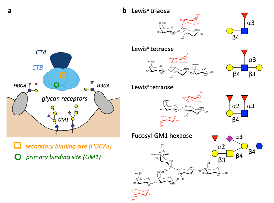
Recent studies have further elucidated the structural basis of the interaction between cholera toxin (CT) and host receptors. For example, researchers have solved a high-resolution crystal structure of the secondary binding site where the CT B subunit engages fucosylated receptors (such as the Lewis x antigen), providing a molecular basis for understanding differences in cholera susceptibility across blood groups. Other research has explored a "virulence-targeting" strategy, using dietary minerals such as zinc, selenium, and manganese to interfere with V. cholerae motility, adhesion, and toxin secretion — opening new avenues for treatment.
Leptospirosis
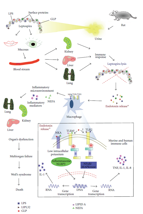
Leptospirosis is the quintessential "water-contact disease" of flood disasters. It is caused by pathogenic Leptospira, transmitted through broken skin or mucous membrane contact with water contaminated by animal urine. Severe cases can involve the liver and kidneys and are accompanied by a bleeding tendency, making this a disease that anyone with prolonged water exposure after a flood needs to watch closely. The surface of Leptospira carries a variety of proteins and lipopolysaccharide components that can be recognized by the human immune system, triggering inflammation and immune responses. Unlike many common bacteria, however, these surface components are not easily and fully recognized or cleared by the immune system, allowing the organism to persist in the body for extended periods — one reason the disease can progress to a chronic, relapsing infection.
Malaria
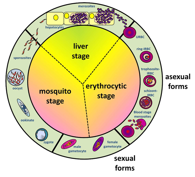
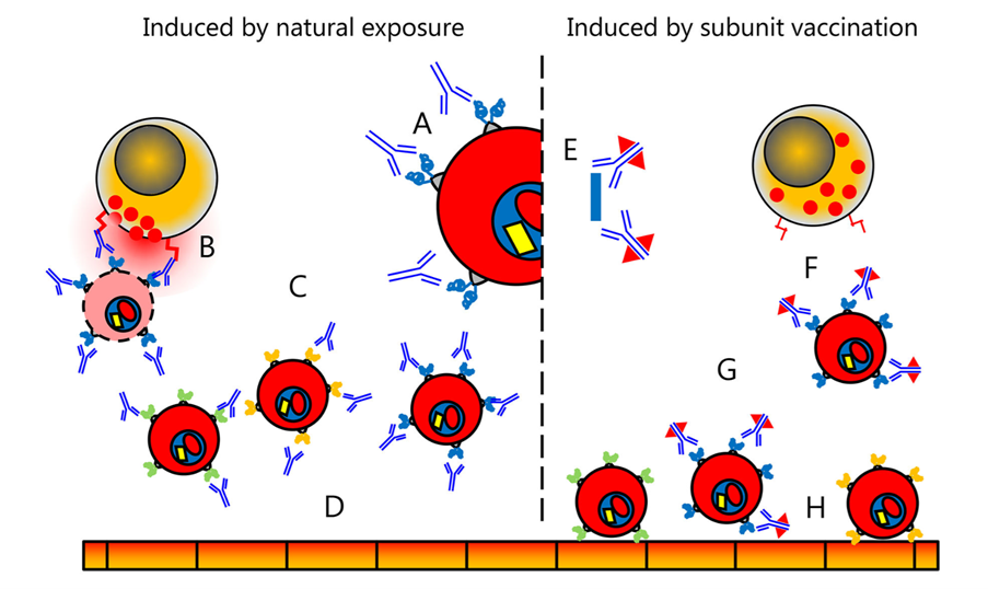
Malaria is caused by infection with Plasmodium parasites transmitted by Anopheles mosquitoes. Extensive standing water after flooding significantly extends the mosquito-breeding window, and historical data show that malaria incidence can rebound in some flood-affected regions — making it a mosquito-borne disease of particular concern in tropical and subtropical disaster areas. After Plasmodium infects red blood cells, it produces a specialized surface protein called PfEMP1 (Plasmodium falciparum erythrocyte membrane protein 1), which causes infected red blood cells to adhere to blood vessel walls. This adhesion can obstruct the microvasculature and impair local blood flow, a key driver of organ damage in severe malaria (particularly cerebral malaria) and also the primary target of the host's immune response.
Over the past decade, the field has accumulated substantial new knowledge on the structure of the PfEMP1 family, var gene regulation, and its organ-specific adhesion specificity, and vaccine development targeting PfEMP1 continues to advance. The latest research from 2025 has further focused on identifying the determinants of anti-PfEMP1 antibodies in acquired protective immunity against cerebral malaria, aiming to clarify which antibody features are truly associated with clinical protection. In addition, vaccines such as RTS,S/AS01 have already been rolled out at scale across multiple African countries, with early data showing a significant reduction in malaria-related hospitalizations and deaths among children under five.
Chikungunya (CHIKV)
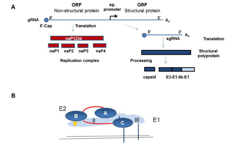
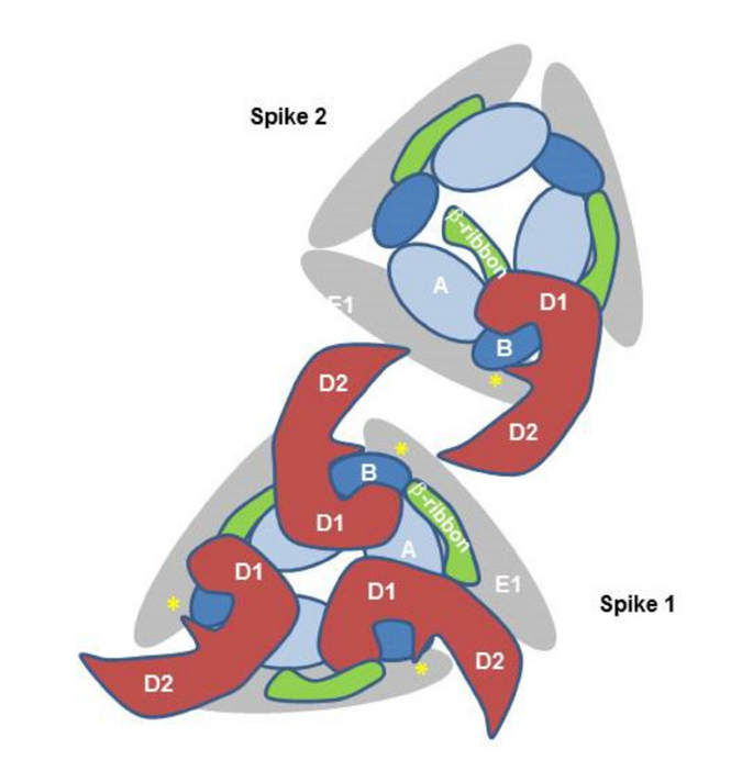
Chikungunya fever is caused by the Chikungunya virus (CHIKV), transmitted by Aedes mosquitoes. It is characterized mainly by fever and severe joint pain, with symptoms persisting for months in some patients. Since southern China is already a region with active Aedes mosquito populations, the increased standing water after flooding significantly widens the window for mosquito-borne transmission, making this a vector-borne viral disease that warrants close monitoring for weeks to months after a flood. CHIKV relies on a surface protein (E2) to recognize and bind to human cells and initiate invasion, and this protein is also the primary target of host antibody responses aimed at clearing the virus. At the same time, the process of viral entry activates inflammatory responses in immune cells, which is thought to be linked to the persistent joint pain seen after infection.
Research on neutralizing antibodies against the E2 protein has been a major focus in the CHIKV field in recent years. Studies have identified a linear epitope at the N-terminus of E2 (E2EP3, amino acids 1–12) as the primary target of early neutralizing IgG responses after infection, and recombinant antigens designed based on this epitope have been shown to induce partial protective immunity. Other groups have developed highly potent neutralizing IgM monoclonal antibodies targeting the E2 N218 epitope, which have demonstrated therapeutic potential in animal models. In addition, small-molecule inhibitors targeting the TLR4 pathway (such as TAK-242) have been shown to significantly reduce inflammatory responses and mortality in CHIKV-infected animal models, offering a new direction for antiviral strategies.
Clostridioides difficile Infection (CDI)
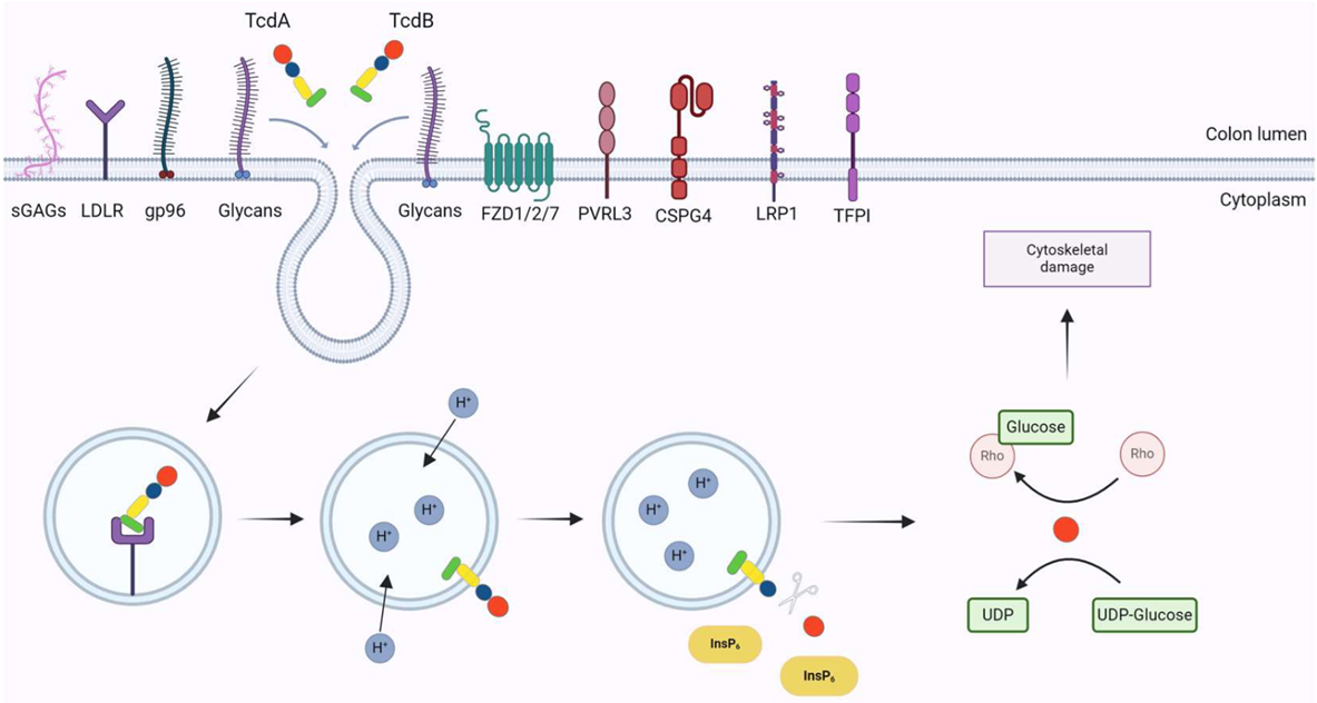
Clostridioides difficile infection is the leading cause of hospital-acquired, antibiotic-associated diarrhea. In settings of densely populated shelters, limited sanitation, and increased antibiotic use after a disaster, the risk rises considerably, with clinical presentations ranging from mild diarrhea to severe pseudomembranous colitis and toxic megacolon. The core of C. difficile pathogenesis lies in the two toxins it secretes — Toxin A and Toxin B. These toxins enter colonic epithelial cells and disrupt normal cell structure and intercellular junctions, ultimately damaging the intestinal barrier, triggering inflammation, and causing cell death — the direct cause of the diarrhea and colitis symptoms.
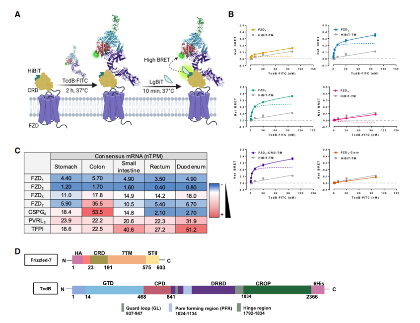
A 2024 cryo-EM study resolved the three-dimensional structure of the TcdB complex with its host receptors from the Frizzled family (FZD1/2/7), revealing the key structural rearrangements required for TcdB receptor recognition. The study also clarified how bezlotoxumab, currently the only approved anti-toxin monoclonal antibody, blocks the TcdB–FZD7 interaction by stabilizing TcdB in its non-binding conformation. In addition, chemical genetic screening has identified small molecules — such as glycyrrhetinic acid — that target virulence-associated metabolic pathways, including adenine deaminase and ATP synthase, to suppress toxin production, offering a new intervention point for "anti-virulence" strategies.
Reliable, Precise Research Tools Are Essential for Post-Disaster Infection Research
Although the pathogens behind these post-flood infectious diseases differ widely, the research pathway is remarkably consistent: resolve the structure of core virulence factors/surface antigens → identify key receptors and pathogenic pathways → develop specific antibodies and detection reagents → support diagnostics and vaccine/drug development. abinScience has long specialized in the R&D and production of research-grade antibodies, recombinant proteins, and detection kits, and offers relevant products for many of the targets discussed in this article to support the research community. Floodwaters recede, but public health vigilance cannot. We wish everyone in the affected areas a swift return to safety, and hope that the power of research can provide stronger support for post-disaster infectious disease control.
References
- Castillo-Macías A, Salinas-Carmona MC, Torres-Lopez E. Immunology of viral infections with a high impact in Mexico: Dengue, Chikungunya, and Zika. Medicina Universitaria. 2018;19:198-207.
- Lee WC, Russell B, Rénia L. Sticking for a cause: the falciparum malaria parasites cytoadherence paradigm. Front Immunol. 2019;10:1444. doi:10.3389/fimmu.2019.01444
- Mahish C, De S, Chatterjee S, Ghosh S, Keshry SS, Mukherjee T, Khamaru S, Tung KS, Subudhi BB, Chattopadhyay S, Chattopadhyay S. TLR4 is one of the receptors for Chikungunya virus envelope protein E2 and regulates virus induced pro-inflammatory responses in host macrophages. Front Immunol. 2023;14:1139808. doi:10.3389/fimmu.2023.1139808
- Montero DA, Vidal RM, Velasco J, George S, Lucero Y, Gómez LA, Carreño LJ, García-Betancourt R, O'Ryan M. Vibrio cholerae, classification, pathogenesis, immune response, and trends in vaccine development. Front Med. 2023;10:1155751. doi:10.3389/fmed.2023.1155751
- Kappagoda C, Senavirathna I, Agampodi T, Agampodi SB. Role of Toll-like receptor 2 during infection of Leptospira spp: A systematic review. PLoS One. 2024;19(12):e0312466. doi:10.1371/journal.pone.0312466
- Gonçalves-de-Albuquerque CF, Burth P, Silva AR, Younes-Ibrahim M, Castro-Faria-Neto HC, Castro-Faria MV. Leptospira and inflammation. Mediators Inflamm. 2012;2012:317950. doi:10.1155/2012/317950
- Pourliotopoulou E, Karampatakis T, Kachrimanidou M. Exploring the toxin-mediated mechanisms in Clostridioides difficile infection. Microorganisms. 2024;12(5):1004. doi:10.3390/microorganisms12051004
- Hviid L, Jensen AR, Deitsch KW. PfEMP1 and var genes — still of key importance in Plasmodium falciparum malaria pathogenesis and immunity. Adv Parasitol. 2024;125:53-103. doi:10.1016/bs.apar.2024.02.001
- Heim JB, Hodnik V, Heggelund JE, Anderluh G, Krengel U. Crystal structures of cholera toxin in complex with fucosylated receptors point to importance of secondary binding site. Sci Rep. 2019;9(1):12243. doi:10.1038/s41598-019-48579-2
- Schnierle BS. Cellular attachment and entry factors for chikungunya virus. Viruses. 2019;11(11):1078. doi:10.3390/v11111078


 中文
中文 English
 한국어
한국어 日本語
日本語 Español
 Français
 Русский