Adherent cells are common samples in flow cytometry experiment. Prior to analysis, cells must be detached from the culture surface and dispersed into a single‑cell suspension using enzymatic dissociation, non‑enzymatic dissociation, or mechanical harvesting. This process must ensure thorough cell dispersion while balancing cell viability and the retention of target surface antigens. Different dissociation methods have varying effects on cells: proteases may cleave certain surface proteins, mechanical action can cause cell membrane damage, and insufficient dissociation tends to leave residual cell aggregates. These changes can further affect antibody binding, staining background, and the cell populations ultimately included in the analysis. Therefore, the preparation of adherent cell samples for flow cytometry requires selecting appropriate dissociation methods and treatment conditions based on the adhesion characteristics of the cells and the target antigens.
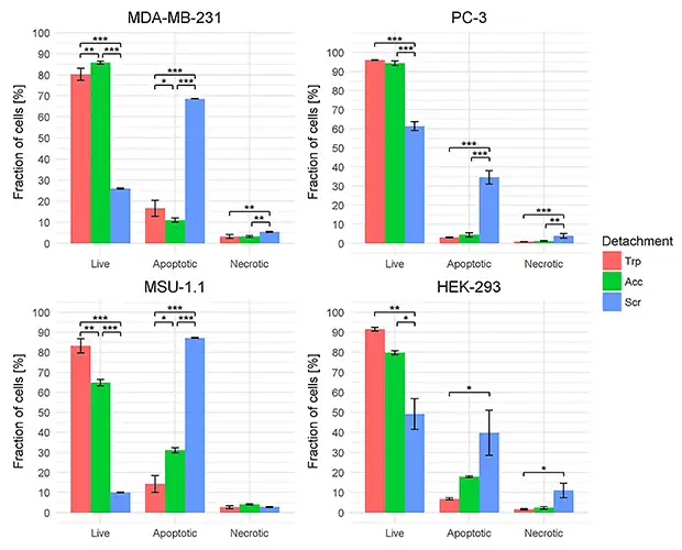
Figure 1. Effects of different dissociation methods on various cell lines (DOI: 10.1590/1414‑431X202010197).
I. Effects of Cell Dissociation on Surface Antigen Detection
1. Proteolytic Effects on Surface Epitopes and Antibody Recognition
Trypsin facilitates cell detachment from the culture surface by hydrolysing adhesion‑related proteins, but its action is not restricted to the junctional structures between cells and the culture surface. The extracellular domains of certain membrane proteins may also be cleaved, leading to a reduction or loss of epitopes recognisable by antibodies.
Consequently, a decrease in signal following dissociation may reflect a reduction in surface antigens recognisable by the specific antibody in use, rather than directly indicating an overall downregulation of intracellular target protein expression. The extent of this effect is also related to the target protein, the antibody recognition site, and the treatment conditions.
For example, in human synovial mesenchymal stem cells, trypsin treatment had a more pronounced effect on CD105 and CD73 staining, whereas CD90 staining remained relatively stable. This demonstrates that normal staining of one surface marker in a given sample is insufficient to prove that other targets remain unaffected.
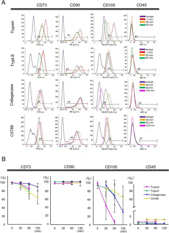
Figure 2. Trypsin treatment affects the detection of CD105 and CD73 in MSCs (DOI: 10.3727/096368917X694831).
2. Cell Damage, Background Staining and Analysed Cell Populations
Mechanical scraping avoids direct protease cleavage but may damage the cell membrane. Abnormal staining of damaged cells, an increase in cellular debris, and population shifts occurring during the subsequent exclusion of dead cells and aggregates can all influence results.
Studies comparing different harvesting methods have found that mechanical scraping is associated with more pronounced cell damage in certain cell models; a method that preserves target antigen signals may still introduce artefacts that affect the assessment of cell viability or apoptosis. Therefore, the evaluation of dissociation methods must simultaneously consider target staining and cellular state.
II. Common Dissociation Methods: Characteristics and Selection Considerations
Dissociation methods disrupt cell adhesion through different mechanisms and can vary in their effects on antigen preservation, cell viability and cell recovery.
| Dissociation Method |
Key Characteristics |
Considerations for Selection |
| Trypsin dissociation |
Promotes cell detachment via proteolysis; frequently used in combination with EDTA |
Sensitivity of the target antigen to treatment and the actual time required to complete harvesting |
| Other enzymatic dissociation reagents, such as Accutase and TrypLE |
Differ in enzyme composition or action characteristics; can serve as candidate alternatives |
Retention efficacy for the target antigen requires specific confirmation |
| EDTA and other non‑enzymatic dissociation buffers |
Weaken cell adhesion by chelating divalent cations, among other mechanisms |
Sufficiency of dissociation, and whether extended treatment or additional trituration is required |
| Mechanical scraping |
Collects cells via mechanical action |
Membrane damage, debris, aggregates, and viable cell recovery |
1. Selecting Enzymatic Dissociation Conditions for Target Antigens
Different surface antigens exhibit varying sensitivities to enzymatic treatment; therefore, when selecting a dissociation reagent, attention must be paid to its impact on the antigen of interest. Priority should be given to reviewing literature related to the target antigen and the specific antibody clone used, noting the dissociation reagents, concentrations, temperatures, and treatment times employed in previous assays as a basis for selecting conditions.
The ability of trypsin, Accutase or TrypLE to preserve a target antigen cannot be determined solely from the reagent name or descriptions such as "gentle". For example, although Accutase is commonly used for milder cell dissociation, it has also been found to reduce the detection levels of Fas and its ligand on the macrophage surface. Consequently, the suitability of a reagent for harvesting cells does not imply that it is appropriate for the detection of all surface antigens.
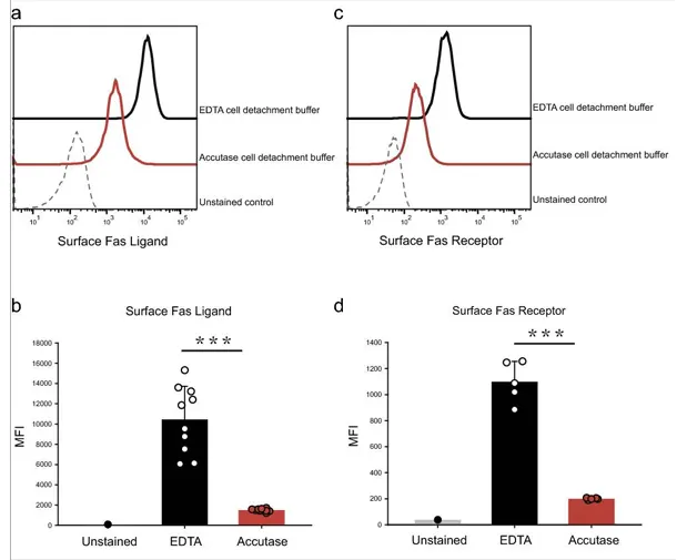
Figure 3. Effects of Accutase treatment on the surface detection of Fas receptor (Fas/CD95) and Fas ligand (FasL) in macrophages. (DOI: 10.1038/s41598‑022‑09605‑y).
When definitive applicability information is lacking, existing conditions can be compared with an alternative candidate dissociation condition, evaluated in conjunction with the target signal, staining background, and cell viability. If the current method adequately balances antigen detection and cell recovery, it may be continued; if the target signal is compromised, shortening the enzyme treatment time or switching the dissociation reagent can be attempted.
2. Antigen Retention and Cell Recovery with Non‑Enzymatic Dissociation
EDTA attenuates certain cell adhesion mechanisms by chelating divalent cations such as calcium and magnesium, thereby mitigating the effects caused by direct protease cleavage. However, for strongly adherent cells, non‑enzymatic methods may struggle to rapidly yield sufficient single cells.
If treatment is persistently prolonged or repeated pipetting is applied solely to harvest cells, the anticipated protective effects may be offset by other forms of damage. In comparisons involving synovial mesenchymal stem cells, Non‑enzymatic reagents required longer treatment times to achieve high cell recovery yields, and prolonged treatment also risked impacting viability. Therefore, method selection requires evaluating antigen retention alongside practical harvesting performance.
III. Assessing the Suitability of Dissociation Conditions
1. Selecting and Comparing Dissociation Conditions
Priority should be given to reviewing antibody datasheets, application notes, and relevant literature to confirm whether the cell model, dissociation reagents, and antibody clones are documented. The previous detection of a specific target on the same cell line can serve as a useful reference.
In the absence of definitive evidence, parallel cultures of cells from the same batch and with similar culture status can be harvested using various feasible dissociation conditions. Keeping the antibody clone, fluorochrome, amount of antibody used for staining, incubation conditions, and acquisition settings consistent, compare the differences in results obtained across the different dissociation methods.
2. Evaluating Antigen Detection and Sample Quality
Dissociation efficacy is assessed through positivity rate, fluorescence intensity, negative background, population resolution, viability, and the recovery of usable single cells.
| Observation |
Key Focus for Assessment |
| Viability and background are comparable, but target signals show consistent differences |
Indicates that dissociation conditions affect target detection; epitope sensitivity should be further investigated |
| Signal enhancement accompanied by increased dead cells and background |
First determine whether damage‑related aberrant staining is present |
| Target staining is satisfactory among viable cells, but overall cell recovery is markedly reduced. |
Examine whether only a fraction of easily detached cells was harvested, compromising sample representation |
| Target remains undetected after method replacement |
Continue verifying cell surface expression and antibody suitability; do not attribute the outcome solely to dissociation |
If another clone successfully detects the target under the same conditions, it can help determine whether recognition region differences exist; however, different clones also vary in affinity and labelling performance, precluding direct comparisons of epitope loss based solely on signal intensity.
3. Use and Limitations of Post‑Dissociation Recovery
Allowing cells to recover after dissociation may restore the surface expression of some proteins affected by the dissociation process, but the recovery process itself introduces new time variables. Particularly when comparing surface expression under conditions such as drug stimulation or inflammatory treatment, supplementary culture may alter the state intended to be captured. Therefore, whether to implement recovery culture should be determined by the assay objectives; once adopted, consistency must be maintained across all experimental groups.
In adherent cell flow cytometry, the dissociation method should be validated and recorded as an integral part of the assay conditions. Once an appropriate method is established, keeping reagents, treatment times, and subsequent staining protocols consistent will minimise variability introduced during the harvesting stage, allowing surface antigen signals to more accurately reflect the state of the cells being analysed.
References
- [1] Tsuji, K., Ojima, M., Otabe, K., Horie, M., Koga, H., Sekiya, I., & Muneta, T. (2017). Effects of Different Cell‑Detaching Methods on the Viability and Cell Surface Antigen Expression of Synovial Mesenchymal Stem Cells. Cell Transplantation, 26(6), 1089‑1102. https://doi.org/10.3727/096368917X694831
- [2] Nowak‑Terpiłowska, A., Śledziński, P., & Zeyland, J. (2021). Impact of cell harvesting methods on detection of cell surface proteins and apoptotic markers. Brazilian Journal of Medical and Biological Research, 54(2), e10197. https://doi.org/10.1590/1414‑431X202010197
- [3] Lai, T. Y., Cao, J., Ou‑Yang, P., Tsai, C. Y., Lin, C. W., Chen, C. C., Tsai, M. K., & Lee, C. Y. (2022). Different methods of detaching adherent cells and their effects on the cell surface expression of Fas receptor and Fas ligand. Scientific Reports, 12(1), 5713. https://doi.org/10.1038/s41598‑022‑09605‑y
Looking for Reliable Flow Cytometry Antibodies to Support your Experimental Workflows?
abinScience Flow Cytometry Antibodies are subjected to rigorous quality control to ensure consistent fluorescent performance and clear discrimination of cell populations, supporting robust and reproducible flow cytometry analyses.
Learn More about abinScience Flow Cytometry Antibodies
About Us
abinScience was founded in 2023 as a strategic venture of AtaGenix (established 2011), dedicated to delivering premium life science reagents that accelerate discovery.
abinScience flow cytometry antibody products cover commonly used detection markers, with a wide variety to meet the research needs of multiple species(Human, Mouse, Rat, Dog, Hamster, Monkey, etc.)We provide stable and reliable support for scientific research.



 中文
 English
 한국어
 日本語
 Español
 Français
 Русский


